
Tissue formation: how cells generate forces
When an organism develops, masses of cells combine to form different types of tissue, all of which have different functions. In order to be able to form and to move, a cell needs to generate mechanical forces by remodelling its cytoskeleton, which consists of various filaments. Filaments from the actin protein, for example, contract and expand. However, not much is currently known about the mechanical forces which are produced by other components of the cytoskeleton. To change this, researchers from Münster and Munich took a closer look at the microtubules, which are tubular filaments that form part of the cytoskeleton. The scientists used genetic, chemical and microscopic methods to show for the first time in a living organism that microtubules play a central role in the cell mechanics and also found out more about the molecular mechanisms that control them. “Our results show how microtubules contribute to collective cell behaviour during tissue morphogenesis,” says biologist Dr. Maja Matis, a group leader at the Cells-in-Motion Cluster of Excellence at Münster University and head of the study. The insights gained could in future help to provide a better understanding not only of the development of organisms, but also of faulty developments. The study has been published in the journal “Nature Cell Biology”.
The story in detail:
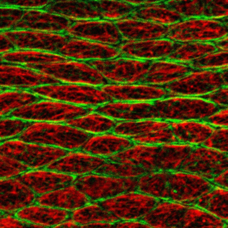
The researchers took a look at the growth process in fruit flies and investigated how the wing epithelium develops. Using a laser beam, the researchers isolated individual epithelial cells from their surroundings to be able to study their mechanical properties during early wing development. As a result, they discovered that at this stage, every single cell appears to be mechanically autonomous and not influenced by external forces. The researchers observed that even without the contact to their immediate surroundings, the cells retained their form. It appeared to be the microtubules which stabilized the cells. They are localized close to the apical surface of the cell, i.e. on the side facing the external surroundings. “We reasoned that this polarized alignment of microtubules could exert compressive forces and counteract the tension in the cell which arises when actin and myosin filaments contract”, says Amrita Singh, a PhD student at the Graduate School of the Cluster of Excellence and the lead author of the study.
The next thing the researchers did was to observe the labelled microtubules in a living organism, using high-resolution microscopy. They found that the microtubules in cells are not static but dynamic. They grow until they reach the cell cortex close to the inside of the cell membrane where they bend. Using a laser beam, the researchers cut individual microtubules and were able to observe how they very quickly lost their curved shape and straightened up – meaning that the microtubules can indeed generate compressive forces. When the researchers inhibited the growth of microtubules using inhibitory drugs, the reverse happened: the cells shortened. “This means that the forces generated by the microtubules are the principal drivers of the initial cell changes during the early phase of tissue development,” says Maja Matis.
By studying genetically modified fruit flies, the researchers also found out more about the molecular mechanisms, which lead to the coordination of forces over the entire tissue. During this process, the so-called Fat Planar Cell Polarity signalling pathway (Ft-PCP) causes the microtubules to arrange themselves in a specific pattern by adhering to the sites of cells where two neighbouring cells form a junction. This leads into the same microtubule pattern in all cells of the tissue causing that the forces produced by microtubules are patterned and as a result the cells jointly form the tissue.
As the Ft-PCP signalling pathway plays a role in numerous processes during the development of organisms, these results could in future help to learn more about disease caused by mutations in the Ft-PCP signalling pathway – for example, neural tube closure, kidney diseases, deafness and skeletal abnormalities.
Funding:
The study received financial support from the Cells-in-Motion Cluster of Excellence at the University of Münster, as well as from the Interdisciplinary Centre for Clinical Research at the University of Münster and from the Priority Programme “Epithelial intercellular junctions as dynamic hubs to integrate forces, signals and cell behaviour” being funded by the German Research Foundation.
Original publication:
Singh A, Saha T, Begemann I, Ricker A, Nüsse H, Thorn-Seshold O, Klingauf J, Galic M, Matis M. Polarized microtubule dynamics directs cell mechanics and coordinates forces during epithelial morphogenesis. Nat Cell Biol. 2018; 20: 1126–1133. Abstract