© CiM - Peter Grewer

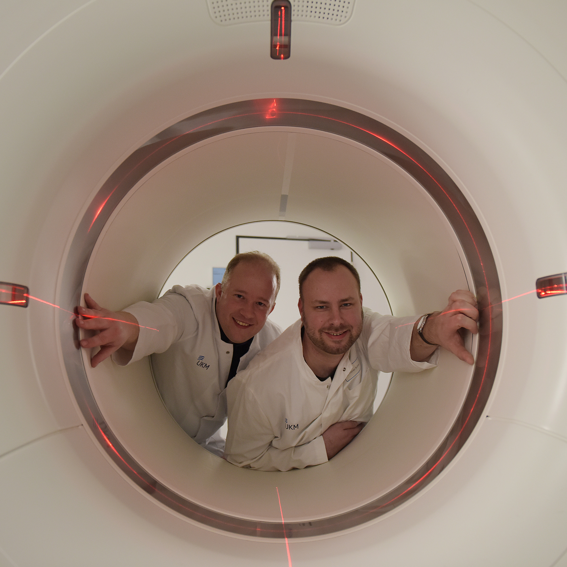
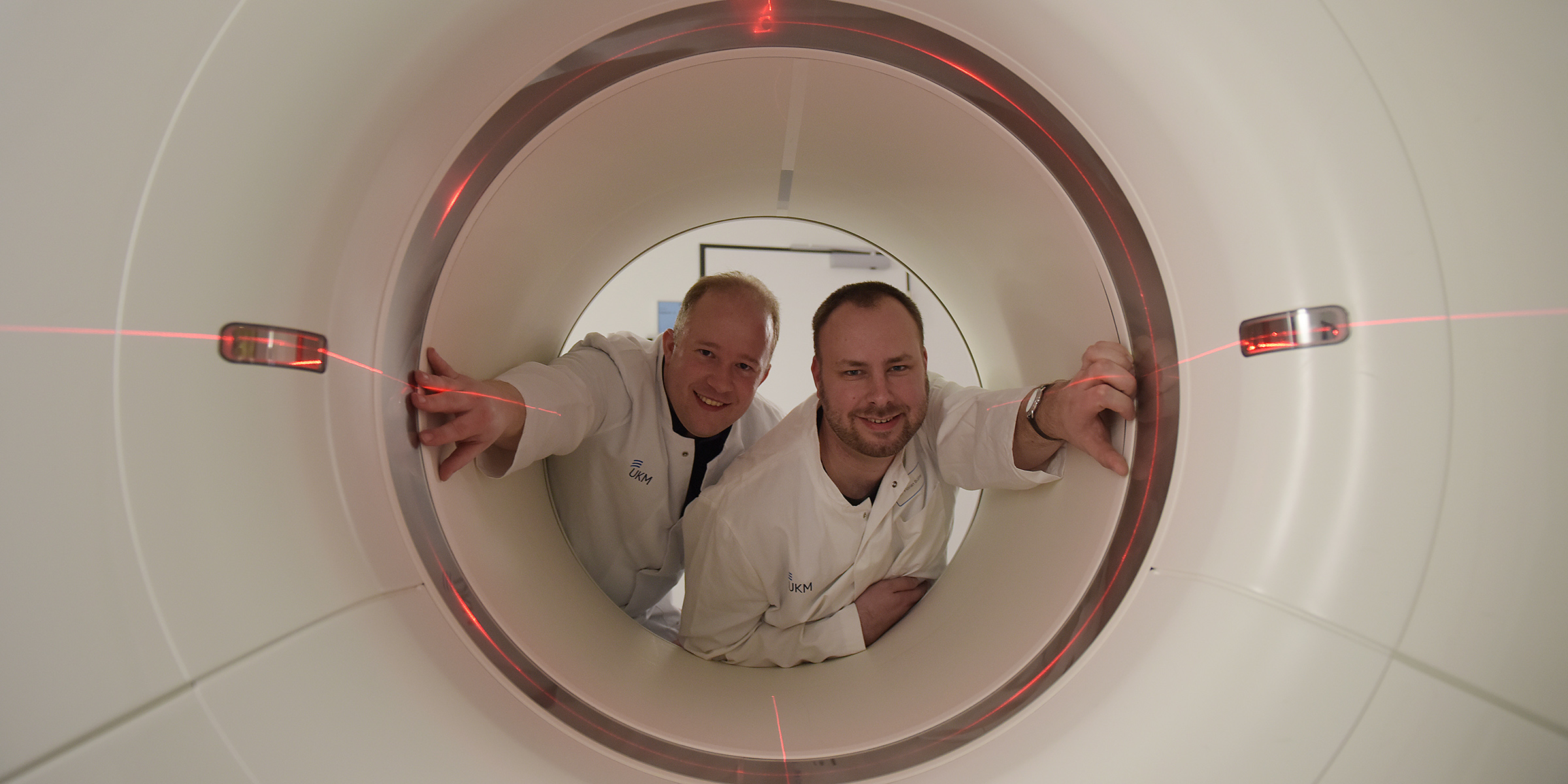
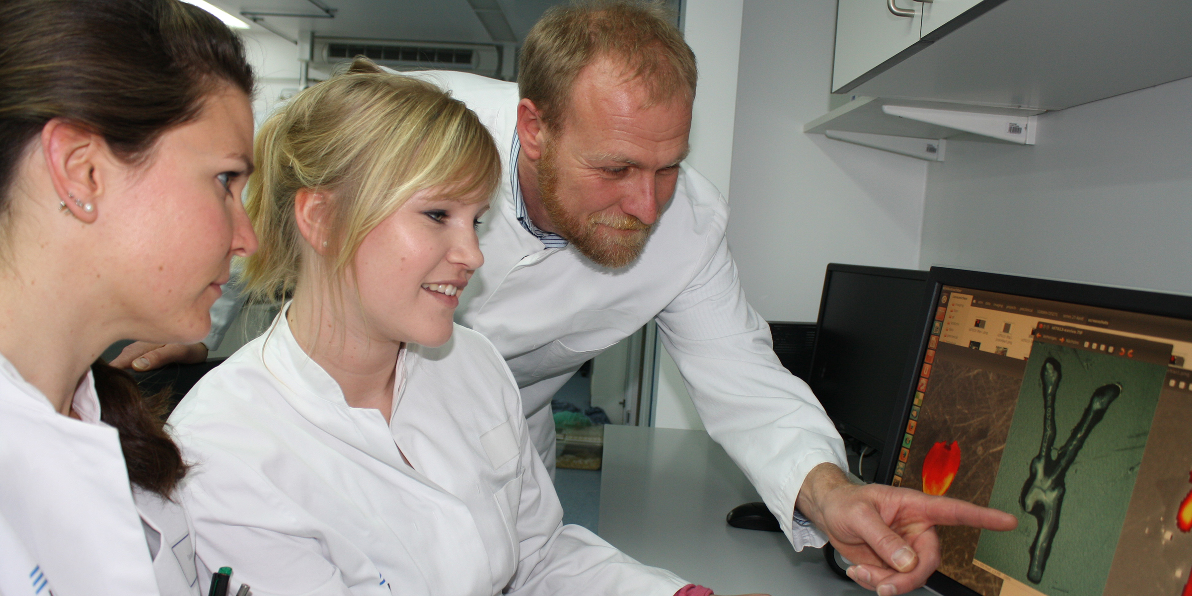
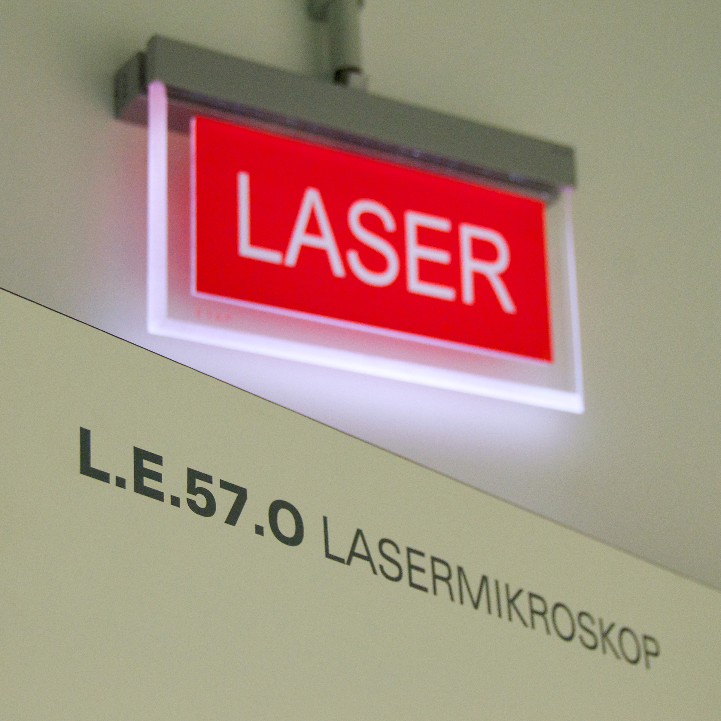
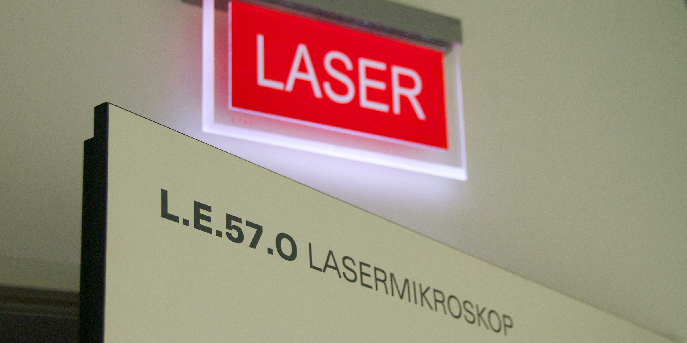
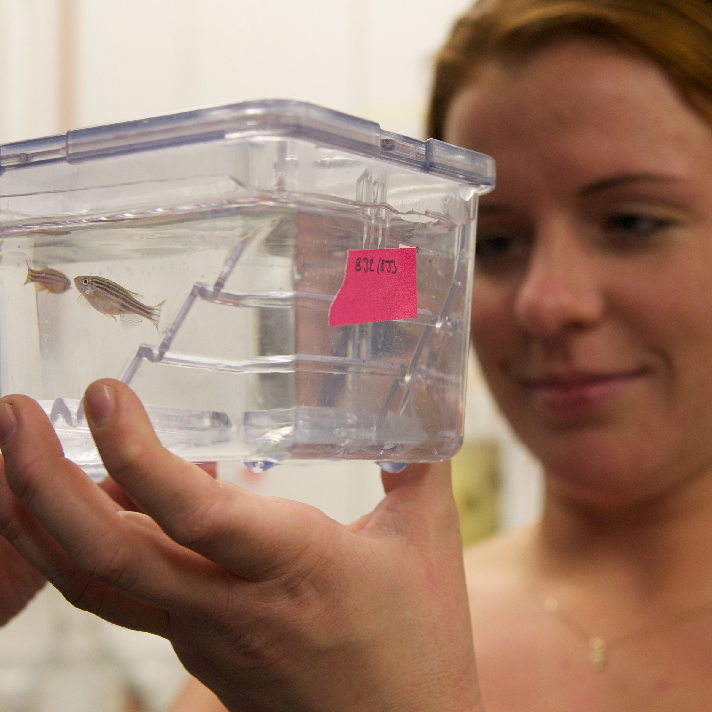
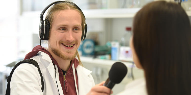
Science to Listen to!

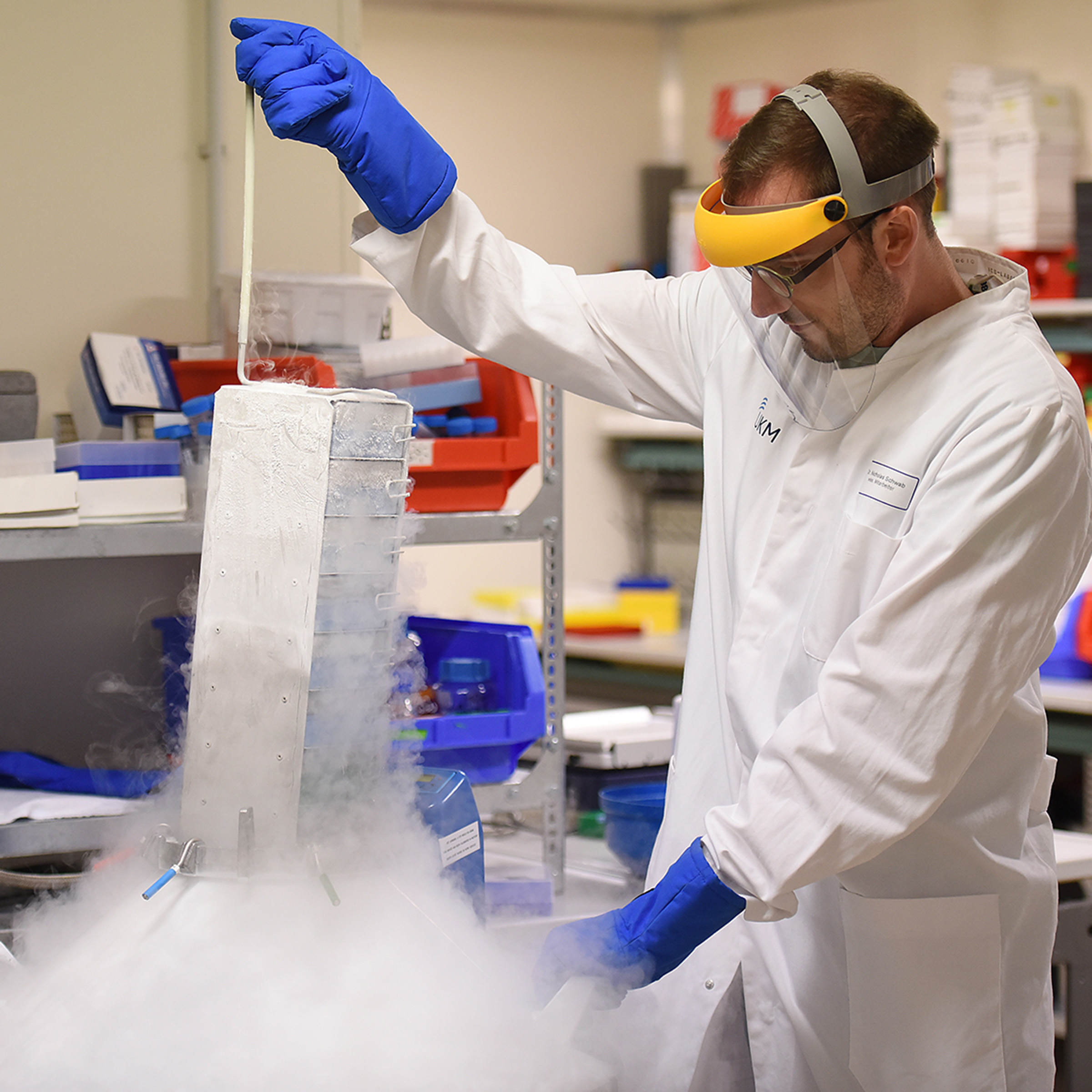
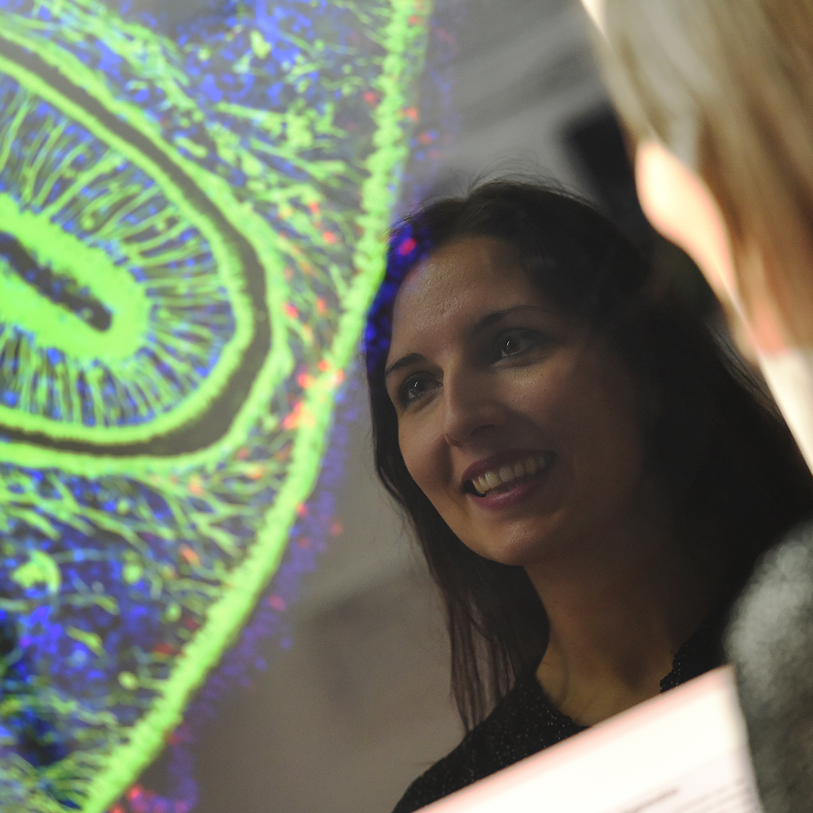
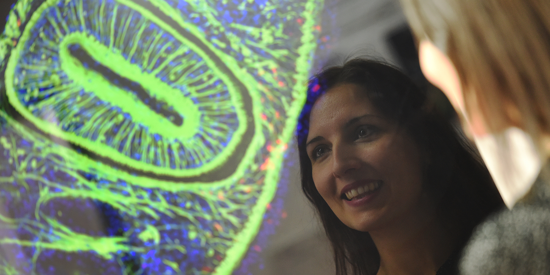
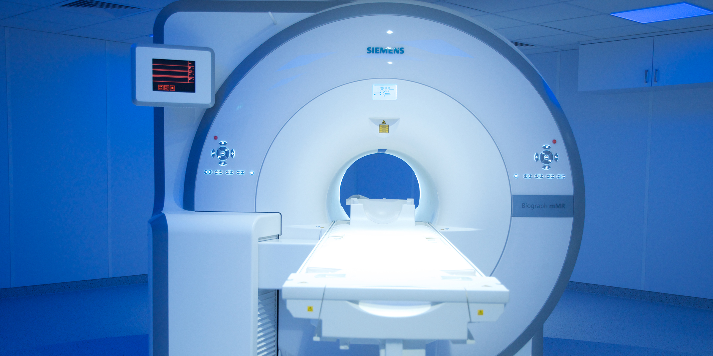
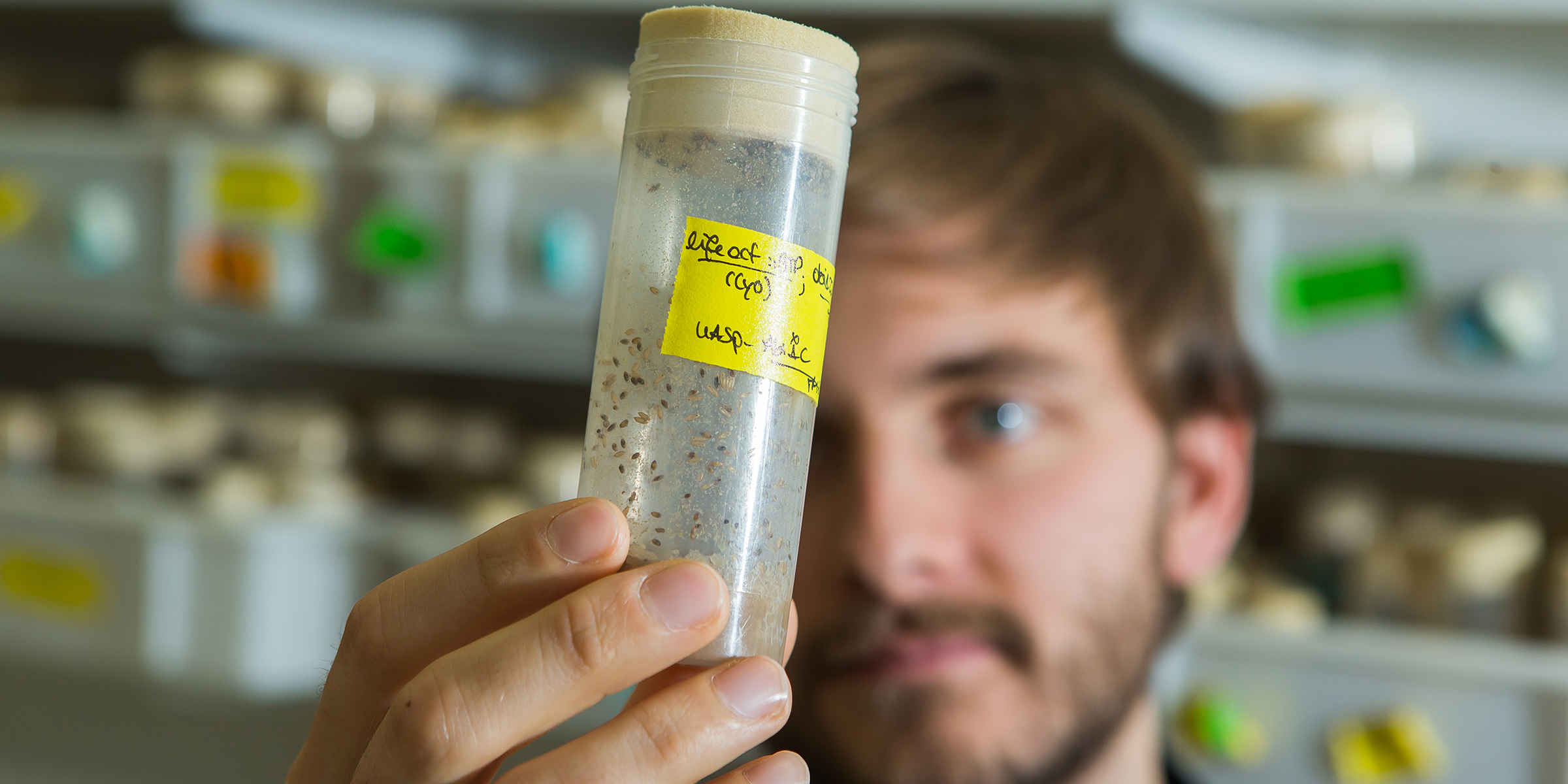
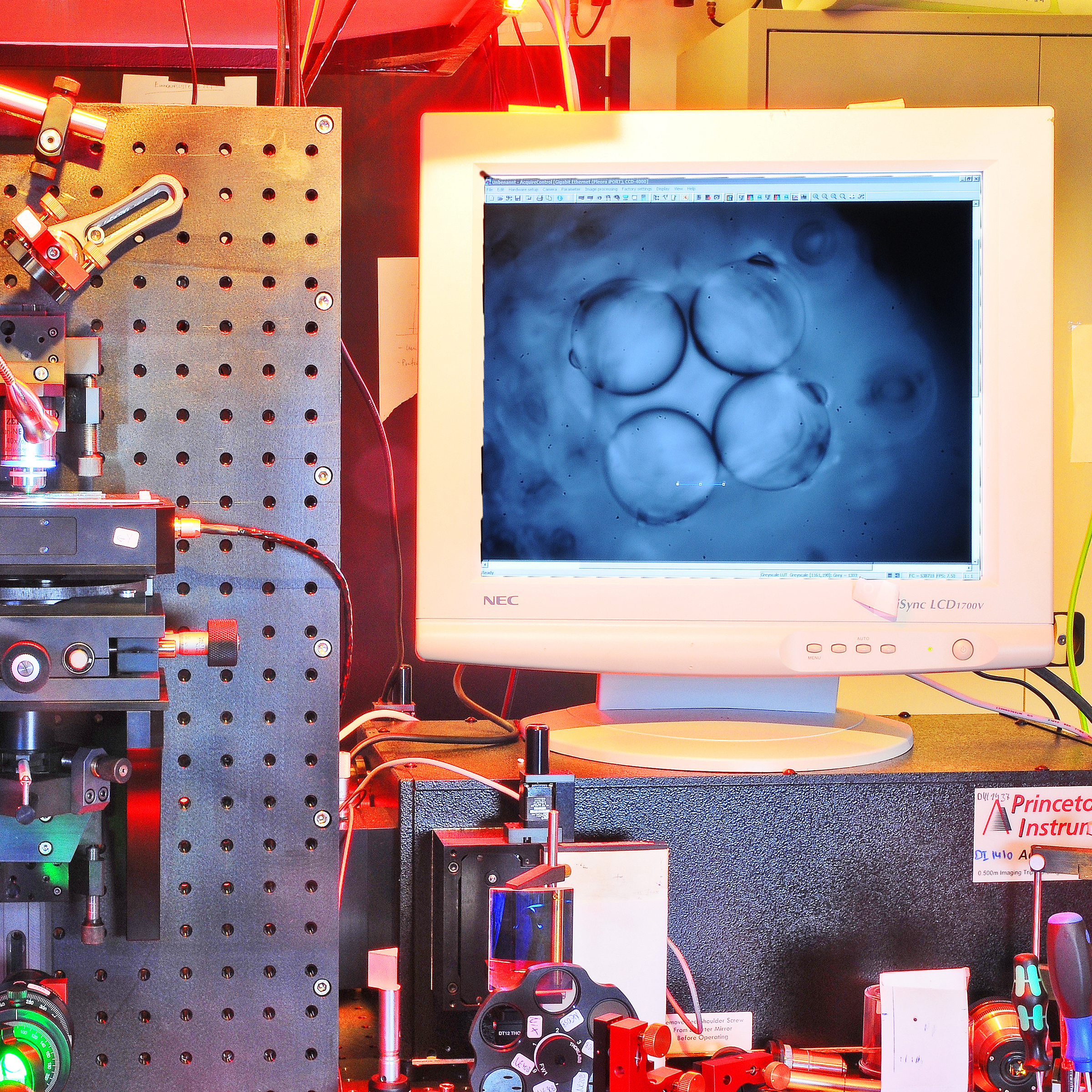
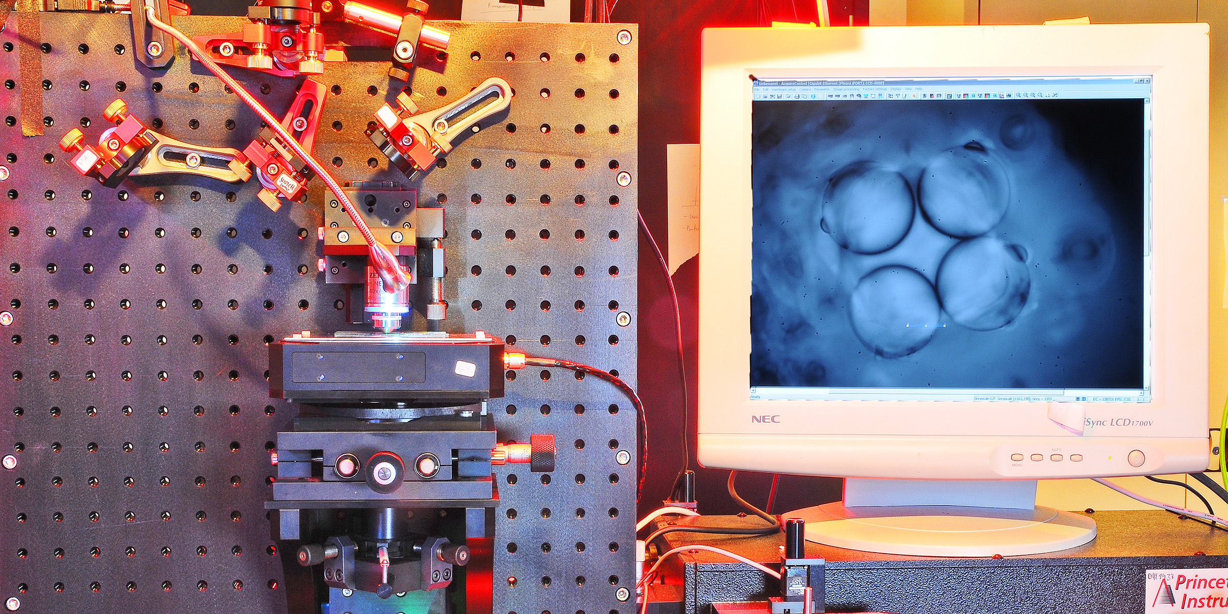
Quick trip through the microcosm: Find out how our researchers gain a close look into organisms, from single cells and their components to larger tissue and organs. Thirteen podcast episodes introduce you to advanced imaging technologies and promising research projects.

© Philipp Bovenkamp, Florian Lindemann, Tobias Brix, Klaus Hinrichs, Cornelius Faber, Verena Hörr

Software Wizards

Cells-in-Motion Podcast | Episode 13

In imaging, scientists have to deal with complex datasets. But sometimes they lack the software to display such a jumble of data clearly. When this happens, computers scientists are in high demand. In Münster, two computer scientists are developing a program that morphs different datasets into vivid 3D-graphics and impressive internal images of the body.

© CiM - Peter Grewer

Risk Signature in the Blood

Cells-in-Motion Podcast | Episode 12

Natalizumab is a miracle drug. It prevents patients with multiple sclerosis from suffering another MS episode. But in rare cases, side effects can lead to death. So neuroscientists in Münster have been searching for a biomarker that can provide information for physicians about if a patient is able to take the drug without great risk.

One-Track Specialists Don’t Get Far ...

Cells-in-Motion Podcast | Episode 11

… because modern scientists have to be open-minded about other fields to achieve progress in their research. That is why the CiM-IMPRS Graduate School trains junior researchers to work in interdisciplinary and international ways. The programme challenges its graduate students with a task they can only complete by working together.

© CiM – Peter Grewer

Indestructible Flatworms

Cells-in-Motion Podcast | Episode 10

It can reproduce itself from a piece of its own tail and can even regrow its head – the regenerative capacity of the flatworm has always amazed scientists and journalists. Now, with the help of modern sequencing technologies, Dr. Kerstin Bartscherer and her team are finally able to explore these complex processes in detail.

© SFB 656 - Peter Grewer

Sharper Images for Medicine

Cells-in-Motion Podcast | Episode 9

If you move during an examination with a whole body scanner the resulting images from inside the body will be blurred. A patient’s breathing is already enough to make tomographic images less precise. That is why medical physicist Dr. Florian Büther and nuclear medicine specialist Dr. Thomas Vehren develop new methods which shall improve the quality of medical images.

© Jahn Müller

Hybrid Imaging with PET/MRI

Cells-in-Motion Podcast | Episode 8

Scanning before dissection – to look inside the human body physicians nowadays use a set of imaging techniques. Just recently, the University Hospital Münster has installed an innovative hybrid system which combines PET and MRI. The high end scanner generates both unique images and challenges for the physicians.

© CiM - Heiner Witte

Wound Closure in the Fruit Fly

Cells-in-Motion Podcast | Episode 7

What tells a cell how and where it should move? At the end of the day genes are responsible. And which gene functions actually regulate a targeted movement of blood cells in order to close a wound is explored by Dr. Sven Bogdan and Prof. Xiaoyi Jiang with the help of both microscopes and algorithms. The perfect model organism for this issue is the fruit fly Drosophila melanogaster.

© CiM - Peter Leßmann

Tracking Cells in Motion

Cells-in-Motion Podcast | Episode 6

The method of tracking allows to follow individual cells in motion over a period of time. With his mathematical models, Dr. Christoph Brune provides great support for Prof. Dietmar Vestweber who studies the specific pathway immune cells take during migration. A targeted blockage of this pathway may help to regulate chronical inflammations in the future.

© SFB 656 - Frieda Berg

Inflamed Coronary Arteries

Cells-in-Motion Podcast | Episode 5

When coronary arteries become inflamed, the vessel walls thicken and may rupture – leading to an acute heart attack. Dr. Thomas Vogl, Dr. Andreas Faust und Dr. Sven Hermann develop substances to visualise inflamed vessel walls with molecular imaging and to assess the individual risk.

© CiM - Michael Kuhlmann

Modern Light Microscopy

Cells-in-Motion Podcast | Episode 4

Cellular structures of different dimensions can be studied specifically by light microscopy – ranging from processes in the whole organism to the smallest components of individual cells. In the last thirty years, microscopes have benefitted from fluorescence and 3D-techniques and are extremely powerful, explain Prof. Jürgen Klingauf and Prof. Friedemann Kiefer.

© CiM - Michael Kuhlmann

Cells on the Move in the Zebrafish

Cells-in-Motion Podcast | Episode 3

How do cells move forward? In cell migration, stiffness and elasticity play an important role. In a new project, Prof. Cornelia Denz and Prof. Erez Raz will measure these biophysical properties for the first time. The embryo of a zebrafish is a very appropriate animal model for that.

© CiM - Elisabeth Deiters-Keul

Looking Inside the Cell Membrane

Cells-in-Motion Podcast | Episode 2

The cellular membrane is an envelope surrounding a cell. It takes over many communicative and controlling functions in central processes of the cell. With novel laser-techniques, Prof. Carsten Fallnich and Prof. Volker Gerke want to decode specific mosaic structures of the membrane that are central to the understanding of cells in motion.

© Westfälische Nachrichten - Jürgen Peperhowe

Inflammation in the Brain

Cells-in-Motion Podcast | Episode 1

As the reasons for multiple sclerosis (MS) are yet not known, Prof. Lydia Sorokin and Prof. Günter Haufe cooperate in a project where they want to bring the progress of MS in sudden inflammatory attacks on pictures. Therefore, they improve and specifiy the method of molecular imaging - this research promises new understanding of the nervous disease MS.