Cells on the Move in the Zebrafish: How Elasticity of Cells Promotes the Zebrafish’s Development
Millions of cells arise in the development of organisms where they migrate to specific regions. Here, they develop to specialized cells of organs. This process is known as cell migration.
On the one hand, cell migration is not only relevant in the emergence of life, but also for our immune system or in wound healing. Cells on the move on the other hand can lead on to diseases. So the mechanisms enabling cell migration are relevant in the field of basic knowledge. Only with this knowledge, the migration of cells will be understood in detail and can be controlled in the future. Prof. Erez Raz from the Institute of Cell Biology and Prof. Cornelia Denz from the Institute of Applied Physics will investigate the mechanisms in their model of the zebrafish – for the first time. For their experiments, they enable an infrared laser beam-based holographic optical tweezers systems incorporated into a high-resolution microscope, and lots of tiny beads, which are incorporated into the embryo of the living fish.
Photos
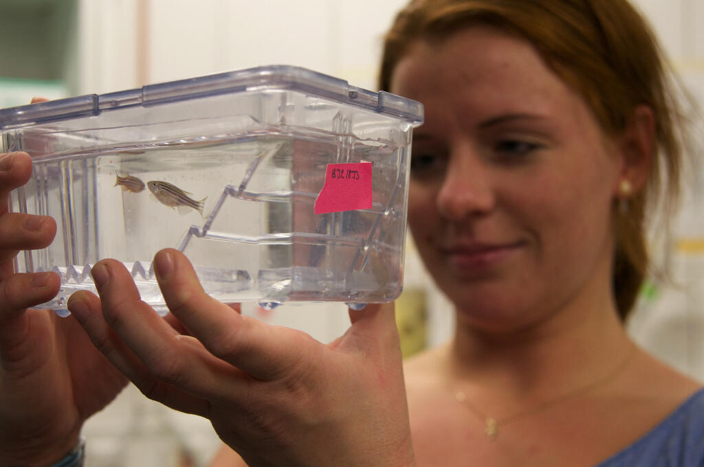
Since a great deal of movement takes place within the embryonic development, the transparent embryo of the zebrafish is particularly well suited to investigate cells in motion.© CiM - Michael Kuhlmann 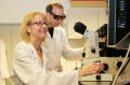
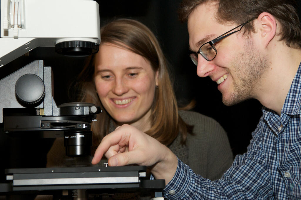
Professor Erez Raz (right) from the Institute of Cell Biology is interested in the factors that influence the movement of a cell. Professor Cornelia Denz (left) from the Institute of Applied Physics can provide an important examination technique.© CiM - Michael Kuhlmann 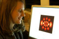
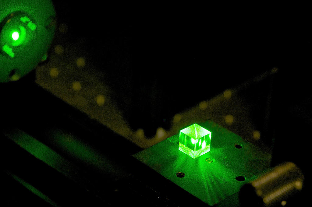
With optical tweezers light can be placed and used in various forms, in order to investigate the zebrafish embryo.© CiM - Michael Kuhlmann 
Christina Alpmann and Christoph Schöler from the team of optical photonics work with optical tweezers to measure the elasticity and stiffness of cells.© CiM - Michael Kuhlmann 
Optical tweezers generate forms of light and can therefore generate pressure and stress in the embryo. This enables producing different movement scenarios.© CiM - Michael Kuhlmann