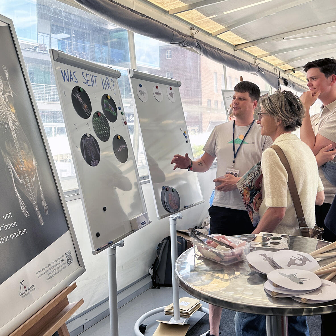
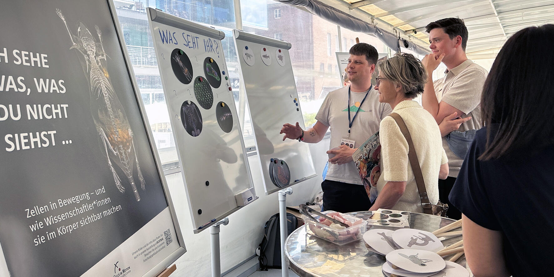
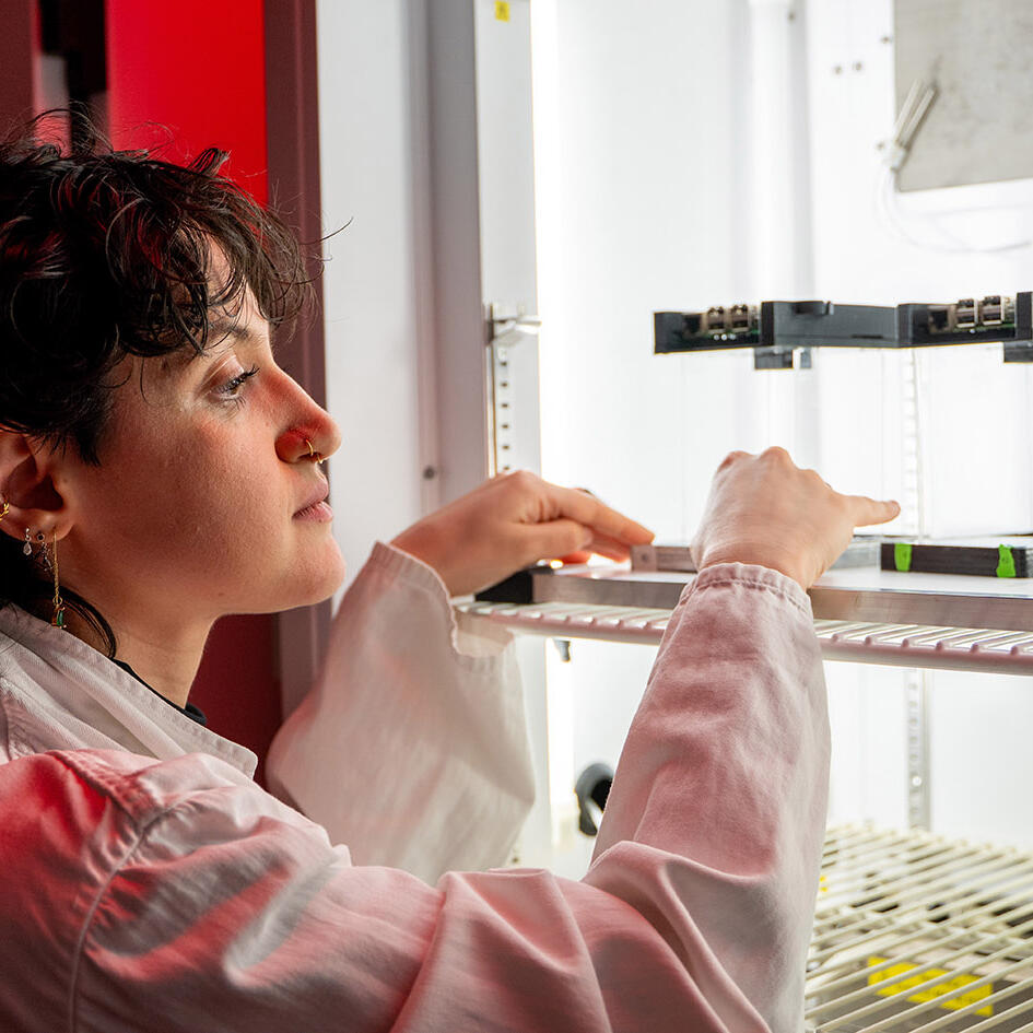
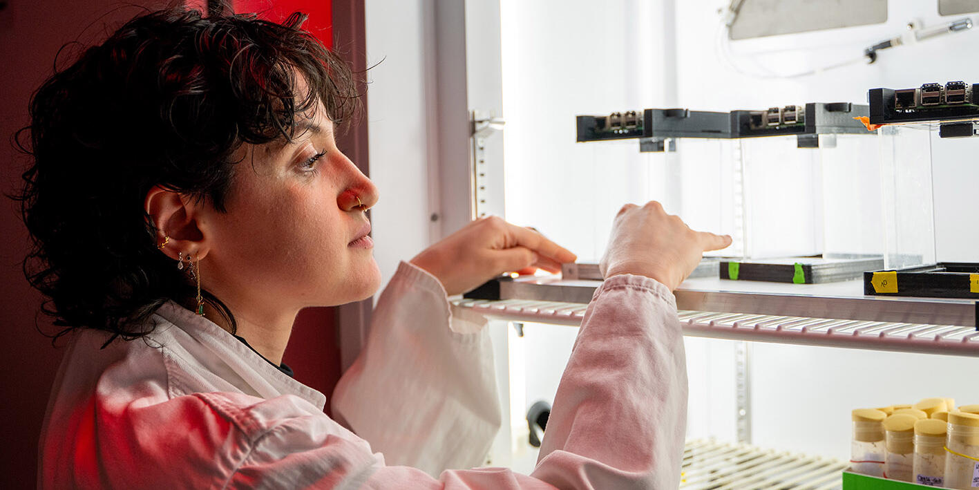
The team from the University of Münster’s Multiscale Imaging Centre (MIC) provided insight into research on bodily processes on board the ‘MS Wissenschaft’ on 4 July 2026. Through images of cells, tissues and organisms, visitors joined the team to explore the world of science. Children slipped into lab coats and playfully mimicked lab work.
In the event of a severe heart attack, immature immune cells are released into the bloodstream from the bone marrow. A research team led by Professor Oliver Söhnlein has demonstrated that the maturity level of neutrophils can be used to determine the short-term risk of death, and this can be assessed through a simple blood test.
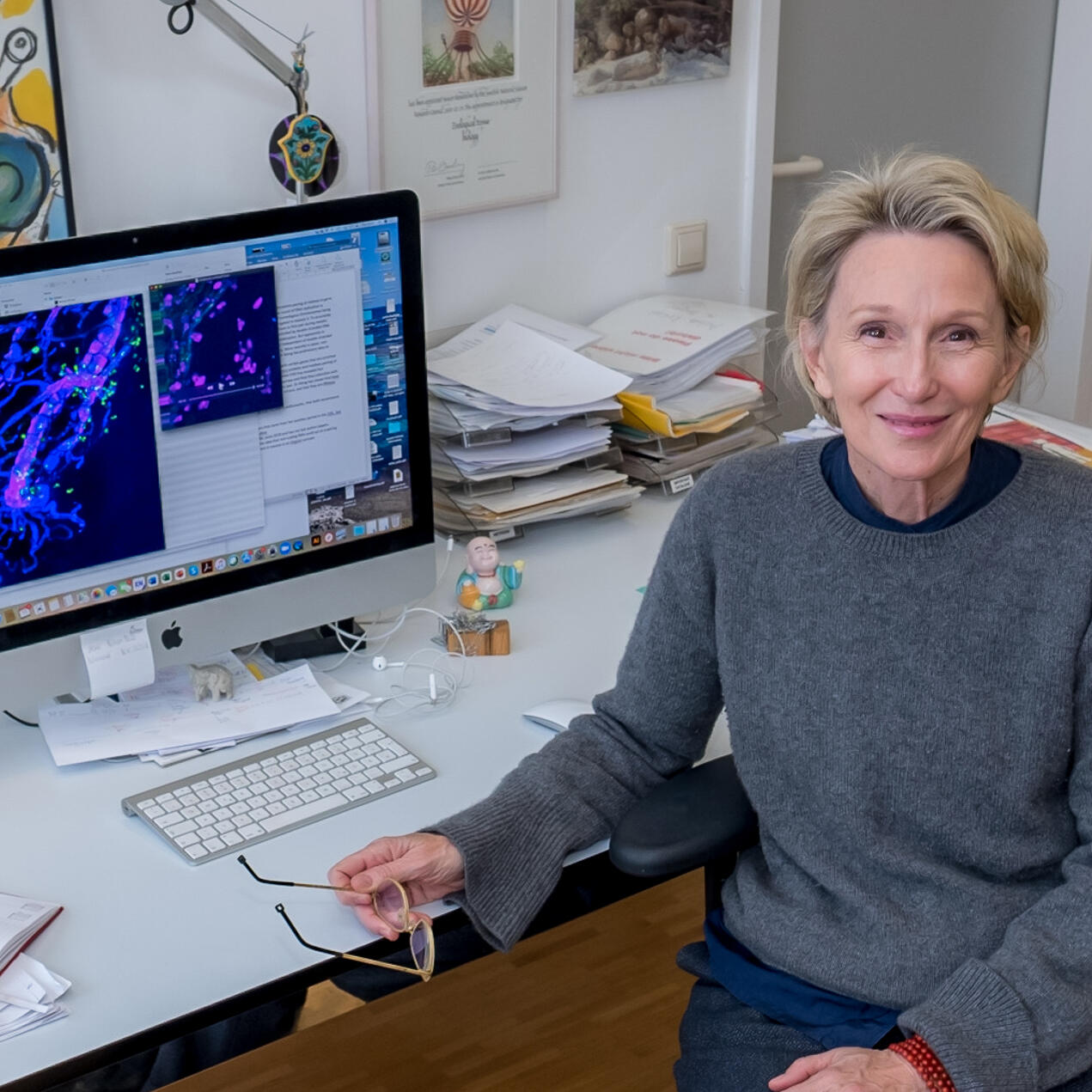
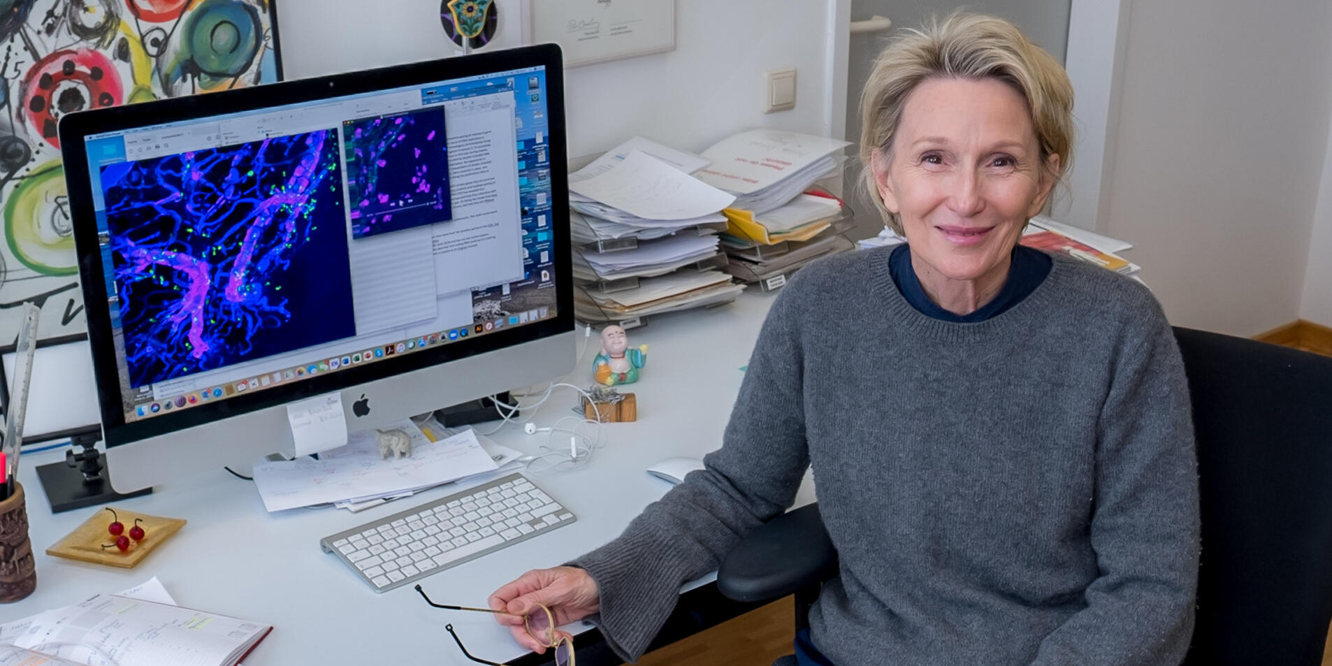
Dermatologist Professor Luise Erpenbeck and her research partners have demonstrated for the first time in real time that the body’s own defence cells use catecholamines – neurotransmitters such as dopamine and adrenaline – to communicate via the same chemical signals as nerve cells. This discovery opens up a new understanding of how the immune system is regulated.
Biologists Dr Angelica Coculla and Professor Ralf Stanewsky are researching the internal circadian clock of fruit flies. Their latest studies show that the insects are able to reset their internal clock. As the molecular clock resets, the sleep-wake cycle returns.
Professor Lydia Sorokin from the Institute of Physiological Chemistry and Pathobiochemistry has been awarded an ‘ERC Proof of Concept Grant’ worth 150,000 euros to further develop a new model of the blood-brain barrier for use in research.
Medical doctor and cell biologist Professor Sara Wickström has been awarded the Körber European Science Prize, worth one million euros, for discovering a mechanism by which cells sense the physical world around them.
In our videos, scientists provide multifaceted insight into their research and everyday work. They talk about current research questions, their new findings and how these findings were generated. They also talk about their personal motivations, the experiences they have had while on their career path and the framework of the scientific system. The videos are in either English or German and many of them have subtitles available in both languages.