Research networks in the field of “cell dynamics and imaging”

Our community is intensively engaged in acquiring external funding, especially for interfaculty research networks in the field of cell dynamics and imaging. By joining forces in the Cells in Motion Interfaculty Centre, we embed the specific scientific topics of these collaborative projects into a larger thematic context, and, at the same time, our network is an incubator for new collaborative initiatives. Together we further develop our scientific field as well as supportive offers and institutional structures.

  • CRC 1450 inSight – Multiscale imaging of organ-specific inflammation

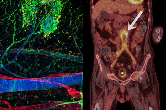
    Imaging of inflammation using microscopy (left) and whole-body imaging (PET-CT, right). At present, there is no methodology available that allows for the compiling of information from these different examinations.
    © AG Kiefer/AG Schäfers

    Researchers in this Collaborative Research Centre (CRC) aim to understand how the body regulates inflammation in different organs and which cellular processes are decisive in determining how an inflammation will progress. They incorporate different imaging technologies into their investigations and develop innovative strategies for the labelling of cells and the evaluation of image data in order to be able to integrate information from single cells to entire organisms, and from mice to men. In the long term, the results could lead to new diagnostic approaches for clinical imaging modalities and help, initially, in the detection of inflammation, then in finding the best suited immunoregulatory therapy for individual patients and, finally, in the assessment of its efficacy.

    Spokesperson: Prof Dr Michael Schäfers (Faculty of Medicine)
    Involved Faculties: Medicine, Chemistry and Pharmacy, Mathematics and Computer Science
    Project term: 01/2021 – 12/2024
    Funding: German Research Foundation (DFG)

  • CRC 1348 Dynamic Cellular Interfaces: Formation and Function

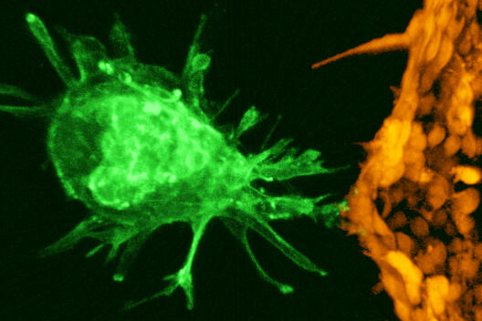
    Microscopy image of an early mouse embryo (green) interacting with endothelial cells (orange) in a novel synthetic cell culture system
    © Niraimathi Govindasamy et al./Dev Cell 2021

    This Collaborative Research Centre (CRC) focuses on the question of how contact points between cells develop and function. Special molecular mechanisms enable communication between cells and their cohesion, thus regulating cell differentiation as well as the development and function of different tissues. Researchers investigate these processes in fruit flies, zebrafish, and mice, combining high-resolution microscopic methods with biochemical and genetic methods.

    Spokesperson: Prof Dr Christian Klämbt (Faculty of Biology)
    Involved Faculties: Biology, Chemistry and Pharmacy, Medicine
    Project term: 01/2018 – 12/2025
    Funding: German Research Foundation (DFG)

  • CRC 1009 Breaking Barriers – Immune cells and pathogens at cell/matrix barriers

    Microscopy image
    Blood vessels consist of a layer of endothelial cells (green) and a basement membrane (violet). When recognizing inflammation, immune cells (red) exit the blood vessel and migrate to the inflamed tissue. Immunofluorescence microscopy
    © Hang Li, Stefan Butz, Dietmar Vestweber

    This Collaborative Research Centre (CRC) investigates inflammatory mechanisms at cellular barriers of complex organisms. Such barriers separate the organism and the outside world but also different tissues and organs from each other and play a central role in many diseases: In addition to the defence against infectious pathogens, these structures also play an important role in regulating inflammatory processes. The research projects use moleculargenetic, cell biological, biochemical, immunological as well as animal experimental and imaging methods to elucidate functional mechanisms that may provide starting points for diagnostic, therapeutic and preventive strategies for infectious diseases as well as autoimmune diseases and allergies.

    Spokesperson: Prof Dr Johannes Roth (Faculty of Medicine)
    Involved Faculties: Medicine, Biology
    Project term: 07/2012 – 06/2024
    Funding: German Research Foundation (DFG)

  • CRC/TRR 332 Neutrophils: Origin, Fate & Function

    Neutrophil granulocytes are the most abundant circulating white blood cells in humans and have been attributed mainly antimicrobial functions. However, recent findings show that they also have underappreciated multiple functions in chronic inflammation and tumour development. Researchers in this Collaborative Research Centre/Transregio (CRC/TRR) aim to understand the role of neutrophils in physiological and pathological immune processes. To this end, they investigate how signals in the tissue influence the production and function of neutrophils and how the intracellular processing of signals regulates neutrophil function. In the long term, the aim is to build a bridge to clinical applications.

    Spokesperson: Prof Dr Oliver Söhnlein (Faculty of Medicine)
    Involved institutions: Universities of Münster, Duisburg-Essen and Munich, associated: TU Dresden, Leibniz-Institut für Analytische Wissenschaften Dortmund
    Project term: 07/2022 – 06/2026
    Funding: German Research Foundation (DFG)

  • CRC/TRR 128 Initiating/Effector versus Regulatory Mechanisms in Multiple Sclerosis

    Microscopy image
    Cell culture model of the blood-brain barrier. A human T-cell (bottom right, blue cell nucleus) loaded with the protein Granzyme K (green), looks for a suitable site to drill a channel through a blood vessel cell (blue cell nucleus top left, red cell margin) for migration. This process does not need inflammation and is part of immune surveillance in the healthy brain.
    © AG Zarbock, AG Schwab

    This Collaborative Research Centre/Transregio (CRC/TRR) focuses on investigating multiple sclerosis. In this autoimmune disease, immune cells attack the very organism they are supposed to protect: They migrate to the brain, where they cause chronic inflammation. In order to develop new therapeutic concepts, researchers in this project want to unravel the changes in the immune system that underlie the disease, the role of the blood-brain barrier and the effects of the immune system's attack on the central nervous system. They examine these processes in rodents and humans and often use innovative imaging techniques.

    Spokesperson: Prof Dr Heinz Wiendl (Faculty of Medicine)
    Involved institutions: Medical Faculties at the Universities of Münster, Mainz and Munich
    Project term: 07/2012 – 06/2024
    Funding: German Research Foundation (DFG)

  • CRU 342 Organ Dysfunction During Systemic Inflammation

    Microscopy image
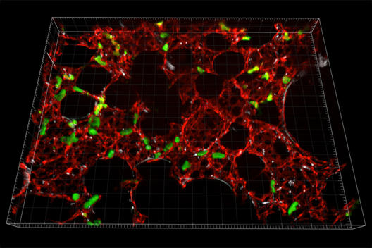
    During inflammation in the lung of a mouse leukocytes (green) migrate into the tissue and interact with platelets (grey).
    © AG Rossaint / AG Zarbock

    This Clinical Research Unit (CRU) focuses on systemic inflammatory reactions such as sepsis which are often fatal. The involved researchers investigate molecular, immunological and cellular signalling pathways that are relevant for such inflammation and subsequent organ failure. They examine these causal disease processes in murine and porcine animal models as well as in studies on patients and want to use their findings to develop new therapy options for patients.

    Spokesperson: Prof Dr Alexander Zarbock (Faculty of Medicine)
    Coordinator: PD Dr Jan Rossaint (Faculty of Medicine)
    Involved Faculties and non-university research institutes: Faculty of Medicine, Max Planck Institute for Molecular Biomedicine
    Project term: 02/2020 – 01/2026
    Funding: German Research Foundation (DFG)

  • CRU 326 Male Germ Cells: from Genes to Function

    Color-coded time lapse overlay depicting one sperm flagellar beat cycle. Patient sperm show reduced amplitude in the flagellar beat compared to healthy donor sperm.
    Color-coded time lapse overlay depicting one sperm flagellar beat cycle. Patient sperm show reduced amplitude in the flagellar beat compared to healthy donor sperm.
    © Timo Strünker

    This Clinical Research Unit (CRU) focuses on unravelling causes of male infertility. 50 researchers of nine institutes intend to decipher genetic and epigenetic factors as well as molecular mechanisms that influence germ cells – from the complete loss to sperm dysfunction. They investigate the interplay of male germ cells in humans as well as in animal models with the aim to improve the diagnostic yield and therapeutic options of male infertility. This will lead to an improved understanding of male reproductive health.

    Speaker: Prof Dr Jörg Gromoll (Faculty of Medicine)
    Research Coordinator: Prof Dr Frank Tüttelmann (Faculty of Medicine)
    Involved Faculties: Medicine, Biology
    Project term: 08/2017 – 07/2023
    Funding: German Research Foundation (DFG)

  • RTG 2515 Chemical Biology of Ion Channels

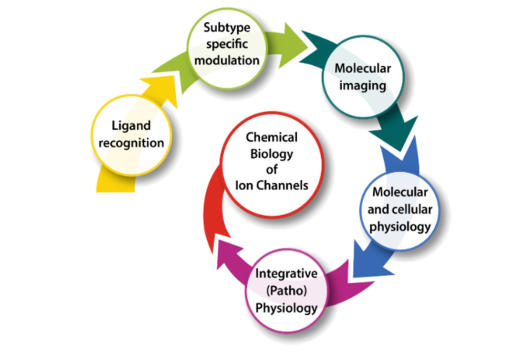
    The five research areas of the Research Training Group
    The five research areas of the Research Training Group
    © Chembion/Heike Blum

    In this Research Training Group (RTG), PhD students investigate the synthesis, modification and biological use of small organic molecules, with which the opening state of ion channels can be specifically controlled and made visible with molecular imaging. Ion channels are proteins which form pores in cell membranes, thus allowing charged particles to pass through the cell membrane and influence processes in cells, tissues and organs. Being able to systematically open and close ion channels could, in the future, open up a range of applications in the treatment of various pathological conditions, such as cancer or neural diseases. Many scientific contacts, which made this joint project possible, were established through the Cells in Motion Cluster of Excellence.

    Spokesperson: Prof Dr Bernhard Wünsch (Faculty of Chemistry and Pharmacy), Prof Dr Thomas Budde (Medical Faculty)
    Involved Faculties: Chemistry and Pharmacy, Medicine
    Project term: 10/2019 – 09/2028
    Funding: German Research Foundation (DFG)

  • Immune-Image

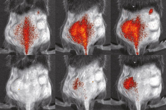
    Distribution of immune cells in the body of a mouse, imaged with optical imaging
    Distribution of immune cells in the body of a mouse, imaged with optical imaging
    © S. Gran & L. Honold et al./Theranostics 2018(8)

    How do immune cells behave in the body? What happens during immunotherapy, which is designed to encourage the body’s own immune system to attack a disease? In this Europe-wide collaborative research project, experts from research and the pharmaceutical industry want to develop biomedical imaging methods for monitoring immune cells before, during and after immune treatment. The researchers will initially look at the processes in animal models for different diseases – with the aim of translating the results and methods to patients mid-term enabling them to establish image-guided immunotherapies with improved efficacy in diseases such as cancer or inflammation.

    Spokesperson: Prof Albert D. Windhorst (Amsterdam University Medical Centre, the Netherlands)
    Involved Faculties at Münster University: Medicine (Coordination: Prof Michael Schäfers, Prof Andreas Jacobs), Chemistry and Pharmacy
    Project term: 10/2019 – 03/2026
    Funding: European Commission (EC)