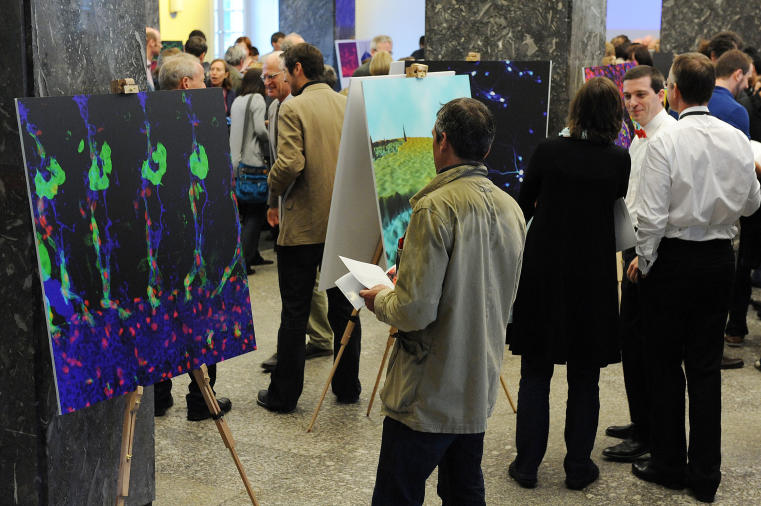
Online exhibition
Exhibition
Morphology of two tumours (top) and their vascular perfusion (bottom). The left panel shows well-perfused tumours (yellow/red). In response to a new vessel-blocking therapy, perfusion of the tumours is significantly lowered (right, blue/violet). MRI© Thorsten Persigehl, Wolfgang Berdel 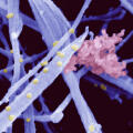
The structure of cartilage can be compared to that of reinforced concrete: The collagen fibres (“steel rods”, blue) and the extrafibrillar matrix (“concrete”, red) are responsible for cartilage’s tensile strength and resistance to compression.© Uwe Hansen, Peter Bruckner 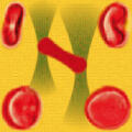
Analysis of blood cell dynamics: Two focussed laser beams trap a single red blood cell (middle). Using this principle, a single cell can be maneuvered and rotated very precisely (depicted in the four corners). “Optical tweezers”© Mike Wördemann, Cornelia Denz 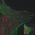
In response to injury, a blood vessel’s cells (endothelial cells) release substances that promote blood clotting and thereby stop bleeding. Those substances are stored in Weibel-Palade bodies (red). Structured illumination microscopy© Inés Pulido, Volker Gerke 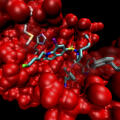
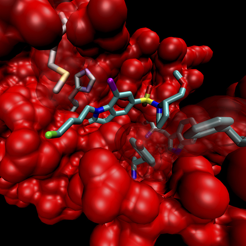
This image illustrates binding of a chemical substance (tracer) to enzymes which are upregulated in inflamed tissues. The green moiety of the tracer is radioactive, which permits imaging. X-ray crystallography analysis© Mark Waller, Panupun Limpachayaporn, Klaus Kopka, Günter Haufe 
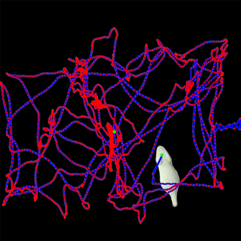
Motion of a mouse, tracked by a fluorescent marker fixed to its head. This detailed tracking information is used to retrospectively correct the movement of objects during image acquisition. Fluorescence reflectance imaging© Mohammad Dawood, Xiaoyi Jiang 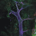
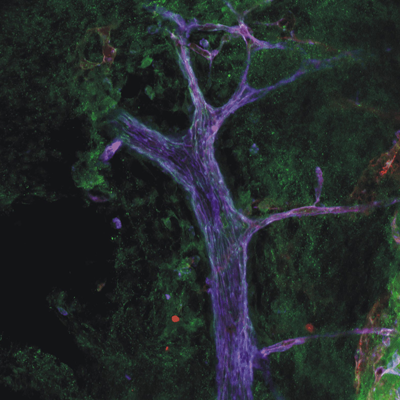
This image shows a blood vessel of the skin. Three proteins expressed on the surface of the blood vessel cells (endothelial cells) were visualised with specific antibodies that are coupled to dyes (red, green and blue). Confocal laser scanning microscopy© Ruben Böhmer, Friedemann Kiefer 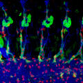
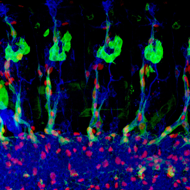
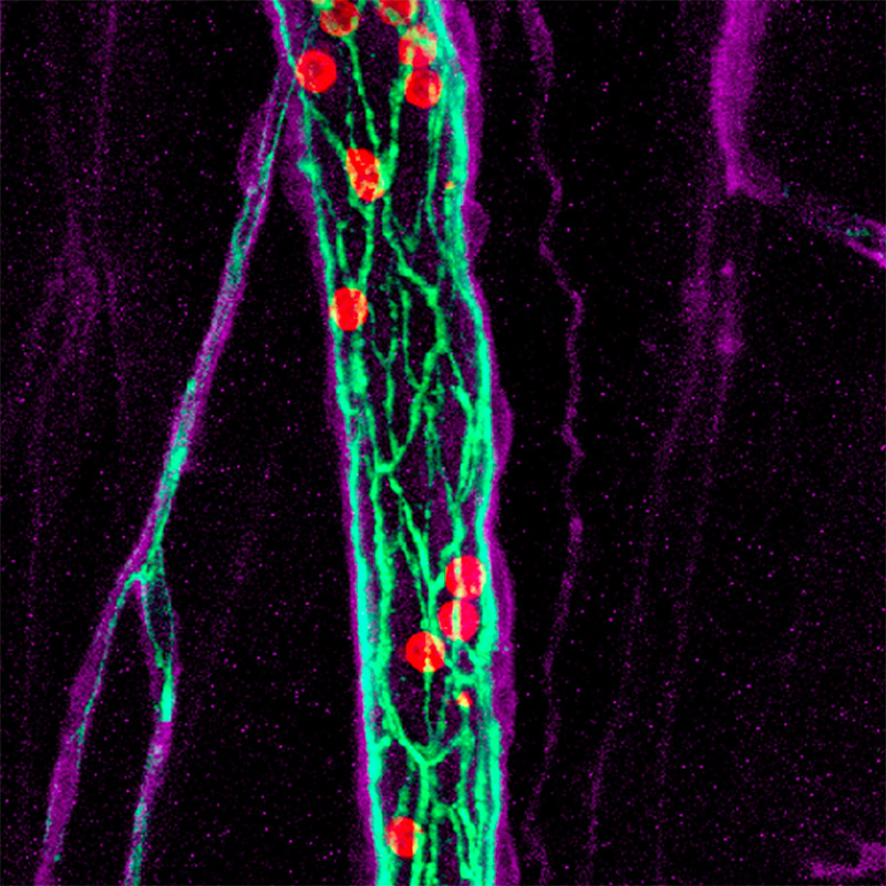
This image shows the peripheral nervous system of a fruit fly (Drosophila). Nerve cells are stained in blue, the surrounding glial cells in green and their cell nuclei in red. Confocal microscopy© Marion Silies, Christian Klämbt 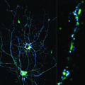
Synapses are the contact and communication interfaces between nerve cells. Innovative microscopy (STED) uniquely visualises the distribution of synapse proteins at a resolution of a few nanometres. © Cora Thiel, Roman Schmid, Jürgen Klingauf 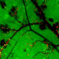
During skin inflammation, immune cells (red) migrate from the blood vessels (black) into the surrounding tissue (green) and cause redness and swelling. Immunofluorescence microscopy© Karin Loser, Thomas Luger 
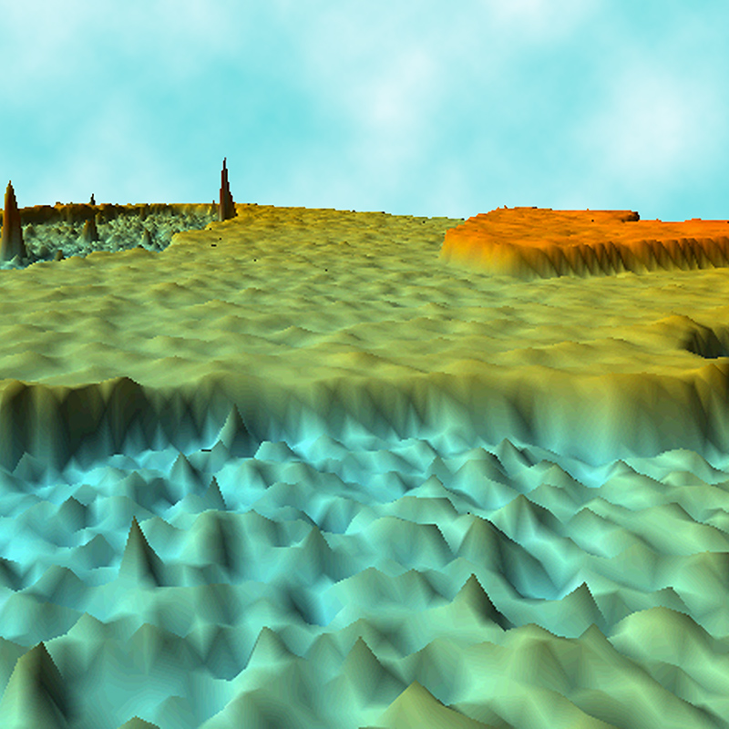
It looks like a landscape, but it is the cell membrane of an egg cell. Two overlapping membranes are depicted (upper right corner). Researchers investigate single protein molecules (cone upper left) in the plasma membrane. Atomic force microscopy© Hermann Schillers, Hans Oberleithner 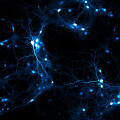
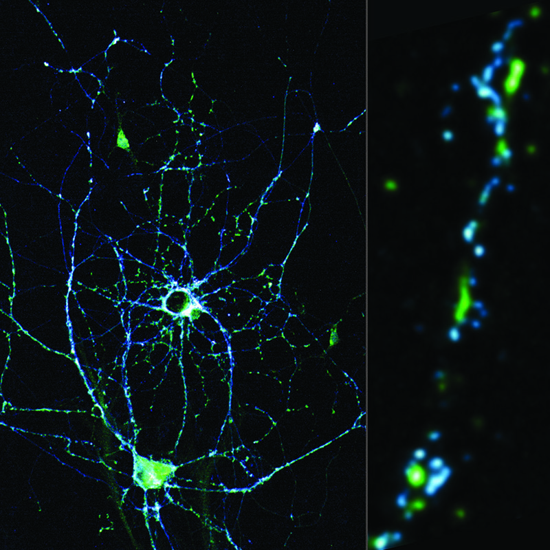
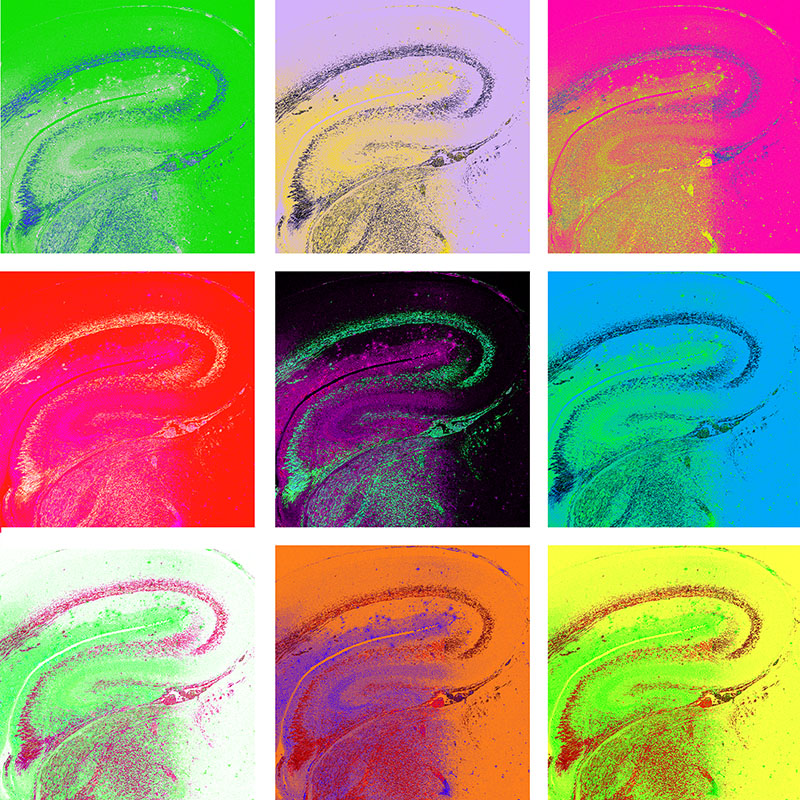
Nerve cells from the hippocampus, an area in the brain which is essential for learning and memory. Each individual cell measures only 1/100 of a millimetre. They form large networks by establishing tight contacts. Confocal microscopy© Frank Erdmann, Hans-Christian Pape 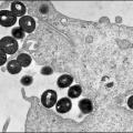
Bacteria (dark) bind to and are taken up by a host cell. If bacteria hibernate in the host cell for a prolonged period, they are hidden from the immune system as well as from antibiotic treatment, thus triggering chronic infections. Electron microscopy© Bettina Löffler, Georg Peters 
This image shows a brain slice stained for various structures of nerve cells. Nerve fibres that connect nerve cells are clearly visible. Immunofluorescence microscopy© Daniel Lutter, Anja Ehrkamp, Andreas Püschel 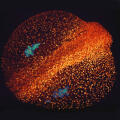
Fluorescently tagged proteins can be expressed in living cells, where they help scientists investigate cellular function. Here, the membranes of zebrafish germ cells express a green fluorescent protein, while nuclei express a blue one. 2-photon microscopy© Azadeh Paksa, Nadine Peyriéras, Erez Raz 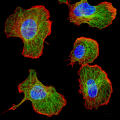
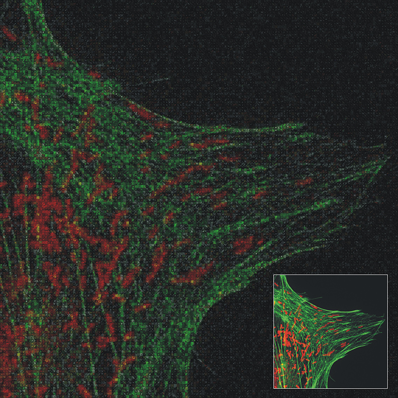
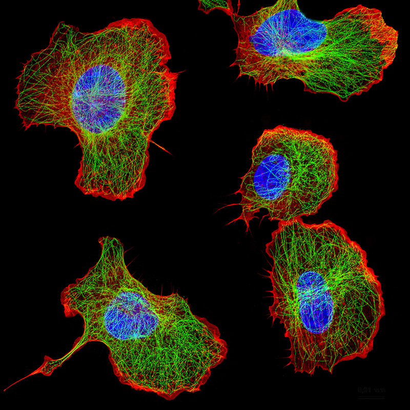
These immune cells respond to a stimulus; they redistribute part of their cytoskeleton (red) to the right. The central portion of the cytoskeleton (green) aligns along the direction of cell migration. Immunofluorescence microscopy© Marc Wolf, Johannes Roth 
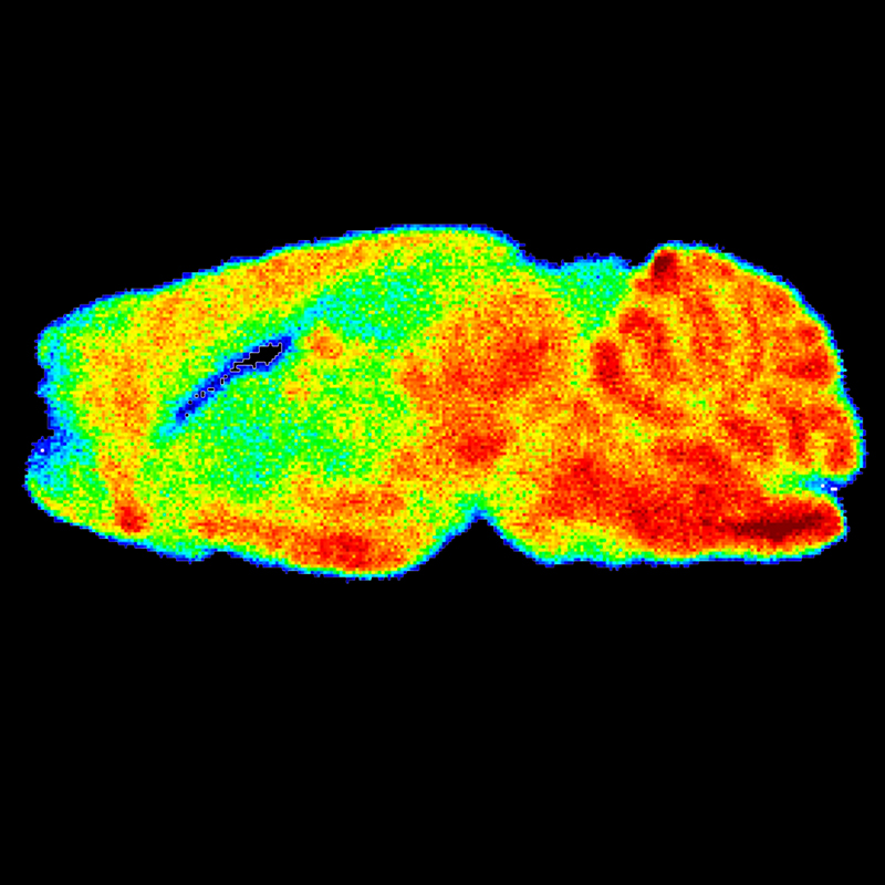
Amongst other sources, cells generate their energy from sugar. Thus, radioactive sugar can be used to image the function of the heart and brain. Here, the accumulation of sugar (red) shows normal function of both organs. PET/CT© Sven Hermann, Michael Schäfers 
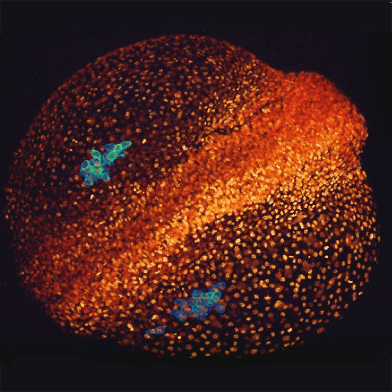
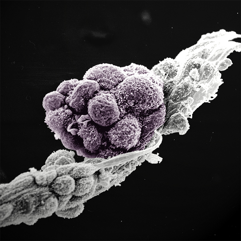
An early germ cell colony (violet) generated from stem cells. This image shows the tight cell-cell contacts between the germ cells, as well as between the germ cells and neighbouring cells (grey). Scanning electron microscopy© Katherina Psathaki, Hans Schöler 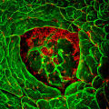
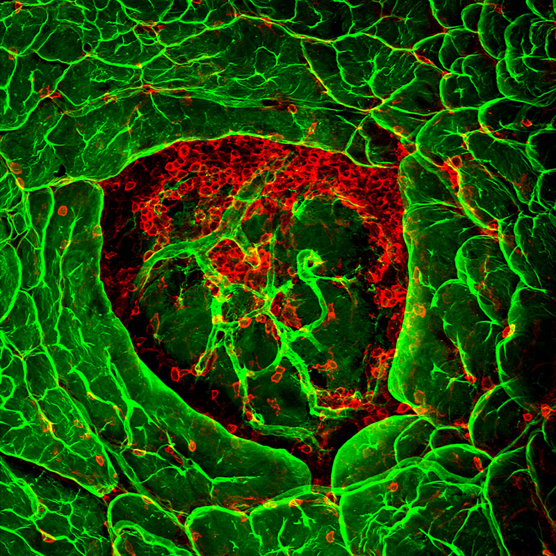
In diabetes, insulin-producing cells of the pancreas are destroyed by a chronic inflammation. This image shows immune cells (red) penetrating the basement membrane (green) of insulin-producing cells. Confocal microscopy© Eva Korpos, Lydia Sorokin 
Blood vessels consist of a layer of endothelial cells (green) and a basement membrane (violet). When recognizing inflammation, immune cells (red) exit the blood vessel and migrate to the inflamed tissue. Immunofluorescence microscopy© Hang Li, Stefan Butz, Dietmar Vestweber 
Receptors in the brain, visualised by a radiolabelled chemical substance. Receptors, which consist of proteins that receive and transduce signals, are important for the maintenance of brain function. Autoradiography© Bernhard Wünsch