To analyse the interplay of cells in the body, researchers have to be able to “see” different cells in tissues and to monitor their behaviour. To do this, scientists of "Cells in Motion" use a broad range of imaging technologies. Interdisciplinary teams are working on further developing these imaging strategies.
How can processes in the body that are normally hidden from the human eye, such as inflammation or disease, be visualized? This requires labelling of cells or molecules that generate signals transmitting information from inside the body to the outside. These signals can be measured and converted into images.
One way researchers can label cells is through genetic modifications. This involves changing the DNA, the molecule in the cell nucleus that carries the genetic information, i.e. the genes. The genes contain instructions for the synthesis of proteins with defined functions in the cell, such as catalysing reactions or becoming building materials. When researchers want to visualize a protein, they link the assembly instructions for the target protein to those of another protein that generates a detectable signal, often referred to as the tag. The genetic information of the so-called green fluorescent protein (GFP), which comes from the Aequoria victoria jellyfish, is the one of the most frequently used signal or tag proteins.
Combining the genetic information of a target protein with a GFP-signal protein gives rise to a protein that emits a fluorescent signal under light. “We’re talking about reporter proteins here,” says Roland Wedlich-Söldner, CiM Professor of Multiscale Imaging and Cell Biology, “because these are purposefully generated fusion products which report to us about which players are located in the cell and which processes are taking place. In a way, we can watch the cell at work,” he explains.
CiM researchers also make processes in the body visible by introducing into the blood circulation chemically generated substances or so-called “tracers” that carry a signal transmitter, e.g. a fluorescent component or radioactive atoms that emit radiation. Such “tracers” act by finding and sticking to their target molecule, which may occur, for example, only in disease. Tracers can perform this “sticking” function because scientists have constructed them like a key that only fits one lock. One challenge in developing tracers is that they must not affect normal processes in the body. “We are placing a lot of hope in chemical carbohydrate compounds, which are natural substances,” explains Ryan Gilmour, CiM Professor of Chemical Biology. He and his team are developing carbohydrates that aid in the study of diseases such as Alzheimer’s or multiple sclerosis. In another project, CiM researchers add minute fluorescent plastic beads into the blood that are recognized and taken up by scavenger cells in the body, the phagocytes, rendering them fluorescent. This permits researchers to analyse how phagocytes contribute to diseases such as atherosclerosis.
From Signal to an Image
Artificially generated signals such as fluorescence, sound or radiation from radioactive substances in cells, tissues and organisms can be measured and converted into images using technologies such as light microscopy, ultrasound or positron emission tomography (PET).
Razor-sharp images of fine structures in individual cells and tissues are obtained using light microscopy. Laser light in such microscopes lights up molecules that are tagged with a fluorescent dye and high-sensitivity cameras pick up even the weakest signals. Computers convert the fluorescent light signals into three-dimensional images, that can represent one moment in time or images can be collected over time permitting changes or movements to be detected. Light microscopy has developed rapidly over the last 10 years. For a long time, 200 to 300 nanometres (one nanometre is one millionth of a millimetre) was thought to be the limit of resolution, now with modern high-resolution microscopes scientists are constantly lowering the limit of the size of structures that can be detected.
Fluorescent signals become highly scattered when they penetrate thick tissues, preventing accurate measurement and sharp images. Using deep penetrating lasers and sensitive detectors, intravital microscopy can analyse cells even deep within organisms. The light scatter effects caused by tissues can also be overcome by photoacoustic imaging. Laser light is used to stimulate fluorescent molecules but, unlike light microscopy where the emitted fluorescence signal is measured, here the resulting molecular vibration is detected by an ultrasound unit.
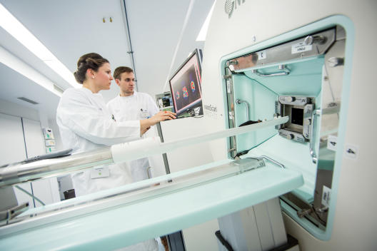
Looking into the whole organism (whole-body imaging), is used both in research but more commonly in clinical practice. PET scanners, for example, make signals emitted by radioactive tracers visible. These tracers emit gamma radiation which, unlike light, can penetrate many tissue layers.
Mathematicians develop computational models for more precise image reconstruction. Physicists contribute their knowledge of the physical properties of light or radiation to such models. Computer scientists develop powerful algorithms, which process and depict the data measured. By combining such skills, CiM researchers can propose unique solutions for difficult experimental conditions. For example, PET scans can be performed on awake, unrestricted mice which avoids any abnormal effects due to anaesthesia. “We record the movement of the mice and correct for it on the image data,” explains Prof. Klaus Schäfers, a physicist. “Without the involvement of various disciplines, such a complex development would not be possible,” adds computer scientist, Prof. Xiaoyi Jiang.