
How the cells’ environment affects their migration
Our body consists of trillions of cells that function together in maintaining life. Cells exist in various types; their different functions and abilities are remarkable: Cells can divide and make copies of themselves. Some cells can produce types of cells that are different from oneself. At the Institute of Cell Biology, we are fascinated by cells that have the ability to migrate within the body. Cells migrate to combat infections, to seal wounds and to replace dead cells. However, when cell migration is misregulated, it can lead to diseases such as cancer. For example, the migration of cancer cells away from the primary tumour is the first step in metastasis, where cancer cells invade other tissues and organs.
Cell migration has been intensively studied in in vitro experiments, experiments conducted outside of a living organism. Such studies provided detailed insights into the basic mechanisms driving cell migration. Inside our bodies, however, the environment is much more complicated. To study the migration of cells within the living organism, we employ zebrafish. Zebrafish are vertebrates whose important organs are similar to those of humans, making them a suitable model for biomedical research. Importantly, the zebrafish embryos are transparent and develop outside of the mother’s body. This allows researchers to examine the movement of single cells and follow the development of an organism “live” under the microscope.
To understand how cells move within the live organism, we exploit the ability of germ cells, the precursors of sperm and egg cells, to migrate through the developing zebrafish embryo. Germ cells are formed early on in the development of zebrafish embryos, when the embryo is only three to four hours old. From this moment on, they migrate through the developing embryo towards their destination, the site where the gonad will form. Thus, germ cells serve as a suitable and readily accessible model to study the migration of single cells in vivo – in the living organism. Indeed, over the last 20 years zebrafish germ cells served as a model for cell migration, revealing important principles and new insights regarding the mechanisms allowing cells to move forward and reach their target. For example, we could recently show that germ cells move by using folds in their membrane. Read the article
My research is primarily focused on understanding how the environment, within which the germ cells migrate, affects their migratory behaviour. Previous research conducted in other labs provided evidence that biophysical properties of the environment – whether the environment is soft or stiff – can influence cell motility. For example, in vitro studies have shown that cells can respond to differences in stiffness of their environment by changing their migration direction and their mode of migration. In contrast to such studies where cell migration is examined in relatively simple two-dimensional environments, we follow the migration process in the context of the live three-dimensional tissue. Specifically, we study the behaviour of the migrating cells as they interact with different cells and physical structures within the living organism, namely the zebrafish embryo. This experimental setup allows us to study cell migration in the context in which it occurs in our body, making the findings of general importance and more relevant for possible medical applications.

Biologists and mathematicians team up
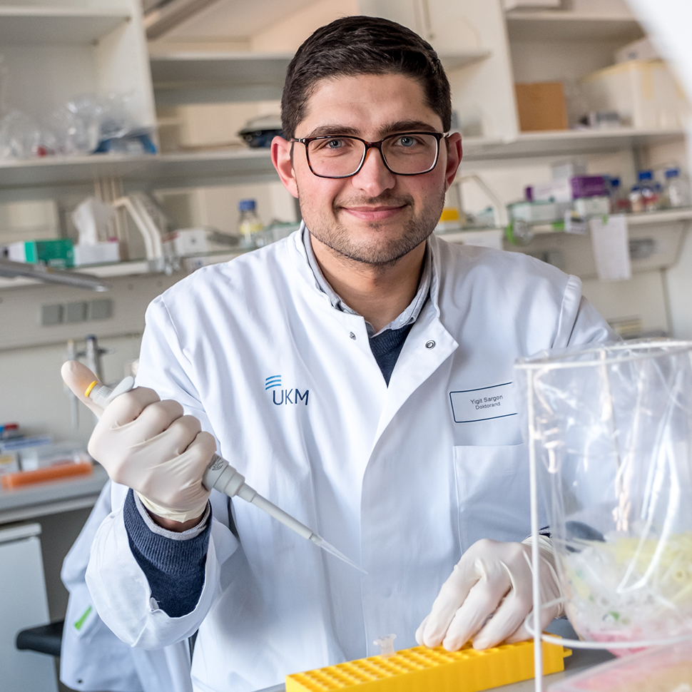
To investigate the interaction of migrating cells with their environment, my colleague Łukasz Truszkowski and I employ a technique called live cell fluorescence microscopy. This type of microscopy allows us to examine the distribution and movement of germ cells in three-dimensional environments. Employing fluorescent dyes that label the germ cells and their environment we follow them under the microscope.
To obtain reliable and statistically meaningful results, we need to study many embryos, usually between 300 and 500. Here, we encounter our first challenge: We have to acquire and analyse a very large amount of data. Furthermore, the microscopy-recorded embryos can vary in their size and in their three-dimensional orientation. At the same time, the imaged data from hundreds of embryos have to be integrated. Together, these challenges complicate the data analysis. Therefore, to process the data, our first step is to “unify” all the recorded embryos, such that we can put all the data on top of each other. Thereby, we will be able to identify areas within the zebrafish embryo where the germ cells preferentially reside. At the same time, we can identify areas that cells tend to avoid.
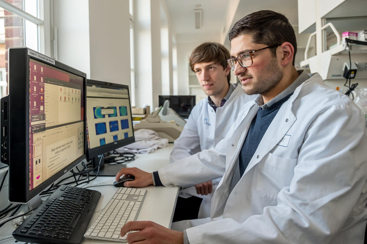
Aligning all the data captured from hundreds of embryos is a challenging task that cannot be solved by employing biological methods. Therefore, we established an interdisciplinary collaboration with the group of mathematicians led by Prof. Dr. Martin Burger, a collaboration funded by the Cells-in-Motion Cluster of Excellence at the University of Münster. Currently, we are working closely with members of this group, Dr. Daniel Tenbrinck, Ramona Sasse and Michael Ryu, on computerized solutions that automatically analyse our large amount of microscopy data sets.
Interdisciplinary work: benefits and challenges
The establishment of such a synergistic collaborative research effort requires the joint work of Biologists and Mathematicians, researchers whose scientific background and “language” differ. Therefore, the first task in the collaboration was to simplify our biological problem such that non-biologists could understand the experimental model and the terminology used. Similarly, the mathematicians explained the formulas and algorithms they developed such that we as biologists could understand them. Over the first few months, an effort was made to explain our technical jargon and introduce each other to the way the different disciplines consider and solve problems. That initial phase of getting to know each other was essential and decisive for our successful collaboration, which turned out to be a long term one.
Through the collaboration, I learned how important it is to avoid being limited to the immediate field, and expand to and incorporate other disciplines in the research. Nowadays, with the Internet of Things, Big Data and Artificial Intelligence permeating our lives, it is becoming increasingly important for experts in different disciplines to come together. When biologists, physicians, physicists, chemists and mathematicians team up on interdisciplinary projects and join their know-how, we will be able to tackle the challenging scientific questions underpinning health and disease better than a single group could alone. I am very grateful to my supervisor, Prof. Erez Raz, for allowing me to work on an interdisciplinary project for my PhD thesis, and I highly encourage colleagues in the biology and medicine fields to integrate other disciplines, such as mathematics and physics in their research, thereby achieving better understanding of the processes they explore.
Background information "Science in a way that everyone can understand"
This article is the result of a communication training for junior scientists. The participants learned techniques for writing an interesting, easily readable text. Subsequently, they wrote an article about their research that also non-experts should understand and translated it into English. The communication team of the Cells-in-Motion Cluster of Excellence initiated the project and supported the participants in individual coaching sessions. The English support office of the University of Münster helped to optimise the translations. The aims: Reflecting terms of content and language when editing their own research topic should benefit the participants in their communication with the public but also within the scientific community. They also gain experience in working with communication departments and photographers.
Further articles resulting from this project:
- How nerve cells communicate in the fear network – Guest contribution by Dr. Lena Goedecke, biologist in the research group of Prof. Hans-Christian Pape at the Cells-in-Motion Cluster of Excellence
- Developmental biology – My research about the coronary vasculature: Guest contribution by Dr. Guillermo Luxán, biologist in the research group of Prof. Ralf Adams at the Cells-in-Motion Cluster of Excellence
- How do neuronal processes degenerate? Guest contribution by Dr. Svende Herzmann, biologist in the research group of Dr. Sebastian Rumpf at the Cells-in-Motion Cluster of Excellence