Interactive and computational analysis of large multiscale imaging data
Principal investigators: Xiaoyi Jiang, Lars Linsen
Project number: CRC 1450 Z01
Project term: 01/2021–12/2028
Within the multiscale imaging strategy of this CRC, the acquired data is large, time-varying, multimodal, multiscale, and represents cohorts. The analysis of such highly complex data requires basic research in image analysis, machine learning, and visualisation. Machine learning will be the key methodological backbone to uncover inherent relationships between patterns at multiple scales. A user-centric analysis of extracted information will be provided based on an interactive visual approach. This project will deliver generally applicable, effective, and efficient methods and tools for multiscale data analysis useful for answering important biomedical questions of this CRC and beyond.
The names of the principal investigators in our network have been bolded. Publications released prior to 2021, when funding for our network commenced, represent previous project-related work.
2023
Eilers F, Jiang X. Building Blocks for a Complex-Valued Transformer Architecture. ICASSP 2023 - 2023 IEEE International Conference on Acoustics, Speech and Signal Processing (ICASSP) 2023: 1-5. Abstract
Kronenberg K, Werner J, Seeba M, Rave H, Linsen L, Steiger K, Jeibmann A, Bohrer P, Paprottka PM, Braren RF, Lohöfer FK, Karst U. A multimodal view at cancerous liver tissue by chemical bioimaging and image segmentation strategies 2023Abstract
Nienkötter A, Jiang X. Kernel-Based Generalized Median Computation for Consensus Learning. IEEE Trans Pattern Anal Mach Intell 2023;45: 5872-5888. Abstract
2022
Drees D, Eilers F, Jiang X. Hierarchical Random Walker Segmentation for Large Volumetric Biomedical Images. IEEE Trans Image Process 2022;31: 4431-4446. Abstract
Nahardani A, Krämer M, Ebrahimi M, Herrmann K-H, Leistikow S, Linsen L, Moradi S, Reichenbach JR, Hoerr V. Time-resolved velocity mapping at high magnetic fields: A preclinical comparison between stack‐of‐stars and cartesian 4D-Flow. Front. Phys. 2022;10Abstract
Schwarz C, Buchholz R, Jawad M, Hoesker V, Terwesten-Solé C, Karst U, Linsen L, Vogl T, Hoerr V, Wildgruber M, Faber C. Fingerprints of Element Concentrations in Infective Endocarditis Obtained by Mass Spectrometric Imaging and t-Distributed Stochastic Neighbor Embedding. ACS Infect Dis 2022Abstract
2021
Bian A, Jiang X, Berh D, Risse B. Resolving Colliding Larvae by Fitting ASM to Random Walker-Based Pre-Segmentations. IEEE/ACM Trans Comput Biol Bioinform 2021;18: 1184-1194. Abstract
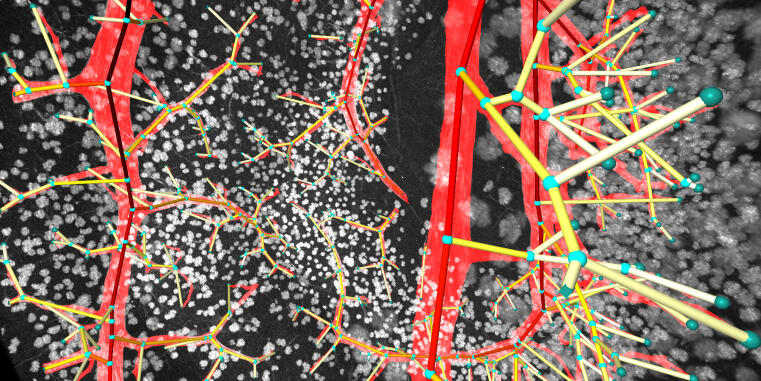
Drees D, Scherzinger A, Hägerling R, Kiefer F, Jiang X. Scalable robust graph and feature extraction for arbitrary vessel networks in large volumetric datasets. BMC Bioinformatics 2021;22: 346. Abstract
Evers M, Huesmann K, Linsen L. Uncertainty‐aware Visualization of Regional Time Series Correlation in Spatio‐temporal Ensembles. Computer Graphics Forum 2021;40: 519-530. Abstract
Kirschnick N, Drees D, Redder E, Erapaneedi R, Pereira da Graca A, Schäfers M, Jiang X, Kiefer F. Rapid methods for the evaluation of fluorescent reporters in tissue clearing and the segmentation of large vascular structures. iScience 2021;24: 102650. Abstract
Nahardani A, Leistikow S, Grün K, Krämer M, Herrmann K-H, Schrepper A, Jung C, Moradi S, Schulze PC, Linsen L, Reichenbach JR, Hoerr V, Franz M. Pulmonary Arteriovenous Pressure Gradient and Time-Averaged Mean Velocity of Small Pulmonary Arteries Can Serve as Sensitive Biomarkers in the Diagnosis of Pulmonary Arterial Hypertension: A Preclinical Study by 4D-Flow MRI. Diagnostics (Basel) 2021;12Abstract
2020
Jawad M, Soltwisch J, Dreisewerd K, Linsen L. Interactive Visual Analysis of Mass Spectrometry Imaging Data Using Linear and Non-Linear Embeddings. Information 2020;11: 575. Abstract
Xiao J, Jia Y, Jiang X, Wang S. Circular Complex-Valued GMDH-Type Neural Network for Real-Valued Classification Problems. IEEE Trans Neural Netw Learn Syst 2020;31: 5285-5299. Abstract
2019
Jawad M, Molchanov V, Linsen L. Coordinated Image- and Feature-space Visualization for Interactive Magnetic Resonance Spectroscopy Imaging Data Analysis. In: Proceedings of the 14th International Joint Conference on Computer Vision, Imaging and Computer 2019;SciTePress: 118-128. Abstract
2018
Klemm S, Jiang X, Risse B. Deep distance transform to segment visually indistinguishable merged objects. In: Proc. of 40th German Conference on Pattern Recognition (GCPR), Stuttgart 2018: 422-433.
Matute J, Telea AC, Linsen L. Skeleton-Based Scagnostics. IEEE Trans Vis Comput Graph 2018;24: 542-552. Abstract
Molchanov V, Linsen L. Shape-preserving Star Coordinates. IEEE Trans Vis Comput Graph 2018Abstract
Scherzinger A, Hugenroth P, Rüder M, Bogdan S, Jiang X. Multi-class Cell Segmentation Using CNNs with F1-measure Loss Function. In: Proc. of 40th German Conference on Pattern Recognition (GCPR) 2018;Springer, Cham: 434-446. Abstract
Sheharyar A, Ruh A, Aristova M, Scott M, Jarvis K, Elbaz M, Dolan R, Schnell S, Lin K, Carr J, Markl M, Bouhali O, Linsen L. Visual analysis of regional anomalies in myocardial motion. In: Eurographics Workshop on Visual Computing for Biology and Medicine. The Eurographics Association 2018: 135-144. Abstract
2017
Hägerling R, Drees D, Scherzinger A, Dierkes C, Martin-Almedina S, Butz S, Gordon K, Schäfers M, Hinrichs K, Ostergaard P, Vestweber D, Goerge T, Mansour S, Jiang X, Mortimer PS, Kiefer F. VIPAR, a quantitative approach to 3D histopathology applied to lymphatic malformations. JCI Insight 2017;2: e93424. Abstract
Ristovski G, Matute J, Wehrum T, Harloff A, Hahn HK, Linsen L. Uncertainty visualization for interactive assessment of stenotic regions in vascular structures. Computers & Graphics 2017;69: 116-130. Abstract
Ungru K, Jiang X. Dynamic Programming Based Segmentation in Biomedical Imaging. Comput Struct Biotechnol J 2017;15: 255-264. Abstract