Research

Picture of a phantom body in a tomograph
© EIMI/P. Leßmann

Technology & Imaging

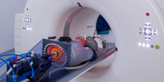
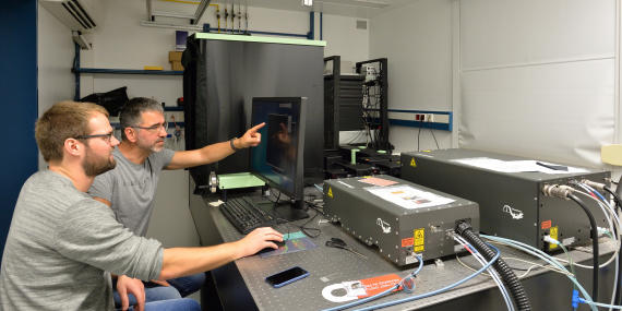
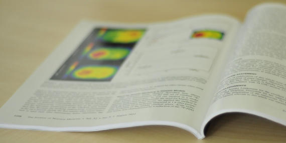
One focus at the EIMI is the design and the chemical synthesis of new target-specific tracers, which will allow visualization not only of inflammatory processes – in particular in vascular, auto-immune diseases and tumors – but also of bacterial infections. To do so, we use scintigraphic and optical imaging methods. In parallel, we continue to improve the technical methods of molecular imaging. Our aim is to translate novel insights and imaging strategies from disease models into patients – for innovative clinical diagnostics and therapies.

Picture of two people in a microscopy laboratory
© EIMI

Intravital Molecular Imaging

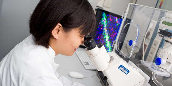
Our team studies the development and pathological scenarios of the vascular system. For example, we investigate how the lymphatic system develops, acquires and maintains its characteristic properties, how tumour vessels grow and how immune cells behave in chronic inflammatory settings. For this purpose, we develop and apply state-of-the-art microscopy, which enable us to generate three-dimensional images of vessels in tissue and to observe processes intravitally, i.e. dynamically in a living organism. Another focus is the refinement of existing and development of novel disease models.

Picture of people in an imaging laboratory
© EIMI/Peter Leßmann

Neuroimaging

We concentrate not only on inflammation which occurs in vascular and autoimmune diseases, but also on inflammatory processes in the brain in neurodegenerative diseases, strokes and tumours of the central nervous system. We develop molecular imaging strategies to localize and quantify disease-specific parameters. Our aim is to use our insights to make better assessments of how patients respond to therapies.

Picture of an opened scientific journal
© EIMI/Michael Kuhlmann

Innovative Ideas and New Insights

Our research is supported by third-party-funding, published in numerous papers in a variety of journals, and has often been patented.

Picture of a scientist working at a microscope
© CiM/Peter Leßmann

Background: Biomedical Imaging

Using imaging methods to investigate processes hidden inside the body is also a central theme of the Cells-in-Motion Cluster of Excellence at the University of Münster. EIMI Director Prof. Michael Schäfers is one of the three coordinators of this research alliance. But how exactly does “molecular imaging” work? This is explained by researchers in an article on the Cluster’s website.

Picture of a mouse in a tomograph
© EIMI/J.-M. Tronquet

Insights into Animal Testing Labs

In November 2017, the EIMI team and further researchers at the University of Münster demonstrated experiments with mice to journalists and gave insights into how different animals are kept at Münster University. The University has compiled an experience report and a collection of links to press articles.