
Imaging training for research
Photos

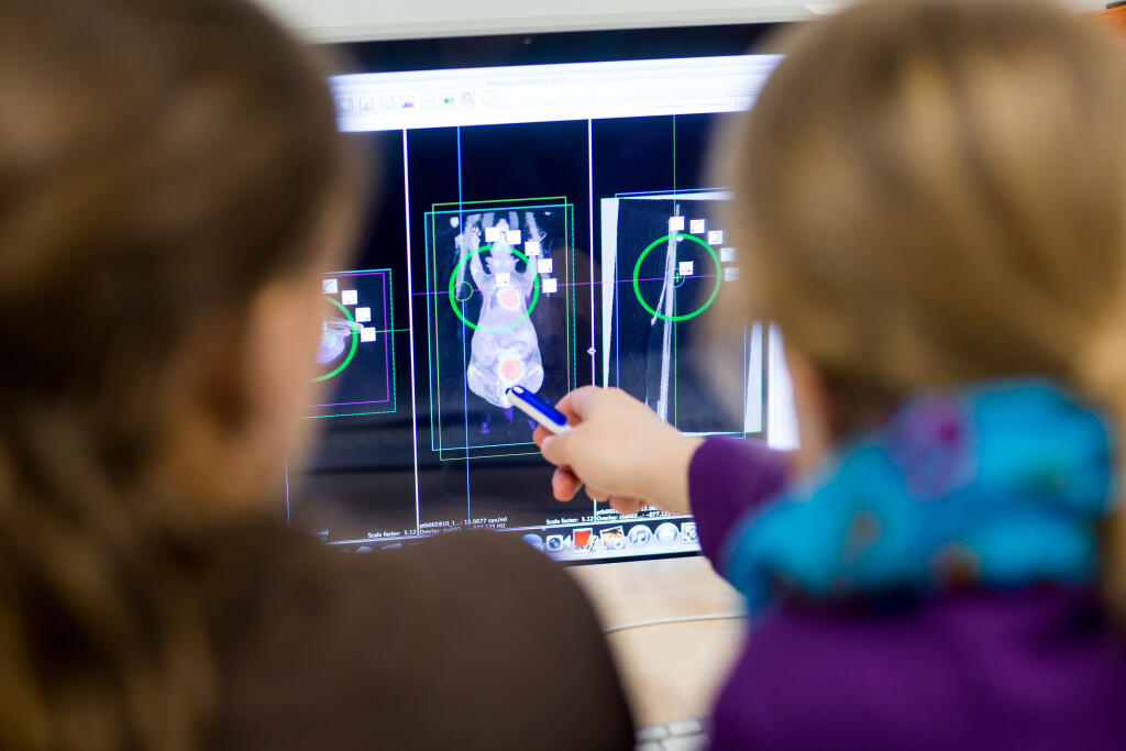
In the visualization laboratory, the participants practice analysing image data from examinations with computed tomography and positron emission tomography.© Uni MS/Peter Leßmann 
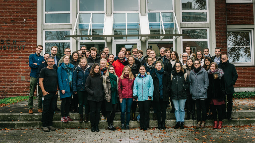
Participants and trainers at the Mouse Imaging Academy in 2019© Uni MS/Peter Leßmann
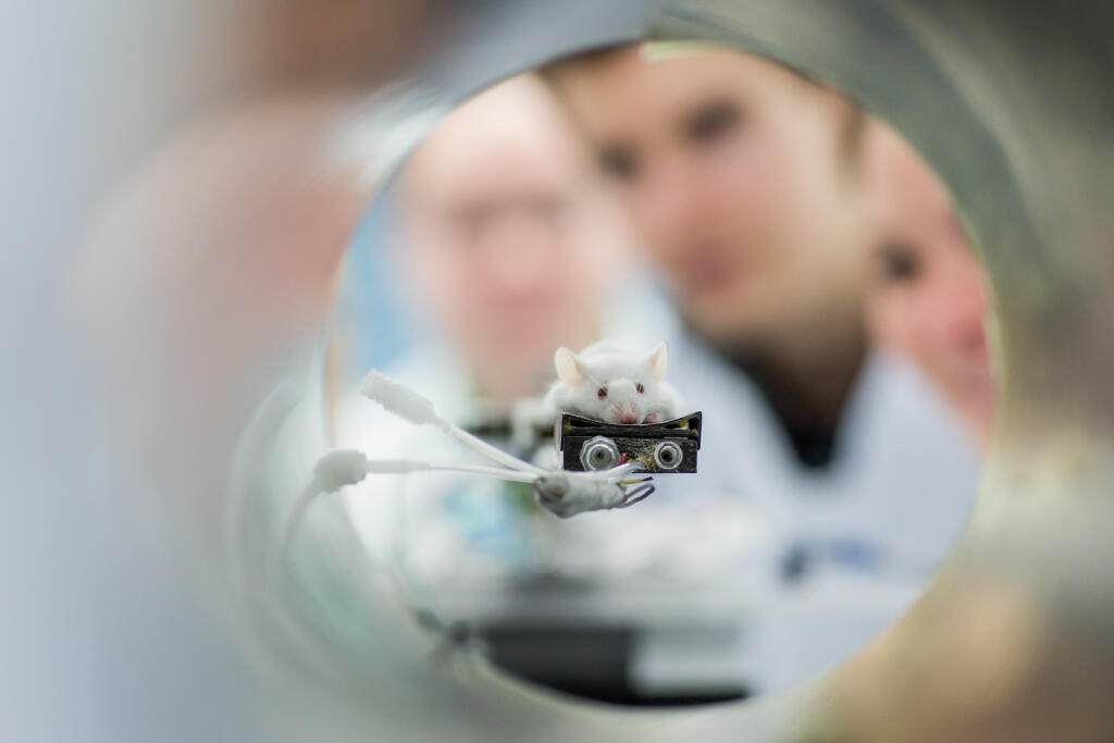
Among the most important tools in medicine are images that make visible what is happening inside the body. Not only do such images provide critical clues for medical care, but they also promote significant advances in biomedical research. Consequently, current research also focuses on the development of innovative imaging technologies. Last week, scientists from the University of Münster shared their knowledge in this field with international colleagues and junior scientists: At the tenth annual “Mouse Imaging Academy” (MIA), they gave a five-day introduction into imaging methods for research with mice.
As the first very fundamental takeaway, participants learned that choosing a suitable imaging technology for an investigation always depends on the question to be answered. “I recently joined a working group which uses optoacoustic imaging,” said biochemist Dr Monika Golinska from Cambridge University. “My colleagues recommended the course to get an overview about different imaging modalities, which is also important for cooperation projects with other labs.” The various technologies trained in the course included computed tomography, magnetic resonance imaging and optical imaging, as well as positron and single photon emission tomography.
In intense practical training sessions and supplementary lectures, the experts equipped the participants with knowledge about how the different modalities work. In addition, they gave insights into current developments in research. In small groups, the participants prepared mice for an examination and reflected on the knowledge they had gained about carefully handling the animals. The experts raised awareness regarding how different anaesthesia methods can influence the results of investigations, and the groups carried out an experiment with each imaging method. Using software developed in Münster, the participants analysed image data and determined the extent of a tumour, for example, or explored the extent to which a substance injected into the body accumulated in different organs over time. With the help of the experts, participants were able to deduce in-depth methodological aspects, which is a key prerequisite for being able to meaningfully prepare and interpret image data.
The almost 250 participants who have trained in Münster in recent years include numerous biologists but also physicians, chemists, physicists and mathematicians. For the workshop, more than 30 percent regularly come from other European countries and even from Asia and the USA. The University of Münster team, acting as trainers during the workshop week, comprises more than 20 imaging experts from nuclear medicine, radiology, biology, medical physics, biochemistry and radiochemistry. “A wide variety of disciplines comes together in the field of imaging – and to be able to select the right tool for a research question, you have to be ready to look beyond your own specialisation,” says Dr Sven Hermann, who is a specialist in nuclear medicine and in charge of coordinating the workshop. “This is why we are working very closely together in research and why we put emphasis on training a broad spectrum of imaging methods in an integrated way in our workshop.”