

From the cell to the patient: researchers in the new MIC building start their work
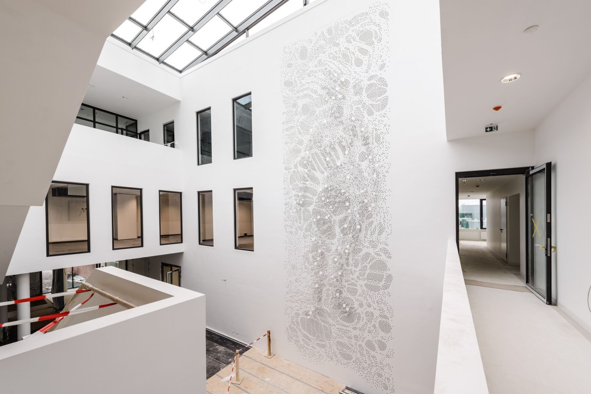
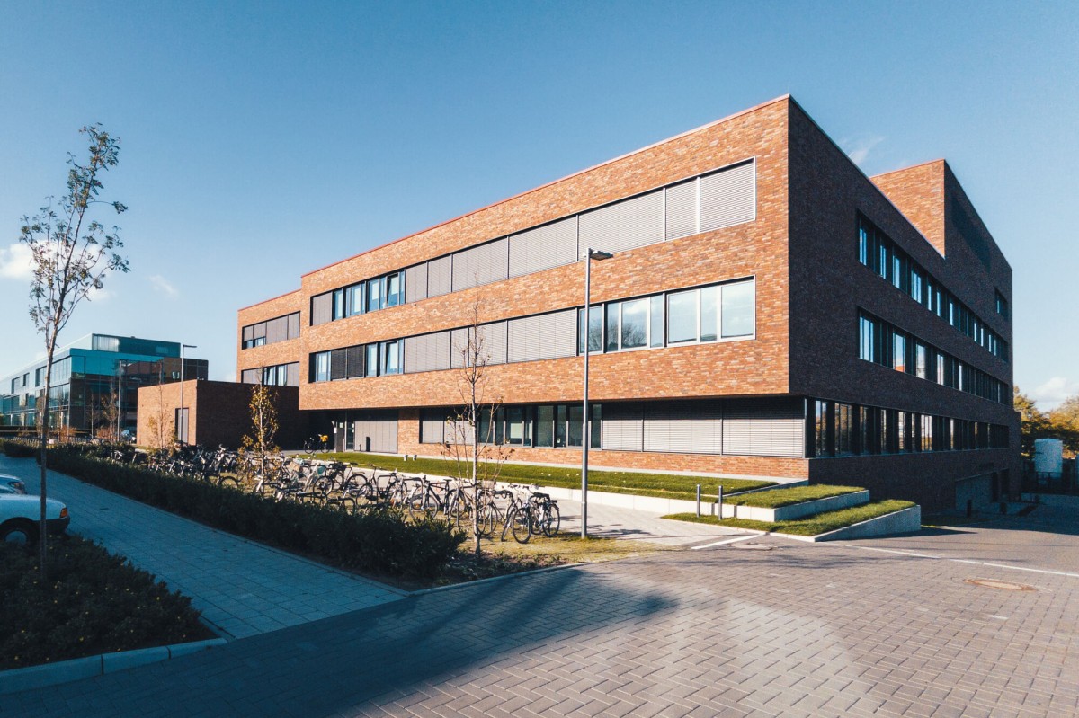
For any visitor walking through the atrium of the research building at the University of Münster – the Multiscale Imaging Centre (MIC) – there are two things which they notice especially: the large window fronts on several sides make it full of light, and on the left side the striking wall installation entitled “Auf|Lösung”. The welcoming entrance area, full of light, fits in well – after all, the MIC is located in the middle of the Life and Natural Sciences Centre of the University. Researchers from the fields of Medicine, Biology, Chemistry and Pharmacy, Mathematics and Computer Science will be working on floor space of 10,000 square metres, over three storeys, looking at how cells in the human organism behave, why they trigger diseases, and what can be done to combat this – and all this on the basis of a variety of modern imaging technologies. The topic of “Cell Dynamics, Inflammation and Imaging” has been one of the University’s research focuses for many years now – and with MIC the researchers now have the perfect infrastructure which they have long wished for.
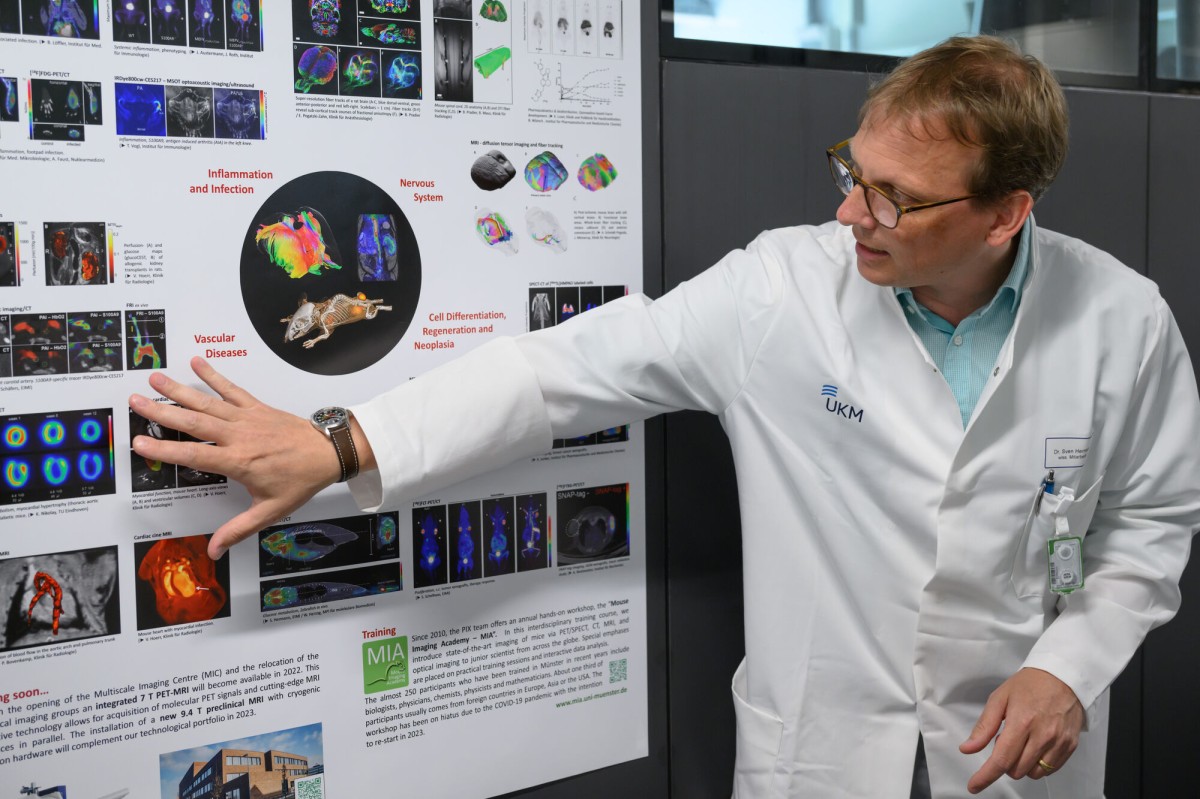
Prof. Michael Schäfers, a specialist in nuclear medicine and the MIC spokesperson, explains the unique nature of the research building: “The building is a lighthouse project, Europe-wide. We combine imaging methods, from microscopy to whole-body imaging, in interfaculty collaboration. This enables us to carry out research on dynamic cell processes at different scales: from the individual cell and tissue structures to organs and the whole body. Another feature is that we can use special imaging methods to visualise cell movements in different time-frames – for example, minutes, hours, days or weeks.” Everything takes place in vivo, in other words, on a living organism.
The aim of biomedical imaging is to conduct research into the behaviour of cells in organisms. The researchers study how organisms develop, how they maintain a healthy balance, and what happens in the case of a disease. They are also developing new possibilities for diagnosing and treating diseases. The University’s Comms and PR Department had an opportunity to take a look behind the scenes and get to know the building – on the basis of three research groups, from small to large.
Insights into research work at MIC
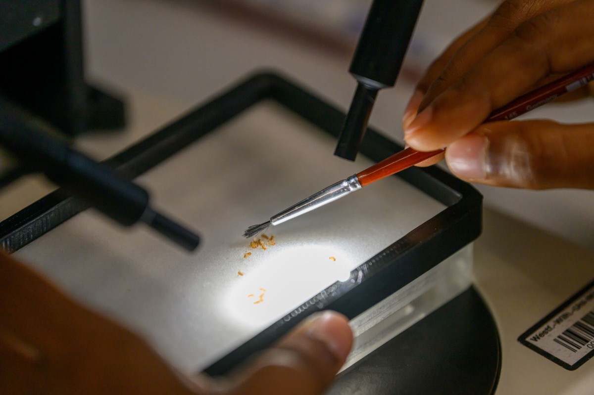
Right at the top, on the third floor, is the smallest model organism at MIC: using high-resolution light microscopy, a team headed by developmental biologist Prof. Stefan Luschnig is examining dynamic contacts between cells in the fruit fly. Luschnig’s aim is to understand how cell connections are produced and dynamically remodelled and how, in this way, cells form barriers which protect tissue and control transport processes. For this purpose, he has in his laboratory around 4,000 strains of fly – 100 flies per tube, with each genetic variant meticulously marked on each tube. Before the images are produced, using microscopy, some delicate preparation is necessary: researchers prepare the flies’ ovaries for the examination while the egg cells are developing. To this end, they mark certain proteins in the fruit fly with fluorescent molecules. In the next-door room, the researchers can then watch the processes in the cells, while they are happening, by means of confocal laser scanning microscopy, which produces razor-sharp images – and they can then analyse the molecular mechanisms behind the processes.
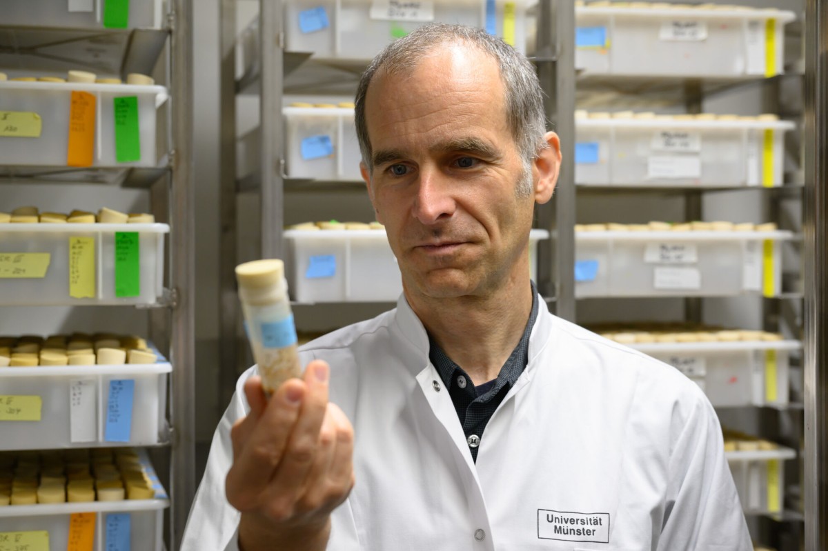
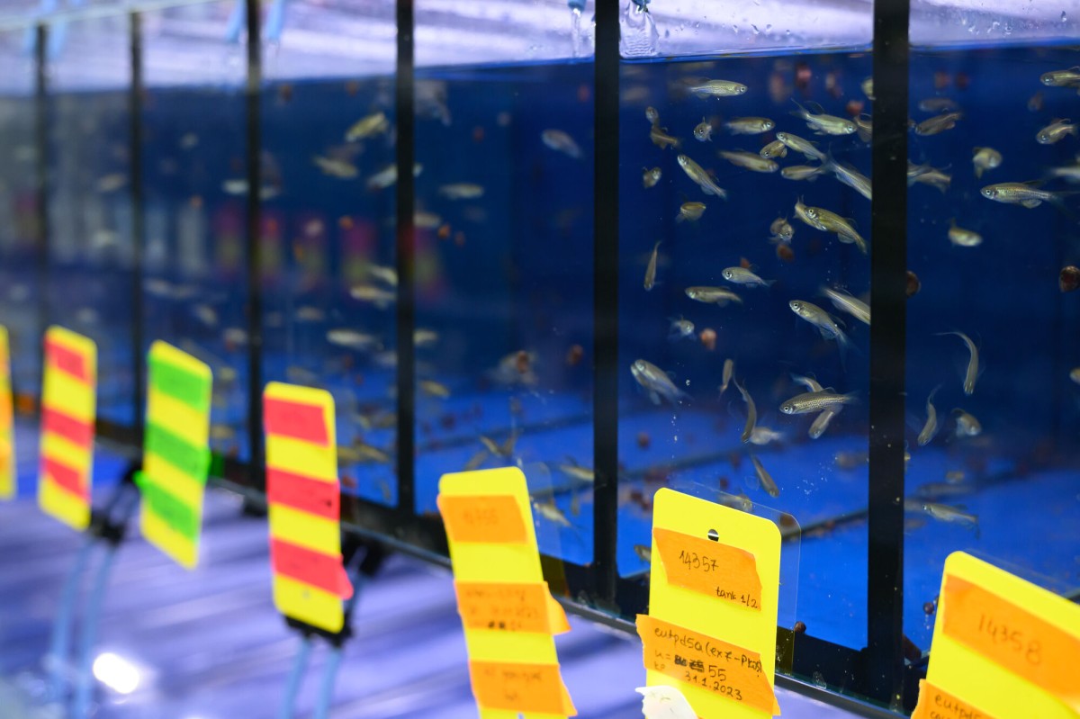
One floor down, Prof. Stefan Schulte-Merker also uses high-resolution light microscopy in his research – but with a somewhat larger model organism: the zebra fish. He has around 20,000 fish he can draw on, with enough room for 60,000. More than 1,260 water tanks, each containing 11 litres of water and 55 fish, are lined up in his lab. Currently, the fish are still getting used to their new surroundings, and, when they have done so, Schulte-Merker’s team can start work. “We’re investigating the development of blood vessels and lymphatic vessels in zebra fish embryos,” the biologist explains. “In the process, we’re looking for genes which are responsible for the individual steps and whose defects lead to diseases in humans.” One big advantage is that the embryos are transparent in the first five days of their lives, and they develop outside the mother’s body. “This means that we can look right inside them and watch the vessels forming.” The researchers watch as the cells form complex structures – from arteries, veins and lymphatic vessels to the entire vascular system.
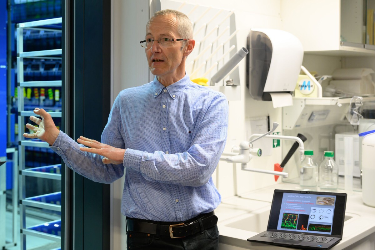
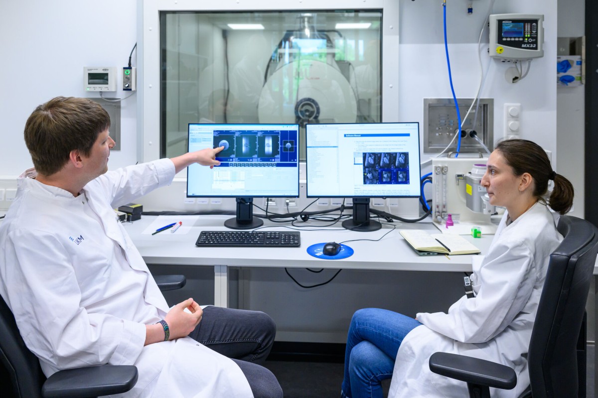
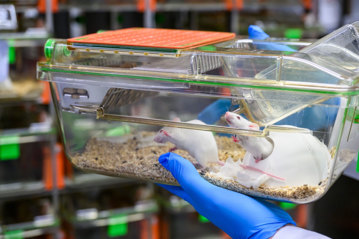
Down on the ground floor, the team headed by Dr. Sven Hermann und Michael Schäfers are examining mice with the aim, among other things, of developing methods for diagnosing inflammations and tumours. To this end they use positron emission tomography (PET), which visualises signals from radioactive tracers in the body and depicts biological processes in the tissue. The equipment used looks like the familiar tomographs used in hospitals – but much smaller: just for mice instead of for humans. One of the devices is a so-called PET-MRI which, in this high-performance design, is found at only four scientific locations. This hybrid device connects highly sensitive PET technology with magnetic resonance imaging (MRI) in a very high magnetic field strength. “With this innovative technology we can observe the course a disease takes – very accurately, and non-invasively over several day and weeks – and, for example, study responses to new therapies,” says Sven Hermann, explaining what the equipment, which cost three million euros, can do. Another special feature is that, unlike other hybrid equipment, the investigations made using the two technologies are undertaken not one after the other but simultaneously. “This is a decisive advantage,” says Michael Schäfers, “as biological processes can change within minutes, or even seconds. As we have no time-lag we can bring together the information from both investigation procedures even better.”
The “Re|Solution” work of art
Whether it is when they are entering the foyer or leaving it, there is one particular eye-catcher that neither visitors nor staff can miss. In the large entrance hall is a twelve-metre high wall installation created by artist Cordula Hesselbarth, Professor of Scientific Illustration. The work of art, entitled “Auf|Lösung” (“Re|Solution”), impressively depicts the field of research in which the researchers at MIC are working. The wall installation explodes the human organism into its component parts, creating an interplay between perceptions of the whole and of its parts.
The MIC profile:
Address
Röntgenstraße 16, 48149 Münster (in the vicinity of the Max Planck Institute for Molecular Biomedicine, the Centre for Molecular Biology of Inflammation (ZMBE), the Center for Soft Nanoscience (SoN) and the Centre for Nanotechnology (CeNTech)
Building commissioned by
Building & Real Estate Management Authority, North Rhine-Westphalia (BLB NRW)
Total floor space
10,000 square metres – of which 5,800 square metres are the main usable area
Lab area: 4,300 square metres
Offices and seminar rooms: 1.500 square metres
Total cost
63 million euros in funding from the “Research Buildings” programme of the German Ministry of Education and Research, plus a grant of eight million euros from the University of Münster
Construction time
Around five years
Beginning of work on building site: January 2017
Handover to the University of Münster: June 2022
First lab groups move in: December 2022
People and functionalities at MIC
18 professorships and junior research groups
Up to 260 staff
Branch of the Central Lab Animal Experimental Unit (ZTE)
Office of the Cells in Motion Interfaculty Centre
Coordination Centre for the Imaging Network