
Scientists receive a boost for research with cutting-edge imaging methods
Many scientists have longed for it and now it is coming to be: following approval of their application to the German Research Foundation’s "Large-scale Research Equipment" funding programme, researchers from the University of Münster will receive equipment for high-performance cryogenic electron microscopy. The equipment will enable the researchers to make molecular processes visible – for example, in human cells – and to examine particles such as viruses and synthetic nanostructures three-dimensionally, down to their individual atoms. The German Research Foundation and the State of North Rhine-Westphalia are providing a total of 7.5 million euros for the preparatory equipment and the latest-generation high-resolution microscope, which will be located in a purpose-built laboratory at the Center for Soft Nanoscience (SoN).
So far, there is no comparable cryogenic electron microscope at Münster University. Indeed, very few German universities have access to cryogenic electron microscopy equipment of this performance class." The University of Münster is considered one of the world's leading universities in the fields of 'cell dynamics and imaging' and nanoscience. The need for this state-of-the-art key technology is therefore immense," emphasises Prof Monika Stoll, Prorector for Research. "The device is a must if Münster University is to remain competitive in the international research landscape and the field of multiscale imaging."


The special structural equipment at the SoN, which was inaugurated in 2018, was a prerequisite for the acquisition of the microscope. For example, the floor in the laboratory where the cryogenic electron microscope will be located is almost perfectly vibration-damped. This is necessary for the operation of the device, otherwise, even a car driving past the building would ruin the complex and expensive measurements. Setting up the necessary IT infrastructure is also a special challenge as the device will record hundreds of terabytes of image data every month. Münster University’s high-performance computer "PALMA II" will be made available to process the data.
Microscopy at low temperatures
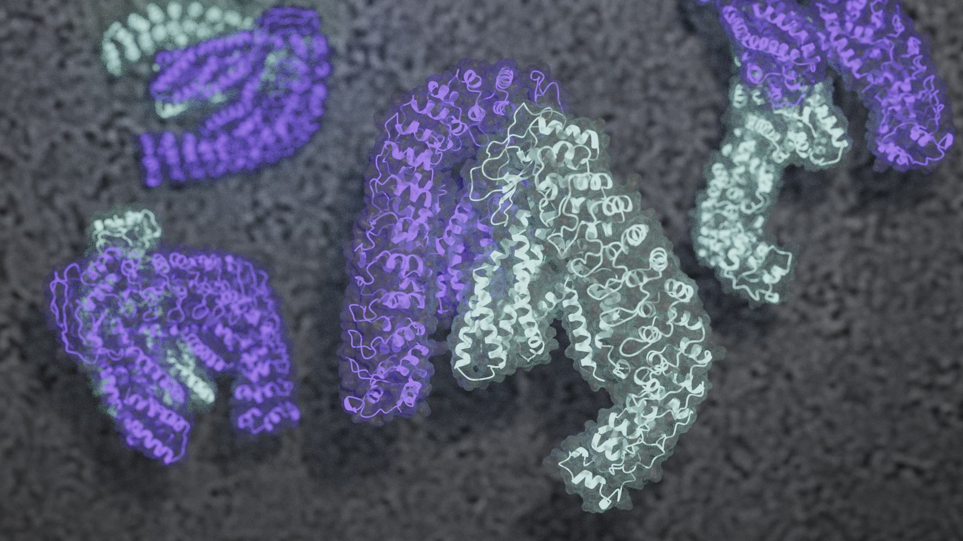
Cryogenic electron microscopy (cryo-EM) is a variant of transmission electron microscopy in which microscopic objects are imaged with the help of electron beams at cryogenic temperatures. In high-performance cryo-EM, the organic molecules to be examined are isolated or visualised directly in the living cell. Using a special procedure, the samples are cooled down to -196 degrees Celsius so quickly that they remain almost intact. In 2017, Jacques Dubochet, Joachim Frank and Richard Henderson received the Nobel Prize in Chemistry for developing this technique. In 2020, a team of researchers succeeded in observing individual atoms in a protein structure for the first time using cryo-EM. This technique makes it possible to understand how proteins function in living cells as well as how they cause diseases.