
Contents
- Transmission Electron Microscope (TEM)- Thermo Fisher Scientific FEI TITAN Themis G3 60-300
- Dual Beam microscope (SEM / FIB)- Zeiss CrossBeam 340
- Scanning Microscope (LSM)- Keyence VK-X
- Scanning Electron Microscope (SEM) - JEOL JSM-IT100
- Scanning Near-Field Optical Microscopy (SNOM) - neaSNOM
- Film thickness measurement - Filmetrics F20
- Time-of-Flight- Secondary Ion Mass Spectroscopy (TOF-SIMS) - Cryo-IONTOF M6 Special Edition
- Ellipsometer - Woolam M-2000
- Film thickness measurement system- Toho Spec 3100
- Atomic Force Microscopy (AFM)- NanoScope Icon, Bruker
- Atomic Force Microscopy (AFM)- Bioscope Resolve, Bruker
- Atomic Force Microscopy (AFM)- Nanowizard 3, JPK-Bruker
- Probe Station- Cascade MPS150
- Optical microscope - Nikon Eclipse LV100ND
Metrology
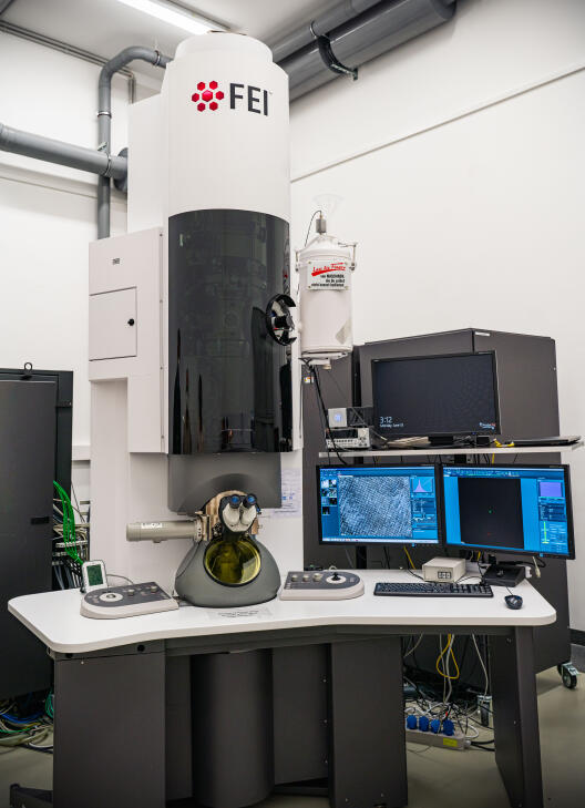
Transmission Electron Microscope (TEM)- Thermo Fisher Scientific FEI TITAN Themis G3 60-300
- Operation voltage 60 kV and 300 kV
- X-FEG field emission gun
- monochromator
- Cs image corrector
- quadrupole EDX-system
- HAADF detector (Fishione Model 3000)
- fast CMOS camera (CETA 4k x 4k)
- high resolution EEL spectrometer (GATAN Quantum 965)
Contact: Harald Rösner
Location: CeNTech II, lab E.05
Dual Beam microscope (SEM / FIB)- Zeiss CrossBeam 340
- SEM/FIB dual beam
- Zeiss Gemini I electron beam column
- Zeiss Capella Ga ion beam column
- Pt gas-injection system (GIS)
- Detectors: In-lens SE, BSD4
- Variable pressure option for biological samples
- Built-in O2- plasma
Contact FIB: Anna Korniushchenko
Contact SEM: Riya Gupta
Location: SoN, FIB zone
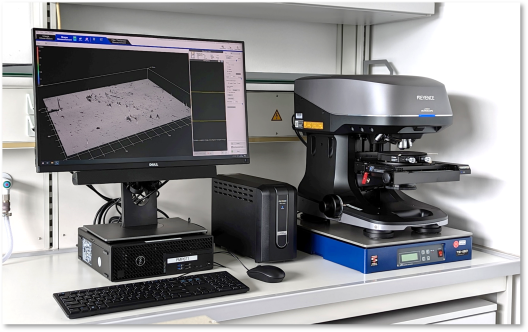
Scanning Microscope (LSM)- Keyence VK-X
- Digital optical Microscope
- 2D Measurements for structures >100nm
- Measure surface topology through:
- focus variation
- confocal laser scanning
- white light interferometry
- Height resolution: (few) mm down to (few) nm (depending on method)
Contact: Stefan Ostendorp
Location: CeNTech II, lab 1.04
Scanning Electron Microscope (SEM) - JEOL JSM-IT100
- Acceleration voltage: 20 kV
- Probe current: 1 pA – 0.3 µA
- Electron source: tungsten hairpin
- High and low vacuum operation: 10-100 Pa
- Detector: secondary-electron and backscattered electron detector
- Max. specimen size: 150 mm diameter
- Specimen movement range: 80 mm x 40 mm
- Focussing range: WD 5-48 mm
Contact: Maik Stappers, Riya Gupta
Location: CeNTech, Development Room, 0.21
Scanning Near-Field Optical Microscopy (SNOM) - neaSNOM
- Scattering-type Scanning-Near-Field-Optical-Microscope
- Ultrahigh resolution imaging with resolution < 10 nm
- Nano-FTIR: Ultrahigh resolution spectroscopy with resolution < 10 nm
- VIS-, NIR-, MIR-spectral region
- Atomic force microscope with resolution < 10 nm
Contact: Ivonne Bente, Daniel Wendland
Location: SoN, SNOM lab, 100.040
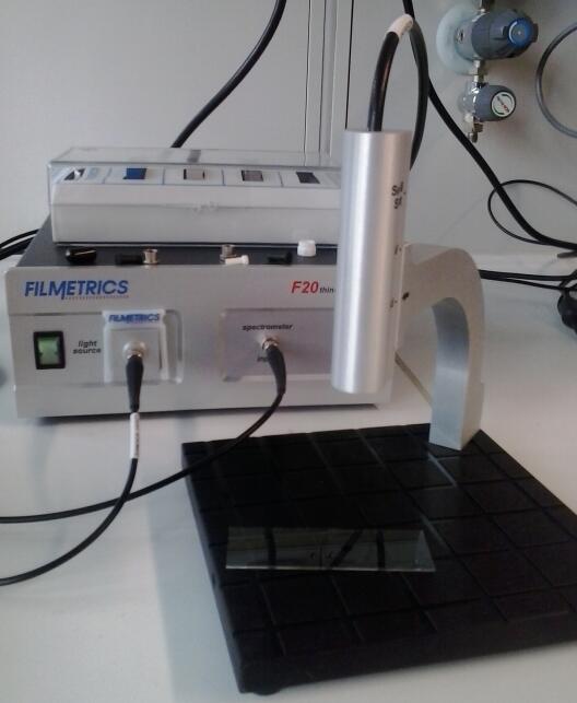
Film thickness measurement - Filmetrics F20
- 15 nm - 70 µm
- Wavlength range 380-1050 nm
- up to 1 µm spot thickness measurements microscope available
Contact: Maik Stappers
Location: SoN, plasma zone, CeNTech I, preparation zone, 0.15
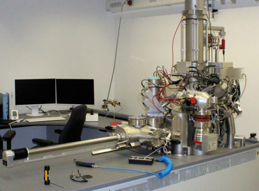
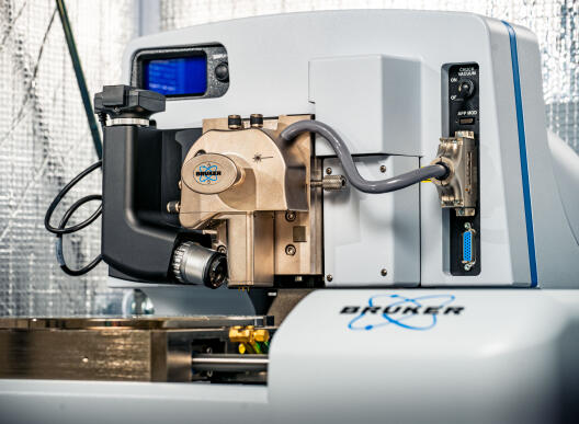
Time-of-Flight- Secondary Ion Mass Spectroscopy (TOF-SIMS) - Cryo-IONTOF M6 Special Edition
- Time-of-flight analyzer for mass resolution up to 30000, mass accuracy of a few ppm
- Bismuth liquid metal ion gun (30 keV) for high resolution (< 70 nm) imaging
- Argon gas cluster ion gun (5 to 20 keV) for analysis and molecular depth profiling
- Dual beam ion gun (0.25 to 2 keV, Ar+, O2-, Cs+) for depth profiling
- Cryogenic sample handling for analysis of hydrated samples
- Programmable sample heating and cooling (-180 to 600 ˚C)
- High speed sample rotation stage for high resolution depth profiling
Contact: Bonnie Tyler
Location: SoN, lab 110.037
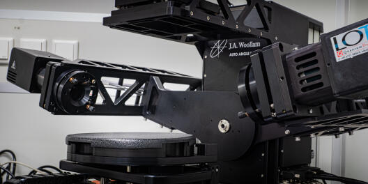
Ellipsometer - Woolam M-2000
- Excels in both general purpose thin film characterization (i.e., film thickness, optical constants) and large-area uniformity mapping
- Covers the wavelength range from 370 nm – 1690 nm
- Measures angle range 55° – 85° with automated tilt stages
- Fine measurement capable with focus probe, with a spot size of 100 um
- Automated alignment
Contact: Riya Gupta
Location: SoN, nanochemistry zone
Film thickness measurement system- Toho Spec 3100
Contact: Peter Lazarowicz
Location: SoN, nanochemistry zone
Atomic Force Microscopy (AFM)- NanoScope Icon, Bruker
- XY scan range: 90μm x 90μm typical, 85μm minimum
- Z range: 10μm typical in imaging and force curve modes, 9.5μm minimum
- Pixel density image up to 5120x5120
- Z range 10μm typical in imaging and force curve modes, 9.5μm minimum
Contact: Riya Gupta, David Lemli
Location: SoN, Bio-AFM lab, 100.042
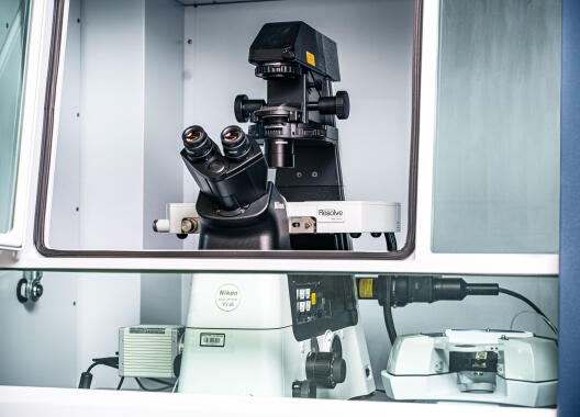
Atomic Force Microscopy (AFM)- Bioscope Resolve, Bruker
- Inverted light microscope
- X-Y Scan Range ≥100 μm, open-loop or closed-loop operation
- Z Scan Range ≥15 μm, open-loop or closed-loop operation
- Deflection Detection IR superluminescent diode (SLD) λ=850 nm
- Baseline Tilt <0.25 nm/μm
- XY Sensor Noise <150 pm
- Height Noise 35 pm (typical with appropriate vibration and acoustic isolation)
- XY Sample Stage Motorized stage with 10 mm x 10 mm range
Contact: Riya Gupta
Location: SoN, Bio AFM lab, 100.042
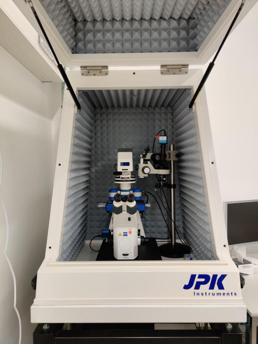
Atomic Force Microscopy (AFM)- Nanowizard 3, JPK-Bruker
- Soft and hard materials, biological samples (in liquid)
- Zeiss inverted light microscope
- Resolution: 1-2 nm
- Comprehensive force measurement from single molecules (suppliers claim) to living cells
- Z sensor noise level better than 35 pm RMS
- Motorized stage with 20mm2 x 20mm2 travel range
Contact: Steffen Lohrmann
Location: SoN, SNOM lab,110.037
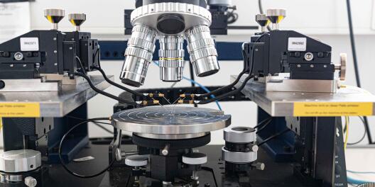
Probe Station- Cascade MPS150
- Manual electrical probing of individual devices
- Contact submicron features
- Chuck ready for single DUT
- Vacuum positioners with 1 μm feature resolution
- DPP450 positioner with nanometer resolution and accuracy
Contact: Connor Graham-Scott
Location: CeNTech II, electronics lab, 2.04
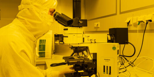
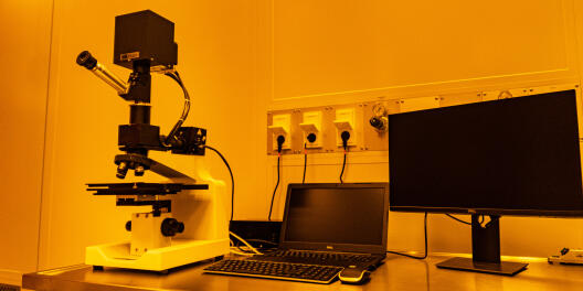
Optical microscope - Nikon Eclipse LV100ND
- Bright & Darkfield
- Magnification: 2.5 - 100 x
- Differential interference contrast (DIC) prism
- Max. sample size: 150 x 150 mm
Contact: Maik Stappers
Location: CeNTech I, preparation zone, 0.15