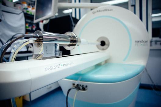
© EIMI Mediso nanoScan PET/MRI
Location: European Institute for Molecular Imaging, Waldeyerstr. 15
Contact Person: Sven Hermann, Tel.: 0251 83- 49303, shermann@uni-muenster.deApplications & Info:
- Integrated Positron-Emission-Tomography (PET) and Magnetic Resonance Imaging (MRI)
- Non-invasive 3D imaging of radiolabeled tracers in living mice combined with anatomical imaging
- PET: Molecular imaging of (patho-)physiological processes, e.g. cell metabolism, receptors, transporters, or cell tracking. Quantitative tracer detection up to few hours after injection, dependent on the half-life of the coupled positron emitting radioisotope (F-18, C-11, Ga-68, N-13), Spatial resolution: about 1 mm
- MRI: Anatomical imaging, Magnetic field strength: 1Tesla
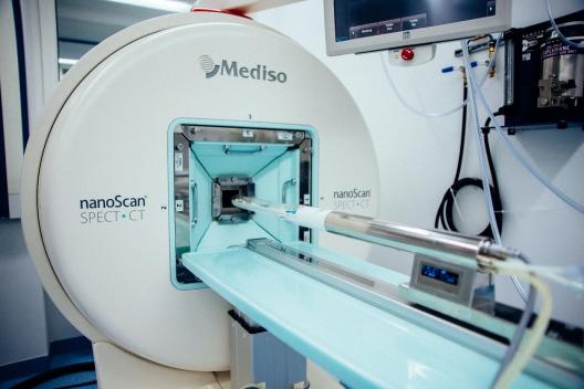
© EIMI Mediso nanoScan SPECT/CT
Location: European Institute for Molecular Imaging, Waldeyerstr. 15
Contact Person: Sven Hermann, Tel.: 0251 83- 49303, shermann@uni-muenster.deApplications & Info:
- Integrated Single-Photon-Emission-Computed-Tomography (SPECT) and Computed Tomography (CT)
- Non-invasive 3D imaging of radiolabeled tracers in living mice combined with anatomical imaging
- SPECT: Molecular imaging of (patho-)physiological processes, e.g. cell metabolism, receptors, transporters, or cell tracking. Quantitative tracer detection up to several days after tracer injection, dependent on the half-life of the coupled gamma emitting radioisotope (Tc-99m, In-111, I-123)
- CT: Anatomical imaging
- SPECT equipment: Multi-Pinhole-Collimators for different applications (APT51, APT52, APT62, APT66), Spatial resolution: 0.5 – 1.5 mm
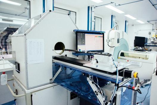
© EIMI quadHIDAC PET
Location: European Institute for Molecular Imaging, Waldeyerstr. 15
Contact Person: Sven Hermann, Tel.: 0251 83- 49303, shermann@uni-muenster.deApplications & Info:
- Positron-Emission-Tomography (PET)
- Non-invasive 3D imaging of radiolabeled tracers in living mice combined with anatomical imaging
- PET: Molecular imaging of (patho-)physiological processes, e.g. cell metabolism, receptors, transporters, or cell tracking. Quantitative tracer detection up to few hours after injection, dependent on the half-life of the coupled positron emitting radioisotope (F-18, C-11, Ga-68, N-13)
- Spatial resolution: below 1 mm
- Field of view: 16 cm x 28 cm
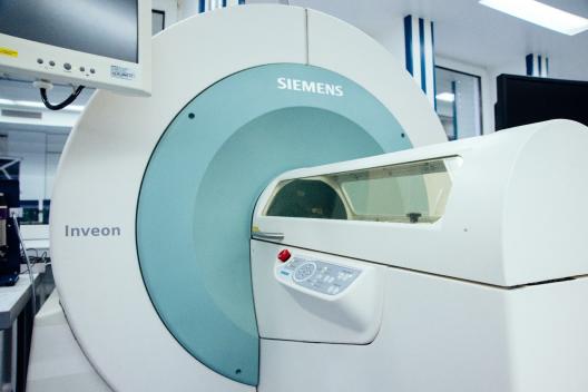
© EIMI Siemens Inveon CT
Location: European Institute for Molecular Imaging, Waldeyerstr. 15
Contact Person: Sven Hermann, Tel.: 0251 83- 49303, shermann@uni-muenster.deApplications & Info:
- Non-invasive computed tomography (CT) of living mice
- Anatomical imaging
- Spatial resolution: 80µm (down to 25µm)
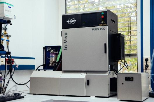
© EIMI Bruker MS FX Pro
Location: European Institute for Molecular Imaging, Waldeyerstr. 15
Contact Person: Sonja Schelhaas, Tel.: 0251 83- 49312, sonja.schelhaas@uni-muenster.deApplications & Info:
- 2D Fluorescence reflectance imaging (FRI) of living mice or ex vivo organs
- X-ray imaging
- Filter specifications: 28 excitation filters (20 nm bandwidth, 410 nm – 760 nm), 6 emission filters (35-40 nm bandwidth, 535 nm, 600 nm, 700 nm, 750 nm, 790 nm, 830 nm)
- Multimodal animal rotation system (MARS)

© EIMI Perkin Elmer IVIS Spectrum
Location: European Institute for Molecular Imaging, Waldeyerstr. 15
Contact Person: Sonja Schelhaas, Tel.: 0251 83- 49312, sonja.schelhaas@uni-muenster.deApplications & Info:
- 2D Bioluminescence and fluorescence reflectance imaging of living mice or ex vivo organs
- Filter specifications: 10 excitation filters (30 nm bandwidth, 415 nm – 760 nm), 18 emission filters (20 nm bandwidth, 490 nm – 850 nm)

© EIMI Visualsonics Vevo2100 LAZR
Location: European Institute for Molecular Imaging, Waldeyerstr. 15
Contact Person:
Sven Hermann
Tel.: 0251 83- 49303
shermann@uni-muenster.deApplications & Info:
- High resolution ultrasound (US) & photoacoustic imaging (PAI)
- US: Non-invasive 2D anatomic and functional imaging in living mice
- PAI: Non-invasive 2D detection of photoabsorbers (e.g. fluorescent dye IRDye800cw) in living mice
Transducers
Frequency Image depth (max) Spatial resolution MS250 (US) / LZ250 (PAI/US) 13-24 MHz 30 mm 75µm (axial), 165µm (lateral) MS400 (US) 18-38 MHz 20 mm 50µm (axial), 110 µm (lateral) MS550D (US) / LZ550 (PAI/US) 22-55 MHz 15 mm 40 µm (axial), 90µm (lateral) MS700 (US) 30-70 MHz 12 mm 30µm (axial), 75µm (lateral) 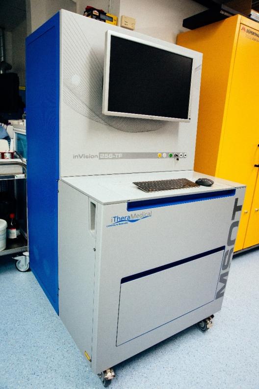
© EIMI iThera MSOT inVision
Location: European Institute for Molecular Imaging, Waldeyerstr. 15
Contact Person: Sven Hermann, Tel.: 0251 83- 49303, shermann@uni-muenster.deApplications & Info:
- Multispectral Optoacoustic Tomography (MSOT)
- Non-invasive 3D detection of photoabsorbers (e.g. fluorescent dye IRDye800cw) in living mice

© EIMI Biospace µ-Imager
Location: European Institute for Molecular Imaging, Waldeyerstr. 15
Contact Person: Sven Hermann, Tel.: 0251 83- 49303, shermann@uni-muenster.deApplications & Info:
- Digital autoradiography camera
- Measurement of radiotracer distribution in tissue samples (cryosections)
Heidelberg Engineering OCT Spectralis
Location: University Eye Hospital, Domagkstr. 15
Group: AG Eter
Contact Person: PD Dr. Peter Heiduschka
Tel.: 0251 83-57532, peter.heiduschka@ukmuenster.de
Applications & Info:- In-vivo Imaging of the retina by infrared scanning laser ophthalmoscopy, optical coherence tomography, and fluorescence angiography
Vectra 3.0 Automated Quantitative Pathology Imaging System, 200 Slide, Perkin Elmer
Location: Gerhard Domagk Institute of Pathology
Group: AG Rössig, in cooperation with AG Wardelmann
Contact Person: Prof. Wolfgang Hartmann
Tel.: 0251 83-58479, wolfgang.hartmann@ukmuenster.de
Applications & Info:- simultaneous detection of multiple markers
- detailed in situ analyses of the cellular composition of individual tissues and tumors
- spatial relationships of immune phenotypes on (tumor) biopsies
