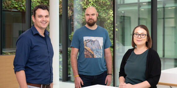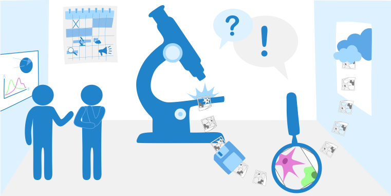
Tools, tips and training for microscopy – we offer individual and comprehensive support to researchers at the University of Münster and beyond in the areas of cutting-edge microscopy, reproducible image analysis, and optimal strategies for data management and publication.
© Münster Imaging Network – Marie Baldenius

Münster Imaging Network – Microscopy

Our team provides a wide range of microscopy systems featuring the latest methods and technologies for biomedical research, and supports users in their application. In addition, we familiarise you with the underlying data management practices – promoting transparency, quality, and reusability in research. We offer tailored advice on experimental design and image analysis, as well as hands-on support for your experiments and for publishing your research. Together with our colleagues in preclinical imaging we form the Münster Imaging Network. We are embedded into the Cells in Motion Interfaculty Centre (CiM), that connects and supports researchers in the field of cell dynamics and imaging across working groups and faculties. Our network is listed in „RIsources”, the research infrastructure registry of the German Research Foundation (DFG). In addition, our microscopy platform is part of NFDI4BIOIMAGE, a consortium within the DFG-funded National Research Data Infrastructure (NFDI).

© Münster Imaging Network – Marie Baldenius

Microscopes

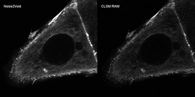
DeepLearning based denoising with Noise2Void

Click on the image to open it with OMERO.iviewer

Noise2Void can be used for denoising your images without the need for a ground truth. Training can be done on single noisy images in Fiji. Noise2Void can also be used if deconvolution is not possible due to unsuitable imaging parameters. Furthermore it can be used for almost all kinds of images, like EM, RGB, photographs etc. Curious what it can do for your images? You are welcome to contact us via @Mattermost or by mail imaging@uni-muenster.de .

Read the paper: Noise2Void - Learning Denoising from Single Noisy Images | Noise2Void on Github

© Thomas Zobel

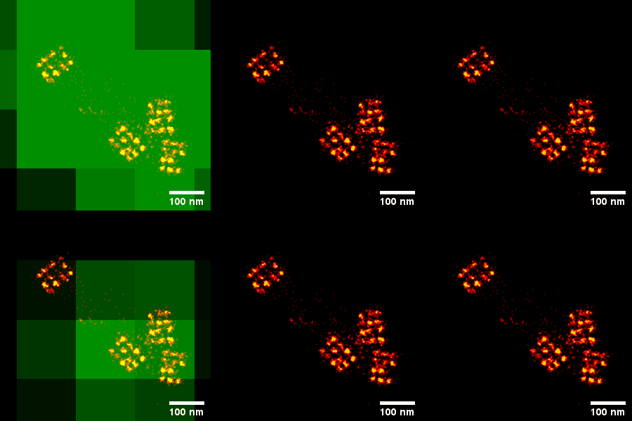
Super-resolution microscopy with DNA-PAINT

Reconstruction of a DNA-PAINT imaging sequence. 20 nm DNA origami nanorulers are shown in red. The original pixelsize during detection is shown in green.

In March 2020, we participated in the "Trends in Microscopy" (TiM-2020) Summer School – an intensive and hands-on event focused on modern microscopy techniques. Among other activities, we carried out DNA-PAINT experiments during full-day practical sessions, achieving an impressive resolution of around 4 nm, despite challenging conditions in a crowded and warm microscopy room. DNA-PAINT is relatively easy to implement, and our group has since gained extensive experience with the method.

Feel free to contact us via @Mattermost or by mail imaging@uni-muenster.de .

Original paper: Super-resolution microscopy with DNA-PAINT Joerg Schnitzbauer, Maximilian T Strauss, Thomas Schlichthaerle, Florian Schueder & Ralf Jungmann Nature Protocols volume 12, pages1198–1228(2017)