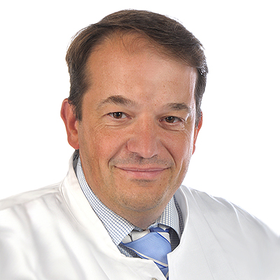
“Will we be able to predict heart attacks one day?”

Prof. Schäfers, what scientific topic are you working on right now?
My research is mainly focused on three research areas. To start with this is imaging of inflammation, which, e.g., is underlying arteriosclerosis in the vessel wall and can trigger myocardial infarction. My team and I develop new technologies that make inflammatory processes visible in tomographs. This needs so-called tracers, in other words substances which upon injection in the blood stream accumulate in inflammatory foci in a patient’s body. These tracers not only enable us to see the location of an inflammation, but also to assess how active the disease is. A second research focus is to follow the movements of cells in the body, as these play a significant role in many diseases. We look at the dynamics of immune cells, for example, in order to gain a better understanding of pathological behaviour. In my third research focus I aim at transferring our research results to clinical applications.
All research in my institute – the European Institute for Molecular Imaging (EIMI) – is centered around imaging, in other words technologies which enable to look inside bodies. What’s special about EIMI is that we’re interested in the entire scale of imaging scales – from microscopy and small animal imaging to applications in clinical practice. We also look at individual cells in tissues as well as in whole organs in mice or in people. This broad scale is a great challenge because every technology functions differently – and also has its limitations. We’re already able to get a good look inside people and mice by using various technologies, but in doing so we look at the whole body and not individual cells. We’re searching for ways of looking at this smallest of dimensions. Given time, we should be able to link up all the different methods.
What characterizes you personally as a scientist?
I like being creative not only in the lab, but also in my private life. I was already making music in my student days, and in the early 1990s some friends and I formed the a capella group “Simple Voices”. Five of the founding members are still singing with us. A few of the current members of the ensemble are co-workers at EIMI. I also like cooking and enjoy little building projects at home. Working with my hands in this way enables me to relax, no matter how much stress there is in the clinic or the institute.
What is your greatest aim as a scientist?
I like translational work, in other words working as a medical doctor and a researcher. This way I can combine basic science and innovative medical applications. What gives me a lot of pleasure is training my staff to go in this direction. After all, both patients and staff alike benefit from this close link between basic research and medical work: we can treat patients better using innovative solutions, and scientists can experience how their research work leads to improvements in clinical processes. That would be my dream, to develop a clinical imaging technology that would be used in clinical practice.
What’s your favourite “toy” for research – and what can it do?
The positron emission tomograph (PET), without a doubt. If this imaging technology is combined with innovative tracers it can have an extremely powerful range of applications. We can look inside a living body from the outside and, with various tracers that we’re developing, specifically study various diseases. When I was a student I got to know PET when it was in its infancy. It was at that time that the first images of glucose metabolism of a human brain were produced using a PET. That must have been around 1990.
Can you remember your happiest moment as a scientist?
I’ve had a lot of good fortune in my research career. My happiest moment was certainly being awarded the Cells-in-Motion Cluster of Excellence, because it was so difficult to get it. We spent a long time working on a coherent strategy which, just from the point of view of its complex structure, was bigger than anything I had known up to then. Apart from that, what makes me happy is working on a scientific publication. When you sit together with colleagues, analyse data and bounce ideas back and forth until you produce story that fosters scientific progress.
What big scientific question would you like to have an answer to?
Will we be able to predict heart attacks one day? It’s a question I am confronted with daily when studying myocardial perfusion in patients. I’d really like to have an answer to this one big question, and – of course – a positive one!

