
Photo gallery: “Lange Nacht der Universitätsmedizin” – Impressions from the Multiscale Imaging Centre
Photos


This is what silicone molds for replicating a beating heart look like. Researchers use a wide variety of materials to build “phantoms” – that is to say experimental laboratory setups that mimic biological scenarios.© Uni Münster - Michael Kuhlmann 
Components of an artificial body: a spine produced using a 3D printer, a heart made of silicone, and tubes in which researchers can simulate blood flow.© Uni Münster - Michael Kuhlmann 
How can we generate images from inside the body? Visitors were given an exciting overview. Here you can see an X-ray image of a hand.© Uni Münster - Michael Kuhlmann 
Visitors were given a heart printed on a 3D printer to take home with them.© Uni Münster - Michael Kuhlmann 
Heading to the microscopy lab!© Uni Münster - Michael Kuhlmann 
Visitors young and old tried their hand at research.© Uni Münster - Michael Kuhlmann 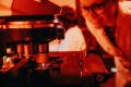
Using a microscope, visitors examined fruit fly eggs and larvae.© Uni Münster - Michael Kuhlmann 
A microscope slide with eggs in which fruit flies develop.© Uni Münster - Michael Kuhlmann 
Do fruit flies have brains? Yes – and this is what a fly brain looks like on a microscopy image.© Uni Münster - Michael Kuhlmann 
All of this is part of a microscopy workstation: not just the microscope itself, but also, for example, a box containing lasers. The laser light makes fluorescently labelled molecules shine. The fluorescence signals are measured by the microscope and translated into images. Researchers can then analyse the images on a computer.© Uni Münster - Michael Kuhlmann 
Visitors also had the opportunity to observe the beating heart of a zebrafish live under the microscope. This is possible because zebrafish are transparent during the first five days of their lives and develop outside the womb.© Uni Münster - Michael Kuhlmann 
The scientists explained what they are researching with the help of zebrafish. They investigate how blood vessels and lymph vessels develop in a healthy body and search for genes whose defects cause diseases.© Uni Münster - Michael Kuhlmann 
A common question: How does research with zebrafish benefit humans? The researchers explained that scientists worldwide have already identified many genes that are also important for vascular development in humans and thus play a role in many diseases.© Uni Münster - Michael Kuhlmann 
Visitors tried their hand at collecting zebrafish eggs from an aquarium using dummies. The researchers breed different genetic strains of fish that they can use to investigate specific questions.© Uni Münster - Michael Kuhlmann 
Ready for the lab! Numerous visitors took the opportunity to gain live insights into different areas of research.© Uni Münster - Michael Kuhlmann 
What can we do using magnetic resonance imaging (MRI)? During the “Lange Nacht”, visitors had the opportunity to watch live as a baby shark fixed in alcohol was examined. Since the procedure is “non-invasive”, researchers can examine such special samples several times without destroying them.© Uni Münster - Michael Kuhlmann 
Here, the shark is placed in the small animal MRI scanner. In everyday research, this device is mainly used to examine mice. It is the same technology that is used in clinical practice on patients, but in a significantly smaller and much more powerful version.© Uni Münster - Michael Kuhlmann 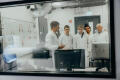
A view from the room where the MRI scanner is located. Visitors could observe the examination from outside through a window. During the examination, the MRI scanner generates powerful magnetic fields and creates images showing how the hydrogen atoms in the body react to them (“magnetic resonance”).© Uni Münster - Michael Kuhlmann 
Here, a group discussed the images that had been created and learned more about how MRI works: During the examination, scientists can vary certain physical parameters (creating “MRI sequences”). Researchers work on testing different imaging techniques and develop new approaches to achieve optimal image contrast for specific questions.© Uni Münster - Michael Kuhlmann 
In the foyer of the Multiscale Imaging Centre, visitors could experience science and art in the exhibition inVISIBLE.© Uni Münster - Michael Kuhlmann 
Visitors explored colorful images from biomedical research and immersed themselves in the impressive stories behind them.© Uni Münster - Michael Kuhlmann 
At dusk, moving light projections illuminated the wall installation in the foyer of the Multiscale Imaging Centre. Cordula Hesselbarth’s work Re | Solution artistically explores the question of how images from inside the body are created.© Uni Münster - Michael Kuhlmann


About 1,000 staff members contributed 220 programme items to open the doors for the public during the “Lange Nacht der Universitätsmedizin Münster” on September 12, 2025. Approximately 14,000 visitors attended the event, which marked the highlight of the anniversary year celebrating 100 years of the Faculty of Medicine and the University Hospital.
Numerous research groups in the field of cell dynamics and imaging were part of the event. Here are some pictures from the research building of the Cells in Motion Interfaculty Centre: the Multiscale Imaging Centre!