
Looking at the mechanisms of translation
Proteins are the workers in the cells of our body, performing all of their tasks. The blueprint for the specific proteins of any given cell is encoded in its genetic information, and they are produced in a process known as “translation”. Because every cell requires specific proteins, translation is highly regulated, but the regulatory mechanisms are still ill-defined. Researchers at the Cells-in-Motion Cluster of Excellence at Münster University (Germany) investigated how translation is regulated during the development of the nervous system. They found that translation is strongly regulated at a time when certain nerve cell processes degenerate during development: “Translation is regulated by so-called translation initiation factors. One of these, which normally plays a key role in growth, is inactivated at this stage” says Dr. Sebastian Rumpf, a junior research group leader at the Cluster of Excellence and head of the study. When this initiation factor, known as elF4F, is inactive, the production of proteins is greatly reduced, thus allowing for the degeneration of nerve processes. However, there are a number of proteins that must be produced at the same time the cell has to ensure that certain genes – those that are indispensable for the degeneration processes – continue to be translated into proteins. The researchers discovered the mechanism behind this phenomenon: the cell initiates a sort of “bypass signalling pathway” in which an alternative initiation factor elF3 is recruited instead of the usual initiation factor elF4F. Even under the low-translation conditions at this stage, eIF3 can still stimulate the translation of proteins that are important for the process. “When nerve cell processes degrade in the course of development, different translation mechanisms appear to be active than when they grow,” says Dr. Sandra Rode, the lead author of the study. The concept of the "bypass signalling pathway" had previously been proposed by other research groups, but in this study it was demonstrated for the first time in living organisms. “We are sure that the mechanism is widespread and could be found in many other contexts,” says Sandra Rode. Biologists, biochemists and molecular biologists worked closely together on the study, which has been published in the journal “Cell Reports”.
The story in detail:
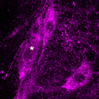
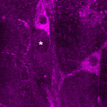
Nerve cells connect through their long processes, the axons and dendrites, collectively known as neurites. Neurites that were wrongly connected in the course of development or did not acquire a specific function, degenerate again and dissolve their connections. A team of Münster biologists led by Dr. Sebastian Rumpf looked at special nerve cells in Drosophila melanogaster fruit flies. These neurons lose all their dendrites at a specific point during development. The team used genetic manipulations – easy in the fruit fly – to study whether translation plays a role during this process.
The researchers took a close look at the functions of various translation initiation factors. These proteins bind to the messenger RNA (mRNA), a biomolecule which transports the genetic information from the cell nucleus to those places in the cell where proteins are produced. Translation initiation factors then cause the translation to begin. First of all, the researchers genetically inactivated one factor – elF4F – that normally plays a central role in translation initiation. Using microscopy they then observed the nerve-cell processes during the fruit flies’ pupal phase and found that they were still degenerating quite normally. However, whenever the researchers inactivated the initiation factors, eIF3 or eIF4A, degeneration was inhibited and there were still dendrites present. “So these two factors appear to be important for the degeneration of cell processes,” says Sandra Rode.
But what are the mechanisms behind this? The researchers examined whether the signalling pathways had any influence on the activity of the MICAL gene. This gene is needed for the degeneration of the nerve-cell processes. The researchers again modified the translation initiation factors and investigated the expression of MICAL in the cell. The results confirmed the connection: whenever eIF4A or eIF3 were inactive, MICAL was expressed in smaller quantities.
“We next asked: which local factors play a role?” says Sandra Rode. The researchers turned their attention to the mRNA, at the start of which (the so-called ‘cap’) the translation initiation factors bind. Previous studies had already shown that translation initiation factors also frequently interact with another region of the mRNA – the so-called untranslated region adjacent to the cap. A team of biochemists led by Prof. Andrea Rentmeister, a professor at the Cluster of Excellence, helped to investigate the role of this region. After carrying out initial experiments in a test tube, the researchers observed the processes taking place in the mRNA of a living organism. To this end, they linked a fluorescent signal with the untranslated area to generate a so-called “reporter gene” and were thus able to measure its activity. When they genetically inactivated eIF4A and eIF3 in the neurons, the fluorescent signal was much weaker. “From this, we concluded that the recognition signals for the initiation factors really are encoded in this region of the MICAL mRNA,” says Sebastian Rumpf.
From their results, the researchers suspected that the two initiation factors eIF3 and eIF4A interact during the translation of MICAL. A team of molecular biologists led by Dr. Sebastian Leidel, also a group leader at the Cluster of Excellence, used biochemical methods to address this question. They discovered that eIF4A and eIF3 are present in a complex connected by the messenger RNA. When they inhibited eIF4A, this also had an effect on elF3 binding to the MICAL mRNA. “In this context, these are very new interactions between the two factors, and we wouldn’t have discovered them without the interdisciplinary collaboration that we had,” says Sebastian Rumpf. In further studies the researchers want to address the precise role played by the cap of the mRNA.
Funding:
The Cells-in-Motion Cluster of Excellence at the University of Münster provided funding for the study as part of two interdisciplinary projects between group leaders as well as between junior researchers.
Original publication:
Rode S, Ohm H, Anhäuser L, Wagner M, Rosing M, Deng X, Sin O, Leidel SA, Storkebaum E, Rentmeister A, Zhu S, Rumpf S. Differential Requirement for Translation Initiation Factor Pathways During Ecdysone-Dependent Neuronal Remodeling in the Drosophila PNS. Cell Rep 2018, DOI: 10.1016/j.celrep.2018.07.074. Abstract