
Learning new methods
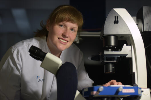
Biomedical scientist Dr. Verena Hofschröer studies malignant melanoma. Her aim is to find out how cells of the primary tumour detach to form metastases. Using atomic force microscopy she first studied how strongly individual cancer cells stick to each other. “I wanted to transfer these findings to networks of cells and to work three-dimensionally,” she explains. She found the solution during a six-month research stay at the University of Nijmegen in the Netherlands, funded by a CiM Train-Gain grant. Through this programme, the Cluster finances travel and accommodation of CiM scientists to undertake specialized research training at renowned international institutions.
From June to December 2015, Verena Hofschröer performed her research in Nijmegen using a confocal laser-scanning microscope in a special set-up, which enables tracking of fluorescently labelled cell clusters within tissues, i.e. three-dimensionally. “I had to learn this method from the ground up,” she says. “Today I feel confident with this technique and that I can use it for my investigations in Münster.” In the future, she wants to combine confocal and atomic force microscopy to study tumour cell clusters in tissues.