
FIM shows fruit flies on a shopping spree
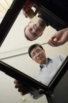
It looks like a simple overhead projector: four legs, a rectangular glass plate, and a light switch. But this FIM table can do much more. The father of the table – now patented – is Benjamin Risse, a formal doctoral student with Prof. Xiaoyi Jiang at the Department of Computer Science. While searching around for a topic for his PhD thesis, Risse – a computer scientist with biology as a subsidiary subject – noticed that previous, internationally used methods for analysing the movements of insects provided inadequate images. In the films the semi-transparent fruit fly larvae were blurred, and reflections or scratches were also visible. “In spite of all the good programmes that our computer scientists had, it wasn’t possible to distinguish automatically between a larva and a non-larva,” explains Nils Otto, a doctoral student at the Institute of Neurobiology and Behavioural Biology. But it is precisely this which is crucial for biologists in their work. They manipulate individual genes and then analyse whether the behaviour of these larvae is any different from that of “normal” animals. Here, even the smallest movement can be important.
So Benjamin Risse, Nils Otto and Dimitri Berh decided to improve the structure of the table and adapt the algorithms behind the process. “First we had to find a common language for biologists and computer scientists,” explains Berh, a computer scientist who is now responsible for the further development of the FIM table as part of his PhD thesis. The advantage, he says, is that the researchers can put new questions to each other and thus open up new possibilities. The interdisciplinary collaboration is not only exemplary, but also successful: the project is being funded by the Cells-in-Motion Cluster of Excellence until the end of 2017.
The method the computer scientists use is Frustrated Total Internal Reflection (FTIR). This gives the project its catchy name: FTIR-based Imaging Method, or “FIM” for short. The infrared light of the LEDs in the edges of the table is captured in the perspex plate. If a Drosophila larva crawls over it, it interrupts this total reflection. Thanks to an infrared filter, the conventional camera under the table can take razor-sharp pictures of the animals even without a microscope. On the images they glow as little white worms on a black background. The modular structure of the table allows it to be expanded as desired, in order for example to use UV light and to incorporate cold, heat or light stimuli.
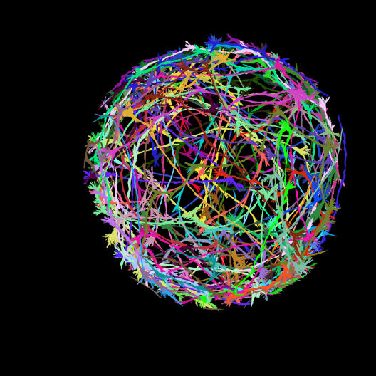
Veritable works of art are produced when the paths taken by the larvae are made visible on the computer by means of multi-coloured traces. The analytical programme developed by the group automatically describes the path and the behaviour of hundreds of animals – in other words, whether the larva is curved to the right or to the left, how fast it is and how far it has crawled.
For Nils Otto and his research on glial cells this is an important step forward, and one which enables him to undertake his PhD thesis with Prof. Christian Klämbt in the first place. Glial cells not only hold the neurones in position, but also provide them with nutrients. Otto is analysing whether the manipulation of certain genes gives rise to any defects in the glial cells. And with the help of the new method the biologist was indeed able to identify one gene that looks after the recycling of metabolic products in the mitochondria of the glial cells – their power plants, so to speak. Without this treatment the neurones can no longer communicate with each other. “Stimuli are not properly processed in the brain and movements change,” explains Otto. A “normal” larva occasionally turns it head from right to left to scan its environment. “By contrast, larvae with the genetic defect stop very often, move their heads in a hectic way and often change direction – just like people in the winter sales,” says Otto. This made it easy to find a name for the gene: “shopper”.
The further development of the FIM table is now well under way. Computer scientist Dimitri Berh is currently working on a prototype designed to make it possible to photograph and analyse the larvae in their natural environment over a longer period of time. One thing is certain, however: this new FIM model will no longer look like an overhead projector …
More information can be found at fim.uni-muenster.de, including assembly instructions and an opportunity to order the table.
This article by Bernadette Winter was published in the university magazine"wissen|leben" No. 2, 20.04.2016.