|
Münster (upm).
Microscopic view of the nervous system of an adult fruit fly. Green fluorescently-tagged channel proteins are shown on the axons of the neurons.<address>© WWU - AG Klämbt</address>
Microscopic view of the nervous system of an adult fruit fly. Green fluorescently-tagged channel proteins are shown on the axons of the neurons.
© WWU - AG Klämbt

Researchers find structures enabling a rapid transmission of nerve impulses in insects

Team of neurobiologists from Münster University publish their findings on the evolution of the glial sheath and rapid transmission of signals in neurons

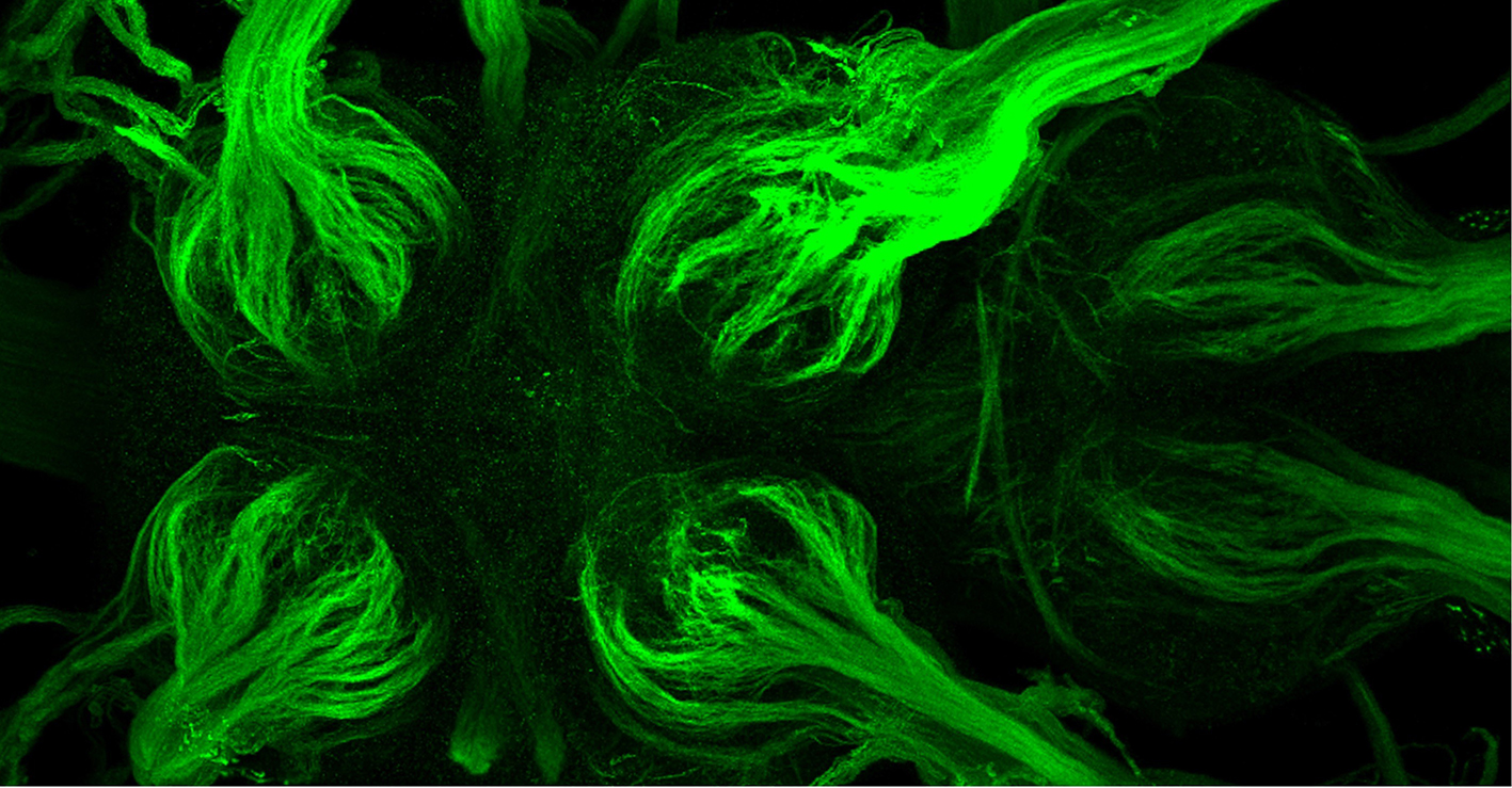
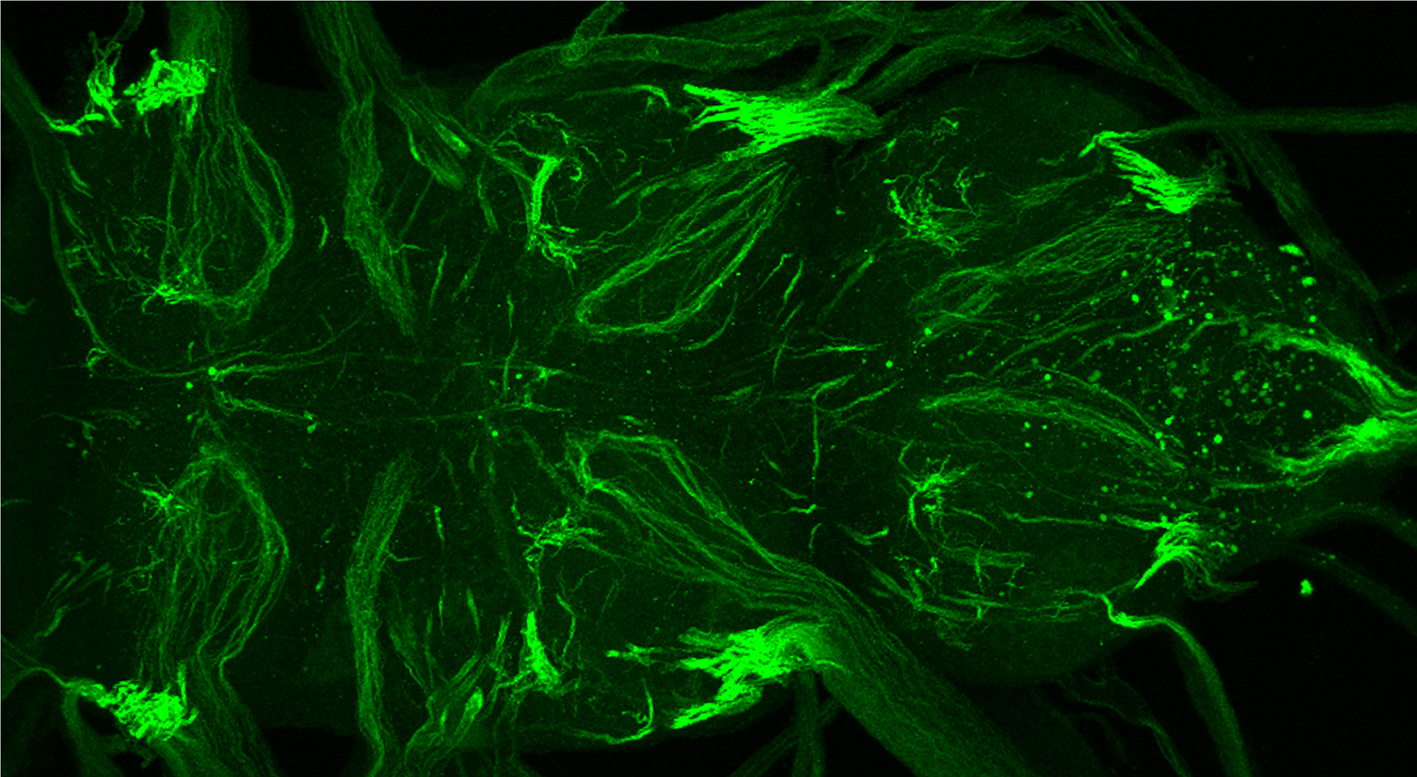
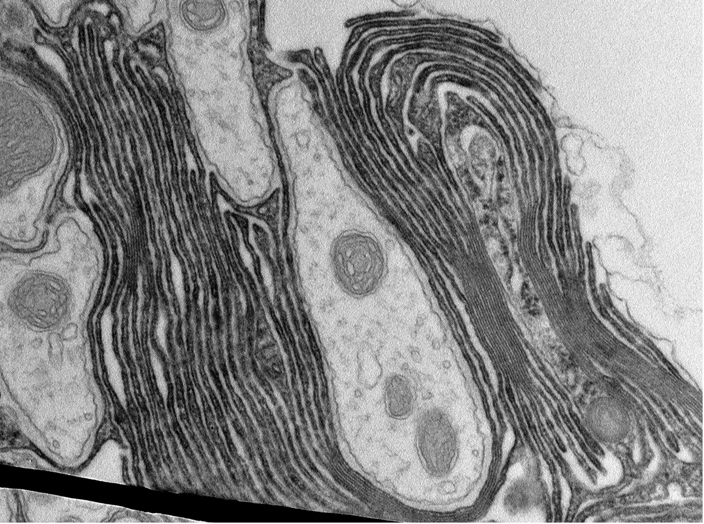
An animal’s brain consists of two different types of cell: neurons, which process and transmit information, and glial cells, which support the neurons in a variety of ways. In 1871, the French anatomist Louis-Antoine Ranvier demonstrated something special about neurons in vertebrates: on the extensions of these nerve cells there are ring-shaped regions which lack a surrounding sheath – the myelin formed by glial cells. Together with the electrically insulating myelin sheath, the so-called nodes of Ranvier form a basis for electrical nerve impulses to be transferred very rapidly over longer distances. They “jump” from node to node at a speed of up to 100 metres per second. This “saltatory conduction” was long seen as being specific to vertebrates. Now, for the first time, taking the fruit fly (Drosophila melanogaster) as an example, a team of researchers led by neurobiologist Prof. Christian Klämbt from the Institute for Neuro- and Behavioural Biology at Münster University has shown that similar structures exist in insects. The study has been published in the journal eLife.

Section of the central nervous system of an adult fruit fly. As the result of a genetic trick, only the channel proteins on motor axons are shown.<address>© WWU - AG Klämbt</address>
Section of the central nervous system of an adult fruit fly. As the result of a genetic trick, only the channel proteins on motor axons are shown.
© WWU - AG Klämbt
For its work, the team analysed fruit flies, looking at the distribution of the proteins necessary for neuronal conduction. Using genetic and microscopic methods, the researchers showed that the ion-conducting sodium and potassium channels underlying the process of transmission are arranged in clusters in a similar way to vertebrates. As recently described in a theoretical paper published by the team, the local accumulation of ion channels necessitates a strict spatial separation of individual axons, the extensions of the nerve cells. In vertebrates, this is ensured by the glial myelin. The team demonstrated that in Drosophila myelin-like structures are also formed around axons close to the plasma membrane region bearing the ion channels. As in vertebrates, the myelin is formed by specific glial cells and is a requirement for rapid, precise conduction.

Image, using an electron microscope, of a cross-section through axons surrounded by myelin-like structures.<address>© WWU - AG Klämbt</address>
Image, using an electron microscope, of a cross-section through axons surrounded by myelin-like structures.
© WWU - AG Klämbt
“We have described for the first time both the dedicated organisation of the voltage-gated ion channels and also the design of myelin-like structures for Drosophila,” says Christian Klämbt. “In addition, we were able to show that glial cells control the genetic activity and the positioning of neuronal ion channels.” The similarities described by the researchers between vertebrates and the fruit flies indicate that the occurrence of ion channel clusters, coupled with increased insulation, is a fundamental concept in the electrical transmission of information.

The work done by the team not only helps in understanding the evolution of myelin, but also enables the biology of myelin formation and regeneration to be investigated in greater detail. This is of major importance in connection with neurodegenerative diseases such as multiple sclerosis. So far, treatment has focused on suppressing the inflammatory reaction, and it has not been possible to promote any effective re-myelinisation. “This means,” says Christian Klämbt, “that our findings will help in discovering alternative forms of treatment for multiple sclerosis.”

For their investigations, the researchers used methods of molecular genetics in combination with various state-of-the-art imaging processes, including the use of innovative electron microscopy for the visualisation of marked proteins, as well as confocal microscopy with especially high resolutions.

The German Research Foundation (DFG) provided financial support (SFB 1348, B5, Kl 588/29).


Original publication

Simone Rey, Henrike Ohm, Frederieke Moschref, Dagmar Zeuschner, Marit Praetz, and Christian Klämbt (2023): Glial-dependent clustering of voltage-gated ion channels in Drosophila precedes myelin formation. DOI: 10.7554/eLife.85752

Further information