


“This degree of detail is astonishing”
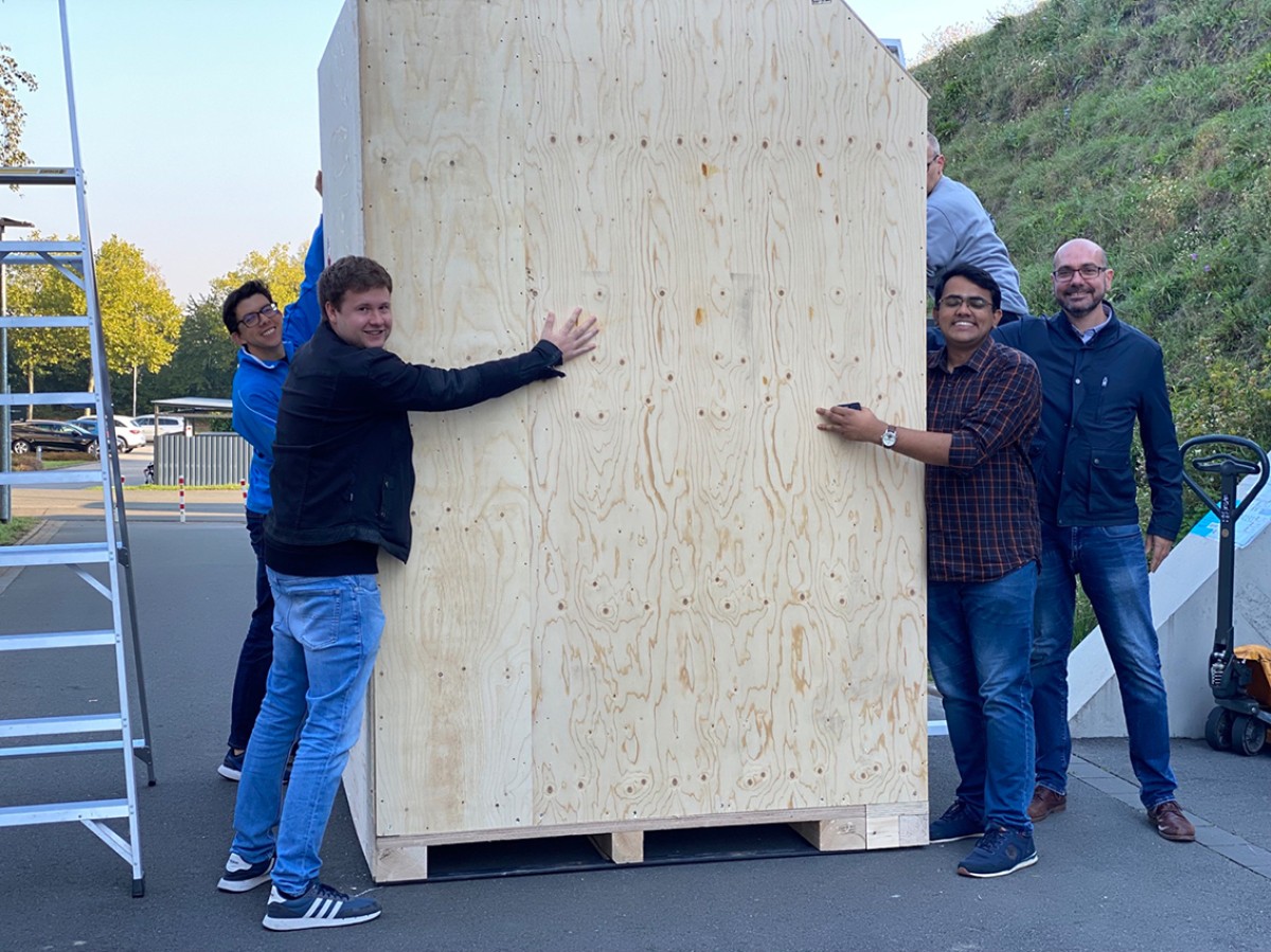
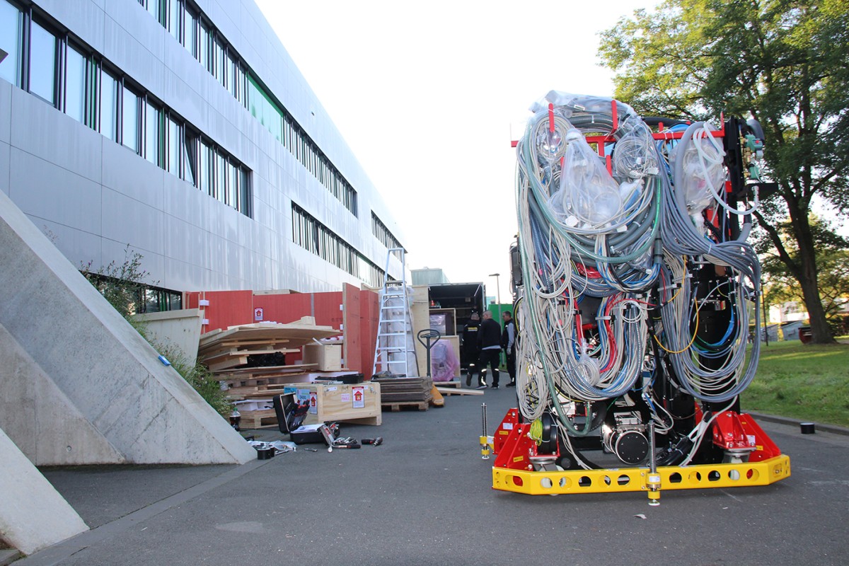
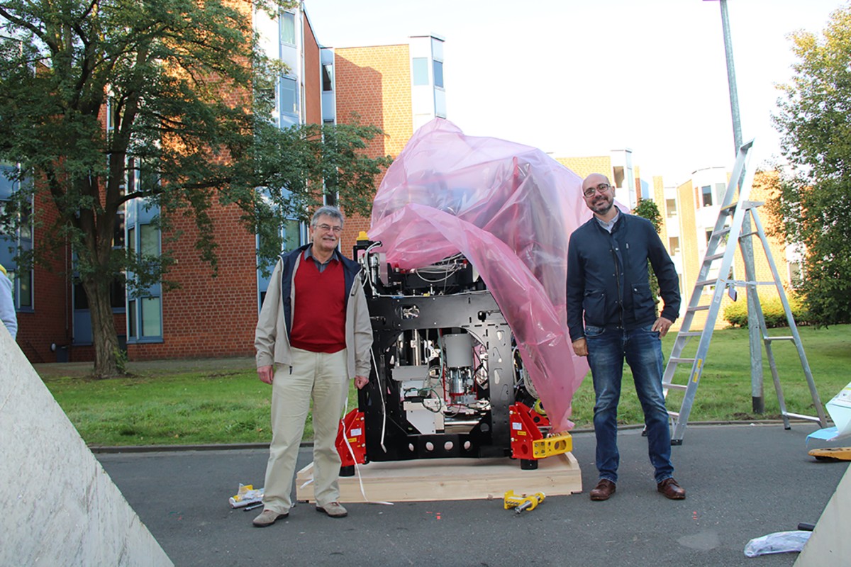
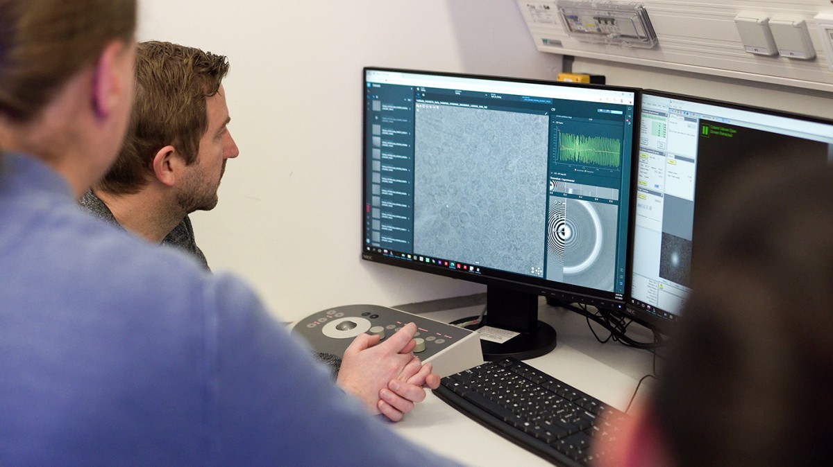
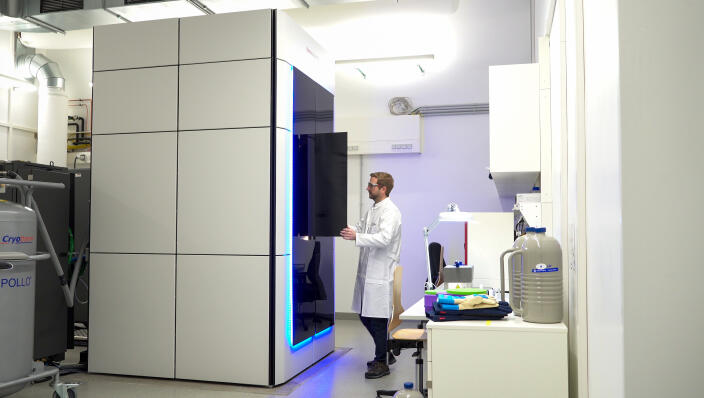
When the big box with this piece of equipment was delivered in September 2022, the members of Prof. Christos Gatsogiannis’ working group at Münster University’s Center for Soft Nanoscience (SoN) gave a joyful welcome to the valuable consignment: “Hug the box!” they declared – a tradition among the “cryo-EM research community”. No doubt about it: the new high-performance cryogenic electron microscope (cryo-EM) really is something special. Today, the equipment has already been installed and put into operation. Around 20 working groups and research alliances from the fields of Medicine, Biology and Chemistry will be using it.
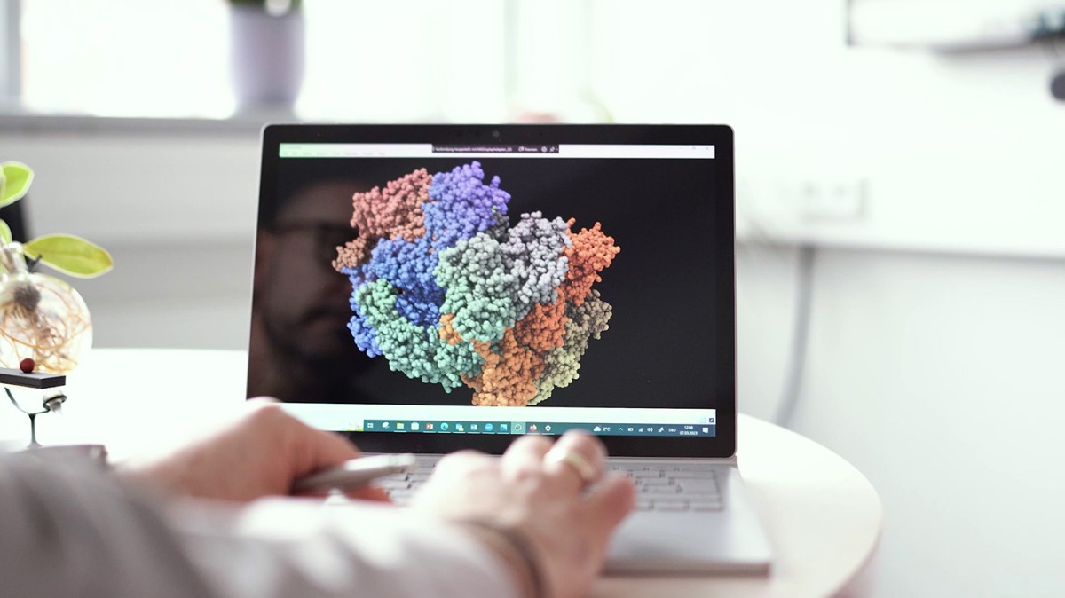
There are only a few of these microscopes in Germany which are in this performance category; at the University of Münster it is the first of its kind. Until just a few years ago, it was inconceivable that minute components of cells could be displayed to this degree of precision – down to individual atoms. “For me too,” says Christos Gatsogiannis, “although I’ve been working in this field for a long time now, this degree of detail is astonishing.” In addition to looking into cells, researchers want to visualise the structures of individual proteins and, as a result, understand the way they function.”
“This state-of-the-art equipment is of enormous importance, both for the University and for Münster as a research centre,” says Prof. Monika Stoll, Vice-Rector for Research. “Our University is one of the leaders in the field of multiscale imaging. This new piece of equipment will make a decisive contribution that we remain internationally competitive in this field.” Access to this key technology will revolutionise numerous interdisciplinary areas of research,” adds Prof. Frank Ulrich Müller, Dean of the Faculty of Medicine. “Cryo-EM is extremely important for the further development of our research profiles at the University and the Medical Faculty.”
The German Research Foundation and the state of North Rhine-Westphalia had made a total of 7.5 million euros available within the framework of the “large-scale research equipment” funding programme. In addition to the high-performance cryo-EM, the “total package” is rounded off by two further pieces of equipment: an automated screening electron microscope which makes it possible to make the optimum preselection of samples, and a cryo-focused ion beam-scanning electron microscope which is needed for the preparation of samples.
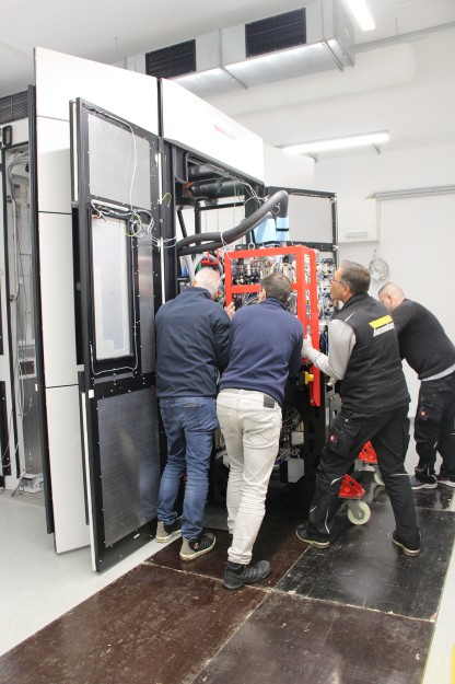
The SoN building’s special construction was a prerequisite for the purchase of the microscope. The room in which the cryo-EM is installed has to be vibration-free. For this reason, experts constructed an almost perfectly vibration-damped floor; also, the room is screened off from disruptive magnetic fields. “Normally, electron microscopes are installed in the cellars of research buildings in order to avoid such disturbances. In our Institute, the equipment is all in a special area on the first floor which is covered with earth,” says Christos Gatsogiannis.
Zum Abspielen des Videos wird dieses von einem Webserver der Firma Google™ LLC geladen. Dabei werden Daten an Google™ LLC übertragen.
Last but not least, the Münster University cloud is also available, providing IT infrastructure which can meet the huge demands of such large-scale equipment. “When the equipment is fully up and running, we expect a data volume of around two petabytes a year, which is about one million gigabytes,” reports Dr. Raimund Vogl, the head of Münster University IT. The University’s high-performance computer PALMA II is available for processing the data.
Christina Hoppenbrock
This article is from the university newspaper wissen|leben No. 2, 29 March 2023.
High-performance cryogenic electron microscopy at Münster University
Cryo-EM is a variant of transmission electron microscopy. By means of electron beams, microscopic objects are imaged at cryogenic – i.e. extremely low – temperatures. The new high-performance cryo-EM reaches a resolution of almost one angstrom, in other words approximately one ten-millionth of a millimetre – which corresponds to the magnitude of atom radii. The digital camera and processing technology can create up to 20,000 individual images in 24 hours and can elucidate up to five protein structures in this time.
Appointment notice
The cryo-EM will be officially inaugurated at a symposium on 19 April.
A quick question: What do you hope for from the new cryogenic electron microscope for your research?

For my group, the new cryogenic electron microscope is a fantastic enrichment for our research. With it, the University of Münster now has outstanding infrastructure for structural biology – by which we mean the structural study of biomolecules in atomic detail. This is an important extension of the scale of biological imaging towards higher resolutions. It means that we can examine questions relating to cell biology and biomedicine at the molecular level. What’s important to us is to understand how the components of cells function and interact with one another. Such findings are important in order to clarify how malfunctions can lead to diseases. With this microscope, we can now advance into new fields. Direct access to this key technology in life sciences will make a decisive contribution to practically all of our projects.

What we hope to be able to do with the new cryogenic electron microscope is depict structures at the kidney filter at a molecular level, both in healthy humans and in the case of genetic diseases. In healthy people, the kidney filter permits toxic products to be excreted and, at the same time, it prevents the loss of plasma proteins. In the case of glomerular diseases, the kidney filter is defective – which leads to a loss of proteins in the urine. This is an important progression factor for glomerular diseases. In order to be able to develop specific treatments for such glomerular diseases, it is very important to understand the molecular mechanisms at the kidney filter. The new cryogenic electron microscopy technology can make a decisive contribution to such an understanding.

The development of cryogenic electron microscopy for the purpose of a high-resolution determination of the structures of proteins is tantamount to a revolution in structural biology. In the past, proteins had to have a crystalline form if we wanted to obtain a structure in atomic resolution by means of X-ray crystallography. Despite all our efforts, it was never possible to crystallise many protein complexes, for example those in biological membranes, and their structures could not therefore be elucidated. As a result of cryo-EM analyses, high-resolution images can be made of such protein complexes in their 3D structure. This has made many spectacular structures visible. But cryo-EM can do even more. Cryo-EM analyses can produce images of dynamic structural changes in proteins. And we can expect much more in the future: we stand on the threshold of a Golden Age, with the development of new kinds of electron cameras and new evaluation software driven by the development of artificial intelligence.