


Measure eye movements when blinking
Scientists from Westfälische Wilhelms-Universität (WWU) Münster have developed a method to measure the full extent of eye movements during blinks for the first time. The new method uses Magnetic Resonance Imaging (MRI), to take many fast measurements of an entire cross-section of the eye, unlike most common eye tracking that measures only the front of the eyeball. This allows measurements even when the eye is closed, additional assessment of the ocular muscles and visualises displacements and deformations of the whole eyeball. The study describing the new method has been published in the scientific journal “eNeuro”.
Eye movements are typically measured using eye-trackers, which are high speed cameras that film pupil position.” We were able to develop a method that captures eye movements in unprecedented temporal resolution using real-time MRI. Using this new method, we could show that the whole eyeball is being pulled back into the eye socket during a blink“, explains author Johannes Kirchner, research assistant at the Institute of Psychology at Münster University. The scientists from the University of Münster developed the method with co-author Prof. Tamara Watson from Western Sydney University.
Method
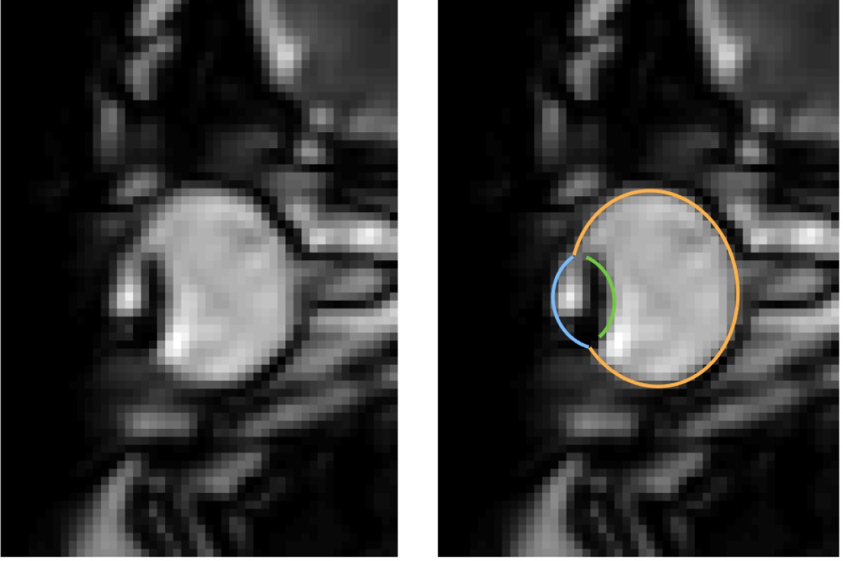
MRI is a relatively slow procedure, the temporal resolution usually ranges between seconds or minutes. Eye movements on the other hand are fast and typically last only a few dozen milliseconds. Recent technological advances allowed the recording of 2-D MR images at a resolution of up to 20 milliseconds. Those ultrafast MR sequences were initially developed for cardiac imaging and haven’t been applied to the recording of eye movements. “We could reach a temporal resolution of 35 milliseconds. Total scan duration of only a few minutes already leads to the acquisition of over 10000 images. For actual eye-tracking, a fully automatic segmentation algorithm to analyse these images was needed”, explains co-author Prof. Markus Lappe, professor for cognitive neuroscience at the Institute of Psychology at the University of Münster. “With ‘MREyeTrack’ we have developed such a segmentation algorithm, which allows the fully-automated analysis of eye position and orientation in every single image.”
The MR-based method is more expensive compared to camera-based methods and also has a worse temporal resolution. “However, the MR-based method might be particularly useful in a clinical context and could be helpful to further the understanding of eye movement disorders. The MREyeTrack algorithm could also be applied to other frequently used MR sequences, where the segmentation fo the eye is of interest. For example, one could use it to circumvent the need of an eye-tracker in fMRI experiments”, emphasises Johannes Kirchner.
Funding
The study has been part of the international „Platypus“ project, which received funding from the EU „Horizont 2020“ research and innovation programme under the Marie Skodowska-Curie grant agreement.
Original publication
Kirchner, Watson, Lappe (2021), Real-time MRI reveals unique insight into the full kinematics of eye movements. eNeuro, DOI: https://doi.org/10.1523/ENEURO.0357-21.2021