
Listening to the molecules in the body
Laser light that cannot be seen, and sounds that cannot be heard: for partygoers this would probably conjure up some boring event – but it brings a sparkle to the eyes of many a researcher. It is precisely this combination that produces something that is all the more visible – images from inside the body that provide information on the processes taking place there. Photoacoustics, also known as optoacoustics, is the name of this method, which has been used for decades now and whose purpose is above all to acoustically record the sounds of molecules. In contrast to ultrasound, however, researchers and doctors do not put energy into the body through soundwaves, but through laser light. Molecules absorb this light and begin to oscillate, and these oscillations, in turn, can be measured using an ultrasound device and then shown as images.
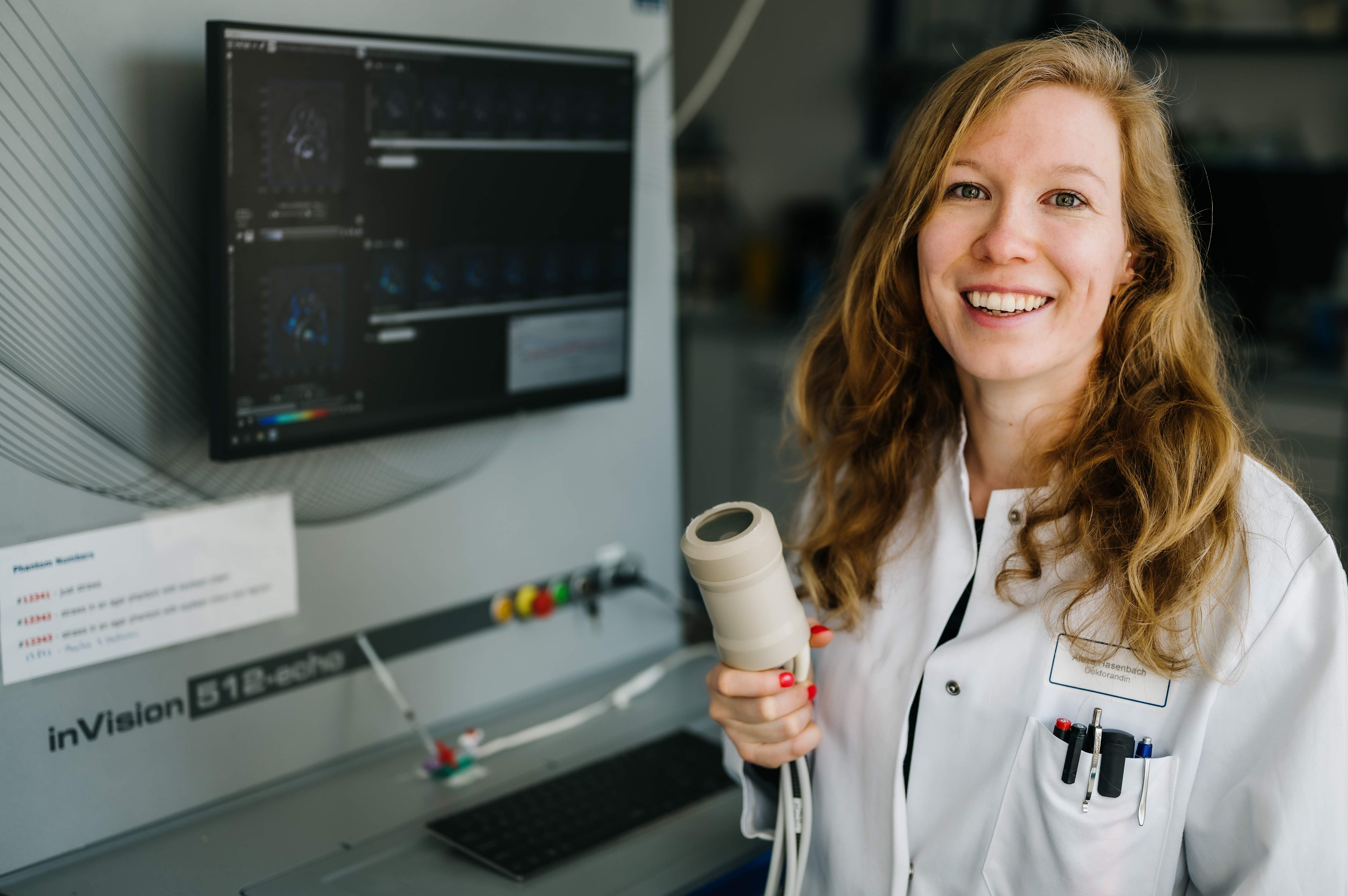
Someone who has already listened to many a sound emanating from the inside of the body is biologist Alexa Hasenbach, who carried out photoacoustic experiments as part of her PhD and was one of the first people who was able to use a special piece of equipment for the purpose at the European Institute for Molecular Imaging (EIMI) at Münster University. Multispectral Optoacoustic Tomography, or MSOT for short, is the method which, for almost 18 months now, has extended the range of preclinical imaging instruments at the EIMI. One of the things that is so special about the equipment is its resolution: it has 512 detectors, twice as many as its predecessor. Another is that the system also emits ultrasound waves in addition to laser light. This means that the researchers get not only molecular but also, at the same time, anatomical information from the inside the body.
“The new system gives us new insights that we did not have before,” says Alexa Hasenbach. She looks at an image which she obtained in the course of her research. It shows the inflammation in the knee joint of a mouse with rheumatoid arthritis. Various colours – blue, green, yellow or red – provide information on the intensity of the inflammation. In addition, as a result of the ultrasound application, skin and bones can also be seen, which means that the knee can be easily marked out.
For years now, researchers at the EIMI, working together with researchers at the Institute of Immunology, have been tracking down inflammation occurring in the case of rheumatoid arthritis. The background to this is that in the case of this autoimmune disease it is the body’s own immune cells attacking the joints and cartilage and cause inflammation. Doctors normally use X-rays and ultrasound to diagnose and monitor these inflammations. It is not possible, however, to examine the activity of inflammatory cells by using these methods. It could be done using nuclear imaging such as positron emission tomography, but such methods take a lot of time and are very costly. If, however, photoacoustic methods could be put into practice, it would be possible to produce images regularly, and at a much earlier stage, providing information on the stages of the inflammation. Doctors would not get reliable images only after cartilage or parts of the bones had been destroyed, and they could begin treatment earlier.
To find out how inflammations in rheumatoid arthritis can be visualised by means of photoacoustic imaging, Alexa Hasenbach focused in her PhD on special inflammation markers – proteins which are secreted by the immune cells. So-called tracers – to which, for example, dyes are attached to track down and visualise. For outsiders, the technology takes some getting used to. Inside the photoacoustic equipment there is a heated water bath to prevent the ultrasound waves from being destroyed by air. The mice to be examined are put under general anaesthetic, given respiration and immersed in the water in a holding device with a thin, transparent wall. Here, they can be exposed to light and ultrasound and the acoustic signals can be recorded. “I am aware of my responsibility towards the test animals,” says Alexa Hasenbach. “I watch intensively to check that they are all right – not only during the experiments, but also before and after them.”
In addition, the equipment has a handheld detector device enabling the researchers to carry out the photoacoustic examination “in the fresh air”. “This is how the method would be applied to patients,” says Alexa Hasenbach, “so what we do here is directly relevant to any clinical application.” The first pieces of equipment of this kind are already being used in hospitals – for example, to examine metastases in lymph nodes.
Further experiments will be necessary before the method can be used on patients with arthritis. As she has almost completed her PhD dissertation, Alexa Hasenbach will not be continuing the work in the lab, but she will be remaining true to “her” subject: she has already started working for the manufacturer of the photoacoustic equipment, where, among other things, she will be working on the further development of the software and hardware. A stroke of luck for the tech-savvy researcher – it means that in future she can continue to listen to molecules.
This article was first published in the University newspaper wissen|leben, No. 2, April 2020.
Further information
- The original article in wissen|leben (in German language)
- All the issues of the University newspaper at a glance (in German language)
- European Institute for Molecular Imaging at Münster University
- Cells in Motion Interfaculty Centre at Münster University
- Research focus “Cell Dynamics and Imaging” at Münster University