
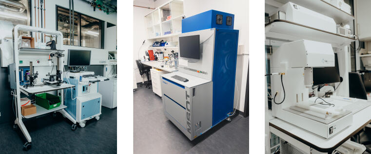
Magnetic Resonance Imaging (MRI)

Bruker BioSpec Maxwell 94/17 MRI © Uni Münster - Michael Kuhlmann 9.4 Tesla small animal MRI scanner
- 2x2 element cryo probe, suitable for mice and rats
- high-power gradient system (Gmax > 900 mT/m)
- 1 kW RF transmitter (dual channel)
- X-nuclei coils 19F, 23Na, 31P
- setup for simultaneous optogenetic (f)MRI, including surface coils with fiber lead-through
- stimulation and monitoring systems for cardiac MRI and fMRI
- animal transport system (ATS) for automated positioning
- application examples: anatomy, relaxometry, cell tracking, diffusion, functional MRI, cardiac MRI, perfusion, chemical exchange saturation transfer (CEST), spectroscopy
Positron Emission Tomography / Magnetic Resonance Imaging (PET/MRI)

Bruker Biospec 70/20 with PET insert Si 103© Uni Münster - Michael Kuhlmann Integrated PET and 7 Tesla MRI: non-invasive 3D imaging of radiolabeled tracers in living mice combined with MR imaging
PET
- spatial resolution: about 1 mm
- application examples: molecular imaging of (patho-)physiological processes, e.g. cell metabolism, receptors, transporters, or cell tracking; quantitative tracer detection up to few hours after injection, dependent on the half-life of the coupled positron emitting radioisotope (e.g. 18F, 11C, 68Ga)
MRI
- 2x2 element cryo probe, suitable for mice
- high-power gradient system (Gmax > 660 mT/m)
- 1 kW RF transmitter (dual channel)
- 13C surface coil
- stimulation and monitoring systems for cardiac MRI and fMRI
- attachment capacities for conventional and MMPF animal beds
- application examples: anatomy, relaxometry, cell tracking, diffusion, functional MRI, cardiac MRI, perfusion, chemical exchange saturation transfer (CEST), spectroscopy
Positron Emission Tomography (PET)

Oxford Positron Systems quadHIDAC© Uni Münster - Michael Kuhlmann Non-invasive 3D imaging of radiolabeled tracers in living mice
- spatial resolution: below 1 mm
- field of view: 16 cm (diameter) x 28 cm
- application examples: molecular imaging of (patho-)physiological processes, e.g. cell metabolism, receptors, transporters, or cell tracking; quantitative tracer detection up to few hours after injection, dependent on the half-life of the coupled positron emitting radioisotope (e.g. 18F, 11C, 68Ga)
Single Photon Emission Tomography / Computed Tomography (SPECT/CT)

Mediso nanoScan SPECT/CT© Uni Münster - Michael Kuhlmann Integrated SPECT and CT: non-invasive 3D imaging of radiolabeled tracers in living mice combined with anatomical imaging
SPECT
- application examples: molecular imaging of (patho-)physiological processes, e.g. cell metabolism, receptors, transporters, or cell tracking; quantitative tracer detection up to several days after tracer injection, dependent on the half-life of the coupled gamma emitting radioisotope (99m Tc, 111In, 177Lu)
- equipment: Multi-Pinhole-Collimators for different applications (APT51, APT52, APT62, APT66),
- spatial resolution: 0.5 – 1.5 mm
CT
- anatomical imaging
Computed Tomography (CT)

Bruker Skyscan 1278 CT © Uni Münster - Michael Kuhlmann Non-invasive computed tomography of living mice
- anatomical imaging
- respiratory gating
- spatial resolution: 80µm
Optical Imaging

Revvity IVIS Spectrum© Uni Münster - Michael Kuhlmann 2D bioluminescence imaging (BLI) and fluorescence reflectance imaging (FRI) of living mice or ex vivo organs
- high-sensitivity BLI and FRI
- filter specifications: 10 excitation filters (415 nm – 760 nm, 30 nm bandwidth), 18 emission filters (490 nm – 850 nm, 20 nm bandwidth)
Ultrasound
Visualsonics Vevo 2100 (left), iThera MSOT inVision512/echo (middle), FUS Instruments RK50 (right)© Uni Münster - Michael Kuhlmann Ultrasound (US)
- Visualsonics Vevo 2100
- high resolution ultrasound
- non-invasive 2D anatomic and functional imaging in living mice
- transducers
- MS400 (18-38 MHz), max 20 mm image depth, spatial resolution: 50 µm axial, 110 µm lateral
- MS700 (30-70 MHz), max 12 mm image depth, spatial resolution: 30 µm axial, 75 µm lateral
Multispectral Optoacoustic Tomography (MSOT)
- iThera MSOT inVision512/echo
- non-invasive 3D detection of photoabsorbers (e.g. fluorescent dye IRDye800cw, gold nanoparticles) in living mice
- multispectral optoacoustic tomography (detector ring setup, 512 ultrasound transducers)
- additional handheld setup
- wavelength: 660-1300 nm, max. power output: 60mJ@710 nm
Focused ultrasound (FUS)
- FUS Instruments RK50
- stereotactic-guided therapeutic ultrasound
- continuous and pulsed exposures
- real-time acoustic feedback integration to regulate exposures via stimulated emissions from circulating microbubbles
- application examples: opening of blood brain barrier, targeted activation of sonosensitizers
Digital autoradiography camera

© Uni Münster - EIMI Measurement of radiotracer distribution in tissue samples (cryosections)
Gamma counter
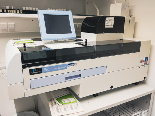
Perkin-Elmer Life Science Wizard2 gamma counter© Uni Münster - Michael Kuhlmann Quantitative measurement of radioactivity in small samples, like ex vivo tissues or samples from cellular uptake assays
Animal housing
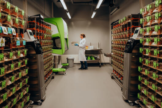
Animal housing© Uni Münster - Michael Kuhlmann - digitally monitored IVC housing (Tecniplast) for mice in S1 and S2 areas, plus respective animal cage changing station
- digitally monitored IVC housing (Tecniplast) for rats in an S1 area
- animal surgery areas (S1 and S2) with various equipment
Lab equipment
Münster Imaging Network – Preclinical Imaging