How does the yolk get into the egg? Studies on the fruit fly
(Research group Prof Stefan Luschnig, Institute for Integrative Cell Biology and Physiology)

What my research is about
Images


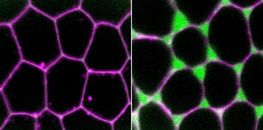
A close-up of connections between three epithelial cells (pink cell membrane) of a fruit fly egg. In such regions, transient openings in the cell layer occur. The green shows a protein (Myosin II) that generates forces in cells. Left: The protein is present on the cell membrane, and no intercellular spaces can be seen. Right: The protein is no longer on the cell membrane, only inside the cell, and an opening can now be seen between the cells.© Jacobs T et al. Current Biology 2025. DOI: 10.1016/j.cub.2025.01.043
In my doctoral thesis, I investigated how cells that build connections, and thus form tissue, allow only certain substances to pass through this tissue. Specifically, I am working with cells that envelop the oocytes (egg cells) of fruit flies. These so-called epithelial cells protect the oocytes from harmful external influences and support their development into mature eggs. The uptake of yolk protein – the egg yolk – into the egg is an important part of the egg development process, as the yolk later nourishes the embryo. The yolk protein is produced in a special organ called fat body and must pass through gaps in the epithelial layer in order to reach the oocyte. These gaps are created when the cells temporarily loosen their connections. I am investigating the role of forces that pull on the connections between the cells from the inside in this process. Through my research, I was able to show that the forces in the cells must be reduced for the gaps between the cells to open.
Why my research is important
Wondering why we should be interested in the egg development of small fruit flies? Gaps in epithelial cell layers also occur in the human body. For example, in the intestine, nutrients, such as building blocks for proteins, are absorbed through similar gaps. Also, in blood vessels, openings are created between the cells when immune cells migrate from the blood vessels into the surrounding tissue in a process that is necessary for the body to combat inflammation. However, pathogens can also exploit the gaps between epithelial cells and enter the body. It is therefore important that we use simple model systems to investigate and understand such processes. The cells of some fruit fly tissues are organized in a similar way to those in human tissue. Thus, the results from examining fruit flies can provide initial clues to understanding the processes in human cells better.
How I conduct my research
In my research, I use the fruit fly Drosophila melanogaster, and I am able to genetically modify these fruit flies in the lab. As cells use genetic information to produce proteins with different functions, when genetic information is altered, this can produce proteins with altered functions or prevent the production of a certain protein. Using this method, I can identify which functions certain proteins have in the formation of openings in the epithelial cell layer. To this end, I am focusing on proteins that are located on the inside of the cell membrane and alter forces in the cell.
To make the changes in the cells visible, I use a special form of light microscopy called confocal microscopy. This allows me to distinguish between the smallest structures down to 200 nanometres (0.0002 millimetres). For example, under the microscope, I can see whether an altered protein is located in a different position in the cell and whether this leads to larger or smaller intercellular spaces. From this information, I can draw conclusions about what functions the protein has in the cell. For example, I was able to show that a specific protein regulates forces at certain points on the cell membrane and prevents the intercellular spaces from opening too early.
Links
- Originalpublikation: Thea Jacobs, Jone Isasti Sanchez, Steven Reger, Stefan Luschnig, Rho/Rok-dependent regulation of actomyosin contractility at tricellular junctions restricts epithelial permeability in Drosophila, Current Biology, Volume 35, Issue 6, 2025, Pages 1181-1196.e5, ISSN 0960-9822, DOI: 10.1016/j.cub.2025.01.043
- Thea Jacobs on Bluesky
- Research group of Prof Stefan Luschnig at the University of Münster
The research project presented here is funded by the German Research Foundation (DFG) within the Collaborative Research Centre (CRC) 1348 “Dynamic Cellular Interfaces”.
This article is the result of a workshop on science communication for junior researchers. The participants learnt about the basic principles, quality criteria and different forms of science communication. Supported by 1:1 coaching sessions, they then created an article about their own research. The training course is a pilot project jointly developed and run by the Cells in Motion Interfaculty Centre and the Centre for Emerging Researchers at the University of Münster.
Article updated on 21-07-2025.

