Breathing Allowed
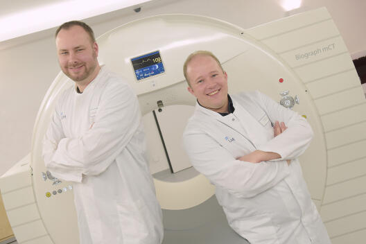
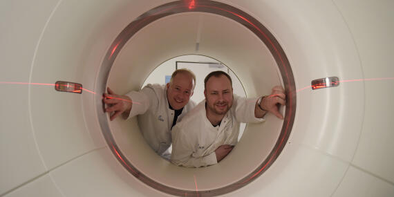
In future doctors will be able to take more sharply defined pictures of their patients’ interiors when making clinical images – and entirely without any bothersome aids. Researchers at Münster University have developed a programme which enables physicians to do away with the so-called respiratory belt in positron emission tomography (PET). Up to now, patients have had not only to keep especially still during PET imaging, but also to put on such a respiratory belt. This belt measures how a patient’s body moves while they are breathing. The information gathered enables the blurring caused by the breathing movements to be eliminated. “Our computer programme provides images which are just as clearly focused as those produced by the system with the belt,” says Dr. Florian Büther, a medical physicist and researcher in Collaborative Research Centre 656, “Molecular Cardiovascular Imaging”, at Münster University. “This simplifies clinical imaging because doctors no longer need the respiratory belt as an aid.”
Most amateur photographers are familiar with the problem of focusing: just one small movement and the holiday photo is blurred – and a wonderful souvenir is ruined. When taken for private use, blurred pictures are only annoying. In the field of clinical imaging, however, blurred pictures can be a problem because doctors need to be able to clearly recognise illnesses on the images they have. Although doctors have learned to make clear diagnoses on the basis of the picture quality available to them, there are times when medical images have to be particularly well focused. “This makes it easier to make a diagnosis – particularly in the case of special illnesses,” says Dr. Thomas Vehren, a nuclear medicine specialist at Münster University Hospital.
The two scientists’ computer programme has already been used in 70 clinical examinations. Florian Büther is today (24 April) presenting his initial scientific results at the annual conference of the German Society of Nuclear Medicine in Hanover. A scientific medical appraisal has not yet been made. However, first impressions confirm Büther’s own assessment.
When Breathing is a Nuisance
During a whole-body scan patients have to follow a variety of instructions regarding their breathing: for example, to breathe as little as possible and to make it as shallow as possible, so that the images made of their interiors are as sharply focused as they can be. “Unfortunately, patients often breathe differently than instructed,” says Thomas Vehren. To counteract this, and in order to be able to record breathing movements, doctors have up to now used a respiratory belt on patients. However, such a belt has only limited uses in the case of people who are seriously ill.
Tomography Data Provide More Sharply Focused Images
Just like the respiratory belt, the Münster scientists’ programme uses a technique known as “gating”, in which images are split up into time phases. Each image is allocated to different breathing phases, enabling a precise allocation to breathing in or breathing out. Conversely, it is possible to recognise to what extent the image of an organ, an infection or a tumour has been changed as a result of the breathing movement. “For the gating process we use the data which are produced during the tomography, which means we don’t need any additional data from the respiratory belt,” explains Florian Büther. “The programme itself splits up the information into breathing phases and converts them directly into more sharply focussed images.” Nuclear medicine specialist Thomas Vehren is using the programme as part of a research project at Münster University Hospital. In the longer term it could be used in more hospitals and clinics.