
Invasion to the Inside
In order to multiply, influenza viruses are dependent on cells of a human or animal body. They board those cells, for example all along the lung surface, and their genetic material migrates into the nucleus, where it is replicated. As a result, new viruses come to life. A team led by scientists from the Cells-in-Motion Cluster of Excellence (CiM), University of Münster, has now, for the first time, succeeded in visualizing structures of the viral genome inside of human cells by light microscopy. With these great insights, the team gains knowledge about how the influenza viruses knock out the immune response of the cells. The results suggest that the virus particle invades the cell "with pulled out weapon" and so suppresses the important immune response of the human body.
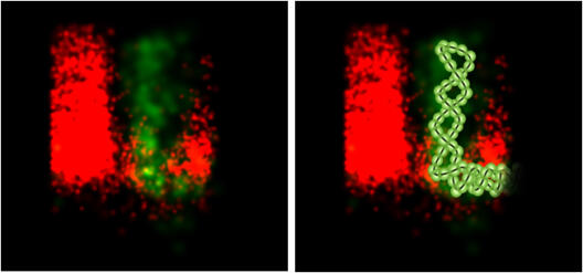
The helical virus genome is split into eight segments that could be imaged by light microscopy now. "We identified viral proteins that are instrumental in helping to suppress the immune response of the cell immediately after the entry of the virus particle. This function was previously unknown", explains Swantje Liedmann from the Institute for Molecular Virology, University of Muenster, who is first author of the study published in the online journal "Nature Communications". Viral proteins, known for their important role in reproducing genome, contribute to this function.
Experts have known for some time that flu viruses carry a protein called "NS1". This protein blocks the immune system very efficiently. "We asked ourselves why there are other proteins for the same purpose. We suspect that 'NS 1' operates somewhat later as our observed proteins", so Swantje Liedmann.
The application of light microscopy as imaging technique played a central role in the study. Together with scientists from Memphis, United States, the team from Münster used a high-resolution microscopy technique also relevant to other issues in the CiM: the "STORM" technology (the acronym stands for "stochastic optical reconstruction microscopy"). This fluorescence microscopy technique opens up many opportunities to further explore the infection biology of influenza viruses and other pathogens. "Above all, the localization of viral components in the cell as well as the studies of the interaction with components of the cell is now possible by this technology," explains Swantje Liedmann.
By precisely answering the question, how viruses inhibit the immune response of their host cells, the researchers hope for finding future approaches to develop new antiviral drugs.