
Joint project on 4D-imaging in medicine gets off the ground
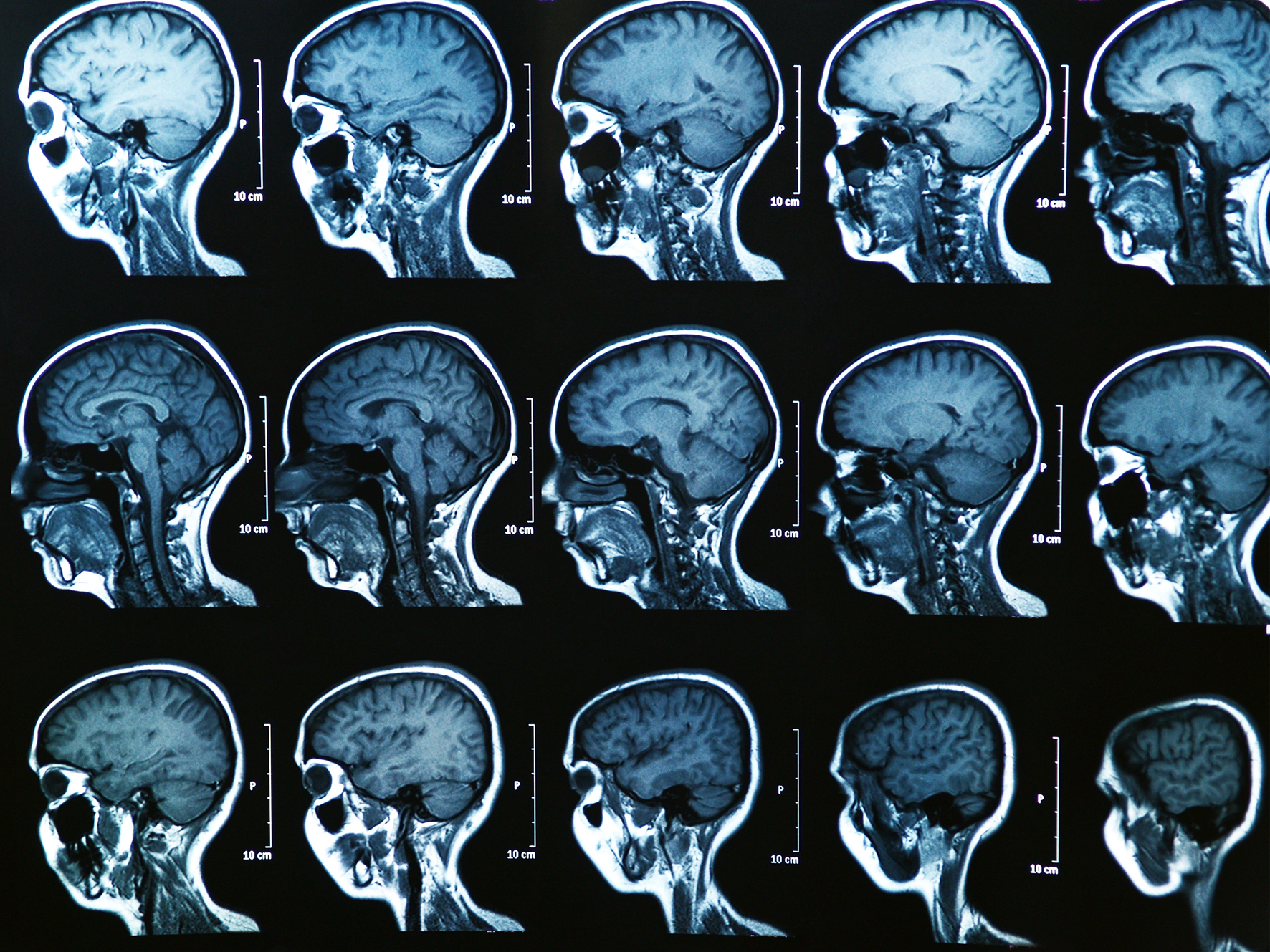
Medical imaging makes it possible to take a look inside the human body from the outside and make hidden processes visible, which makes it important for the diagnosis and treatment of a variety of diseases. Something that is becoming increasingly important is so-called four-dimensional imaging. In this process, three-dimensional images are generated and, in addition – as a “fourth dimension” – changes that occur over time can be reconstructed and made visible. This makes it possible, for example, to record movements resulting from the heartbeat or from breathing and produce sharper images in doing so. Mathematics plays a key role in this. A new joint project being undertaken nationwide in Germany is now aiming to improve the methods of 4D-imaging – making it possible, for example, to analyse movements and simultaneously measure molecular processes quantitively. “Quantitively” in this case means, for example, that it can be measured how much sugar the cells of an organ or tissue consume in a certain period of time (glucose metabolism). The project is being coordinated by Prof. Martin Burger, a mathematician at the University of Münster.
The four-dimensional imaging process takes place using magnetic resonance imaging (MRI), for example, or positron emission tomography (PET). In these cases, signals from inside the body are measured. However, these signals are not present, initially, in the form of images. Only by using mathematical models can the three-dimensional images generated over time be reconstructed from the measurement data. “It’s a really fascinating challenge, not only to record the movements of organs or of patients during a dynamic measurement, but also to use these movements to improve the quality of the images,” says Martin Burger. “This makes it possible to see more precisely how contrast media spread out in the body.” These new methods are important for example in examinations of the cardiovascular system or of kidney functions, he adds, and the aim is that they should help in enabling physicians to make clearer assessments of functionality and to draw up more precise diagnoses of diseases. The project is being funded to the tune of 836,000 euros, for three years, by the Federal Ministry of Education and Research out of the “Mathematics for Innovations in Industry and Services” programme.
Prof. Martin Burger heads the “Imaging” working group at the Institute of Numerical and Applied Mathematics at Münster University. He is a group leader at the CiM Cluster of Excellence and heads a sub-project within CRC 656. Other researchers from Münster are Dr. Frank Wübbeling and Dr. Hendrik Dirks (Münster University/Mathematics), Prof. Klaus Schäfers (Münster University/European Institute for Molecular Imaging) and Dr. Stefan Reuter (Münster University Hospital).