

A focus on the microalgae ‘Chlamy’
Protozoa of the genus Chlamydomonas are microalgae. They are at the bottom of the food chain and make a major contribution to global carbon dioxide fixation, making them extremely valuable from an ecological perspective. But the small green algae, which were first described by the German naturalist Christian Ehrenberg at the end of the 1830s, are also essential for research. Around 250 scientists from all over the world will be meeting at the University of Münster from 24 to 29 August for the ‘Chlamy 2025’ conference to discuss the latest research findings.
Research is focussing on the species Chlamydomonas reinhardtii. As the scientific name sounds complicated, the researchers call the algae ‘Chlamy’ for short - which sounds like a beloved pet. In some respects, Chlamy is actually a bit like an animal: it swims with the help of its two flagella. It can perceive light with its ‘eye’. So it swims towards the light in order to have optimum conditions for photosynthesis. If it gets too bright, it swims away. Another remarkable feature: under certain conditions, the microalgae can abandon photosynthesis and instead absorb and metabolize carbon compounds like acetate from their surroundings, a behavior typically associated with animals, fungi, and bacteria, and highly unusual for vascular plants.
The genome of the alga, first sequenced in 2007 and continuously refined over the past 15 years, revealed an astonishing total of over 17,000 genes – remarkably close to the approximately 20,000 genes found in humans. The genes also include many that are not found in vascular plants, but in animals. These include genetic information that is responsible for the formation of the two flagella that Chlamy uses to swim.

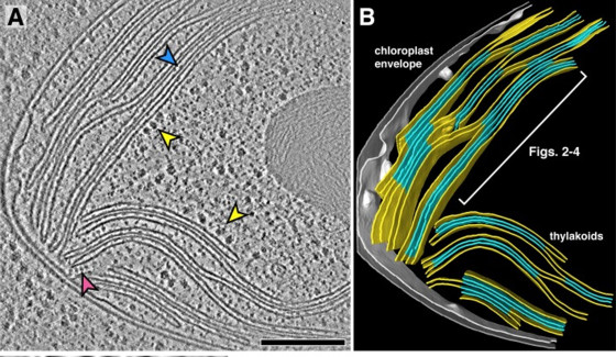
Another research trend: cryo-electron microscopy and tomography can be used to visualise protein structures down to atomic detail and understand molecular processes inside the cell. Here, too, research at Chlamy is particularly advanced.
After decades of intensive research, are there still many unanswered questions about Chlamy? ‘Of course,’ emphasises Michael Hippler. ‘The devil is in the details. We still have a lot to learn about the processes inside the cell.’
About the conference
The Chlamy conference takes place every two years – alternately on a different continent. Scientists from various disciplines such as structural biology, physiology, biochemistry, biophysics and medicine come together to network and share their latest research findings. The last time the Chlamy conference was held in Germany was in Potsdam in 2012. This year’s conference is being organised by Gaia Pigino from Human Technopole (Milan, Italy), Michael Schroda from the RPTU Kaiserslautern-Landau and Michael Hippler from the University of Münster.