
Quantitative Microscopy – Lifetime Imaging and Tension Sensor Multiplexing
Intracellular FRET signals can be analyzed by different microscopy methods (Cost et al. 2015). However, only a few techniques such as single photon counting fluorescence lifetime imaging (TCSPC-FLIM) allow the direct calculation of FRET efficiency, a value that is required to calculate molecular forces. We have therefore established microscopy protocols and custom written data analysis software to acquire and evaluate live cell TCSPC-FLIM data (Austen et al. 2011). In combination with the FL-based module and bi-exponential fitting of fluorescent lifetime decays, FLIM can be also used to estimate how many molecules of a given population are mechanically engaged.
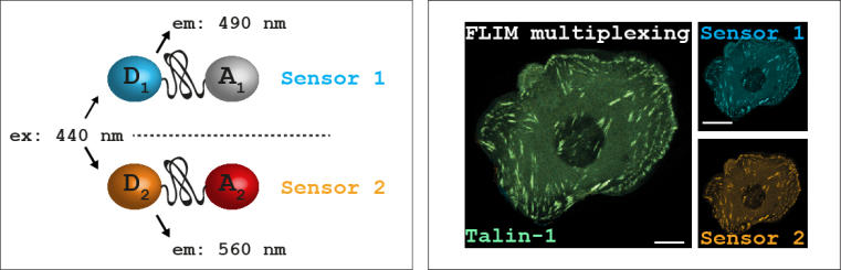
In addition, we have developed a tension sensor multiplexing technique to analyze two co-expressed tension sensor proteins simultaneously. By adjusting the excitation and emission spectra of donor and acceptor fluorophores, we generated a pair of orthogonal tension sensor modules that can be excited with the same wavelength (440nm) but analyzed separately in distinct emission windows. As a result, two FLIM signals originating from the same cell (and even the same subcellular structure) can be quantitatively analyzed (Ringer et al. 2017). We have successfully used the method to evaluate mechanisms of force propagation across the essential cell adhesion molecule talin-1.
Austen K, Kluger C, Freikamp A, Chrostek-Grashoff A, Grashoff C. Methods Mol Biol. 2013; 1066:169-184. >>
Cost AL, Ringer P, Chrostek-Grashoff A, Grashoff C. Cell Mol Bioeng. 2015; 8(1):96-105. [Epub 2014 Dec 2] >>
Ringer P, Weißl A, Cost AL, Freikamp A, Sabass B, Mehlich A, Tramier M, Rief M, Grashoff C. Nature Methods 2017 Nov; 14(11):1090-1096. [Epub 2017 Sep 18] >>