FIM Imaging to Visualise and Quantify Internal Organs
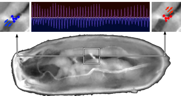
The importance of studying model organisms such as Drosophila melanogaster has significantly increased in recent biological research. Amongst others, Drosophila can be used to study heart development and heartbeat related diseases. We have established Frustrated Total Internal Reflection (FTIR) to improve the analysis of small animals like insects. This FTIR-based Imaging Method (FIM) results in an excellent foreground/background contrast and even internal organs and other structures are visible without any complicated imaging or labelling techniques. For example, the trachea and muscle organizations are detectable in FIM images. We demonstrate that FIM enables the precise quantification of locomotion features namely rolling behavior or muscle contractions by performing cluster analysis using histogram-based statistics. We also demonstrate that these imaging techniques enables automatic in vivo heartbeat detection of Drosophila melanogaster pupae based on morphological structures which are recorded without any dissection. Our approach is easy-to-use, has low computational costs, and enables high-throughput experiments.