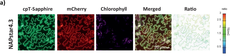
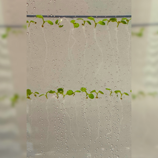
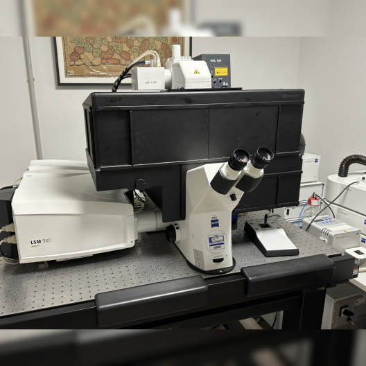
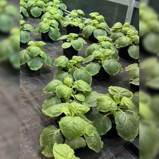
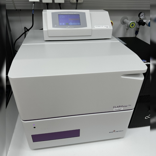
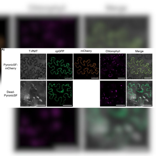
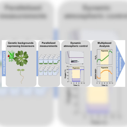
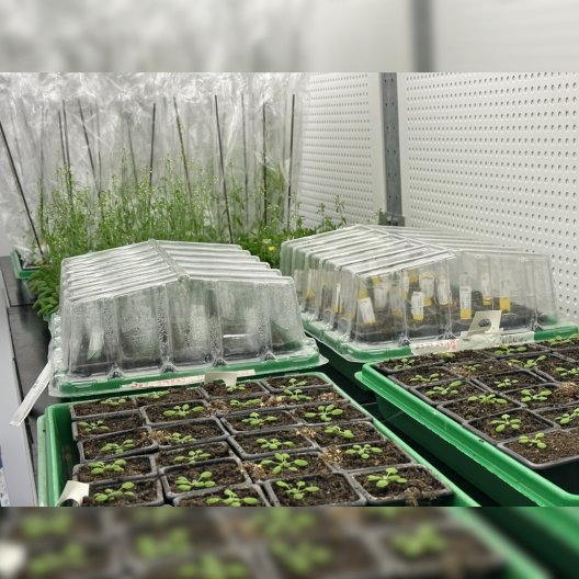
Bewegung von Mitochondrien (grün) gegenüber eines Plastiden (rot) innerhalb einer Epidermiszeller eines Blattes. Aufgenommen mit einem CLSM. Confocal microscopy images of NAPstar4.3 expressed in the cytosol of Arabidopsis thaliana plants.© Nature - Scherschel, Niemeier et al. Selektion von Arabidopsis thaliana Pflanzen auf Agarplatte© Uni MS-AG PEBMit Hilfe der konfokale Laser-Scanning-Mikroskopie können die wichtige Fragestellungen der Arbeitsgruppe beantwortet werden.© Uni MS-AG PEBTabakpflanzen im Gewächshaus für Infiltrations-Versuche© Uni MS-AG PEBDer Plate Reader misst selbst kleinste Veränderungen durch optogenetischen Biosensoren. Viele wurden hier in der Arbeitsgruppe entwickelt.© Uni MS-AG PEBConfocal microscopy images of the subcellular localization of the pyruvate biosensors in epidermal cells of transiently infiltrated N. benthamiana leaves. © Journal of Experimental Botany, Multhoff et al., 2024Workflow for live monitoring of plant hypoxia physiology using fluorescent protein-based biosensors.© Plant Physiology - Panicucci et al., 202Arabidopsis Pflanzen in verschieden Wachstumsstadien in der Klimakammer.© Uni MS-AG PEB