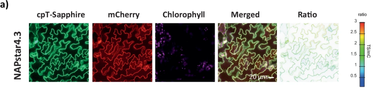
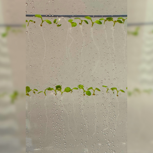
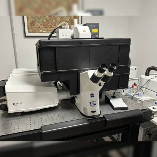
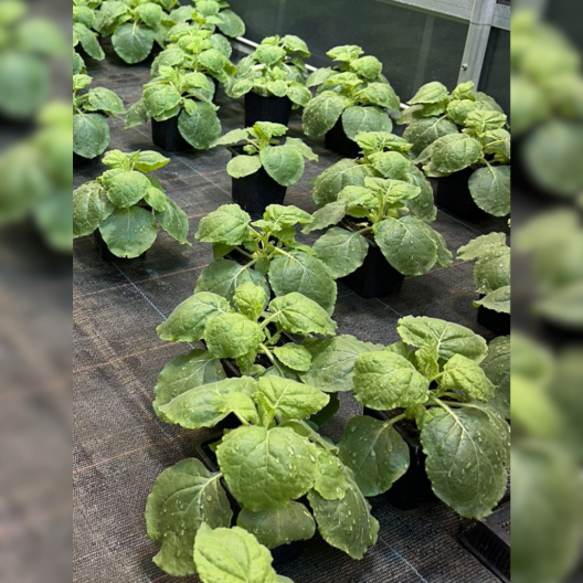
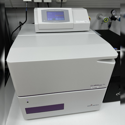
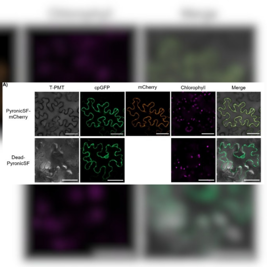
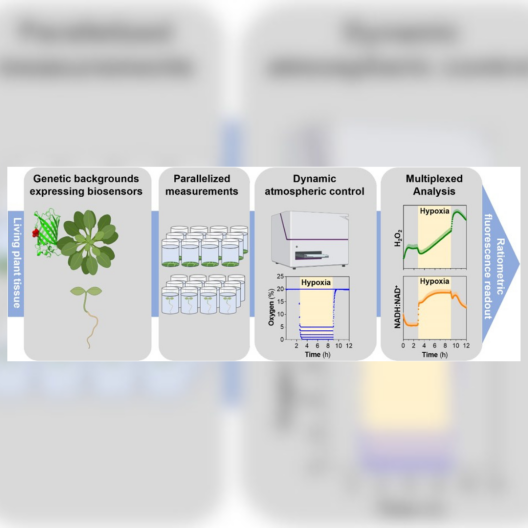
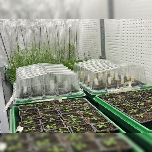
141 / 5.000 Movement of mitochondria (green) relative to a plastid (red) within an epidermal cell of a leaf. Imaged with a CLSM.Confocal microscopy images of NAPstar4.3 expressed in the cytosol of Arabidopsis thaliana plants.© Nature - Scherschel, Niemeier et al. Selection of Arabidopsis thaliana plants on an agar plate© Uni MS-AG PEBConfocal laser scanning microscopy can be used to answer the important questions of the research group.© Uni MS-AG PEBTobacco plants in our greenhouse for infiltration experiments© Uni MS-AG PEBThe plate reader measured even the smallest changes using optogenetic biosensors. Many of these were developed here in the research group.© Uni MS-AG PEB Confocal microscopy images of the subcellular localization of the pyruvate biosensors in epidermal cells of transiently infiltrated N. benthamiana leaves. © Journal of Experimental Botany, Multhoff et al., 2024Workflow for live monitoring of plant hypoxia physiology using fluorescent protein-based biosensors.© Plant Physiology - Panicucci et al., 202Arabidopsis plants in various growth stages in the climate chamber.© Uni MS-AG PEB