Modern Light Microscopy: How Cellular Structures Come to Glow
The most important tool in modern biology is clearly the microscope. Revolutionary technical improvements made it so efficient that it can help to detect structures from micrometer (1/1.000 mm) to nanometer (1/1.000.000 mm). Acting on the scale of nanometers, smallest components of cells can be visualized, e.g. synapses. Synapses are communication interfaces between nerve cells, that transfer signals. In order to do so, they need a pool of transmitters. Prof. Jürgen Klingauf, expert in cellular biophysics, investigates with light microscopy how cells re-fill this pool again and again. Therefore he uses fluorescent dyes.
Prof. Friedemann Kiefer uses fluorescent dyes as well. In the chemical laboratory, these dyes are generated in a way, that they bind to selected structures in the organism and glow from there. Sensitive cameras capture the fluorescent light and produce images of the glowing structures that can be assembled three dimensionally in the computer. In the area of micrometers, Prof. Kiefer investigates specimen of tumor tissue. In recent times, so-called intravital microscopy (lat. during life) allows to observe even living, unfixed organisms on the scale of centimeters.
Photos

Professor Friedemann Kiefer (left) and Professor Jürgen Klingauf use light microscopes in their daily work. Due to the continuous improvement both are offered new opportunities to investigate the behaviour of cells in organisms.© CiM - Michael Kuhlmann 
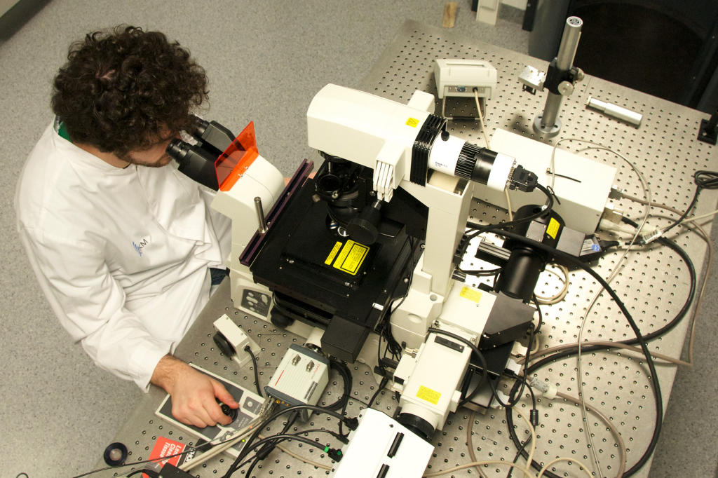
Jeroen Terwort, PhD student in the working group of Jürgen Klingauf, uses a light microscope, which overcomes the natural diffraction limit by a trick. The development of this STED microscope was awarded with the Nobel Prize in 2014. © CiM - Michael Kuhlmann 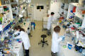
The working group of Friedemann Kiefer also works with light microscopy.© CiM - Michael Kuhlmann 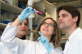
Dr. Vivien Schütz (left), Dr. Cathrin Pollmann and Dr. René Hägerling give fluorescent dyes into the tissue, afterwards the sample is scanned under the ultra-microscope layer by layer by a laser beam.© CiM - Michael Kuhlmann 
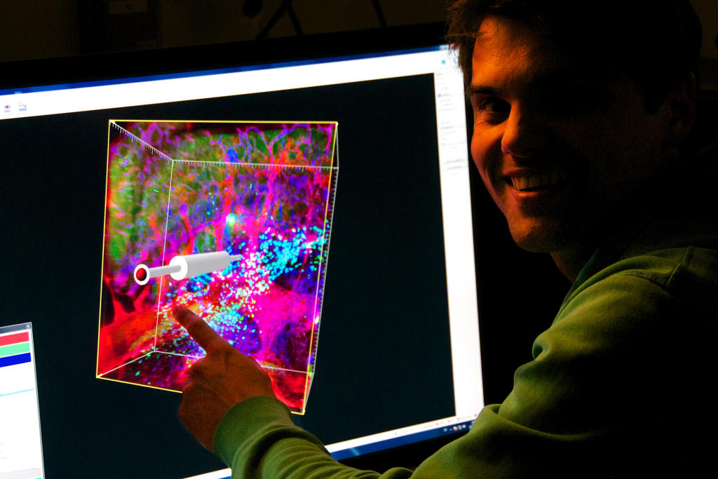
René Hägerling shows the result: On the computer the individual layers of ultra-microscopic tissue examination are assembled and can be analysed from all perspectives.© CiM - Michael Kuhlmann