Risk Signature in the Blood – Long-standing Lab Work Makes Possible What Clinics Wish for MS Therapy
In the autoimmune disease multiple sclerosis (MS), the immune system attacks endogenous nerve fibres and destroys their protective layer, which may cause something called “MS relapses”, often resulting in impaired vision or palsy. Nowadays though, these MS relapses can be suppressed by a particular medicine. But in rare cases, death can occur as a result of one severe side effect: brain inflammation.
Multiple sclerosis is a focus of the Department of General Neurology at the University Hospital Münster, and its physicians are working on ways to reveal an MS patient’s risk of brain inflammation before they take this medicine. According to physician Prof. Heinz Wiendl, information about how high the risk of brain inflammation is for an individual patient can be provided by a biomarker. Dr. Nicholas Schwab and his team had been searching for such a biomarker, and they finally found one. However, until the biomarker finally becomes part of a doctor’s checklist, it must first undergo years of laboratory research at both the federal and the EU-level.
Photos

Professor Heinz Wiendl wants to bring biomarkers into clinical practice. To optimize MS therapy, he has to coordinate both therapy in the hospital and research in the labs.© UKM 
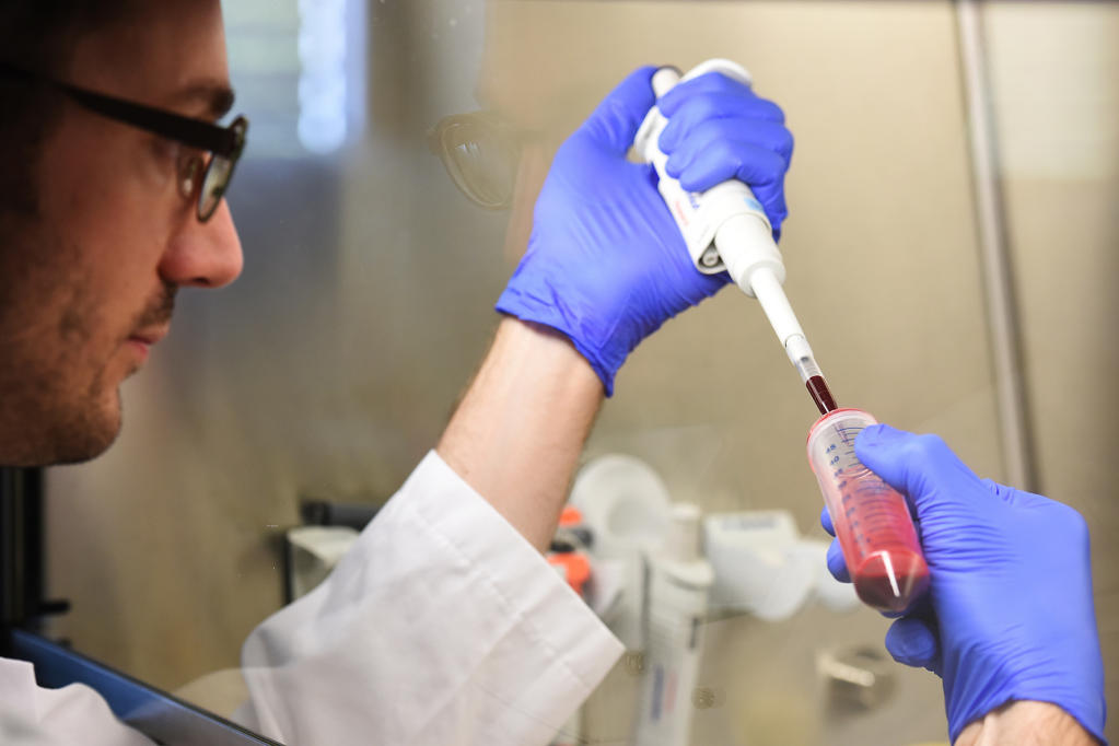
Dr. Nicholas Schwab’s working group gets blood samples directly from the university hospital. The first step is to isolate the immune cells from the blood samples for further analysis.© CiM - Peter Grewer 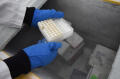
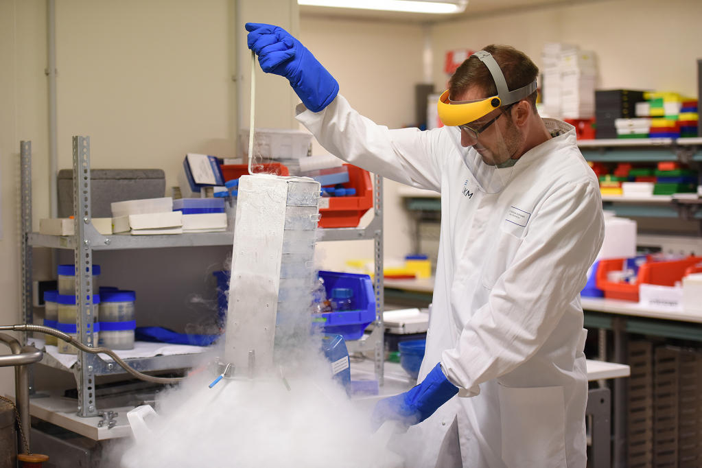
Because researchers must compare huge numbers of samples, isolated immune cells are imported from all around the world. To store the cells, they first have to be cooled down slowly and step by step. Otherwise they would be destroyed by ice crystals.© CiM - Peter Grewer 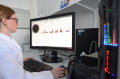
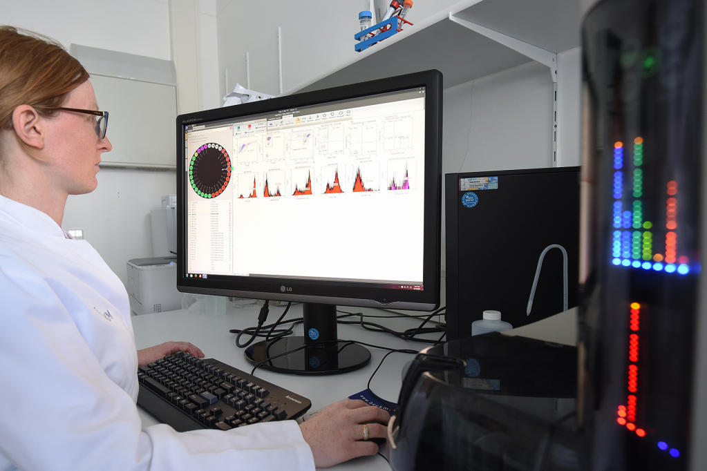
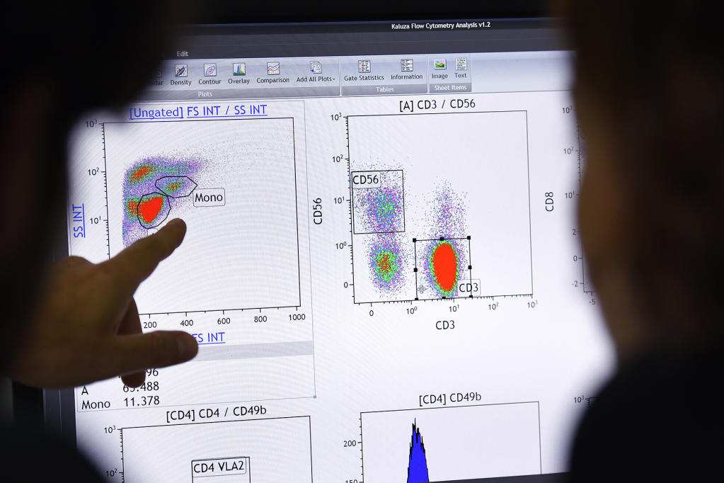
Flow cytometry not only analyses several thousand immune cells every second, but it also sorts them. The fluorescence-based technology enables researchers like Dr. Susann Pankratz to see them on the monitor.© CiM - Peter Grewer 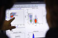
In this way, Schwab’s team can compare samples from different patient groups in detail to find out if they have the same biomarker.© CiM - Peter Grewer