Sharper Images for Medicine – How Medical Physics Helps to Optimise Clinical Scans
To locate metastases in the body or to diagnose heart diseases patients are usually examined with whole body scanners. Positron emission tomography (PET) is a technique which takes several minutes. But as a patient has to breathe and thus moves during the examination the resulting images show blur effects – like a blurred holiday snap. Physicians have learned to cope with that. But the better the image, the easier to make a precise diagnosis, especially in difficult cases.
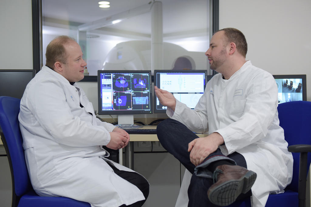
That is why medical physicist Dr. Florian Büther (European Institute of Molecular Imaging, University of Münster) and nuclear medicine specialist Dr. Thomas Vehren (Department of Nuclear Medicine, University Hospital Münster) develop new methods which shall improve the quality of medical images. The most promising techniques are called ‘gating’ and ‘motion correction’. They measure a patient’s movements to reconstruct images without any motion blurring. Instead of using external hardware like a respiratory belt, Büther and Vehren want to solely work with computer programmes in order to save time and to generate data with high statistic quality.
Photos
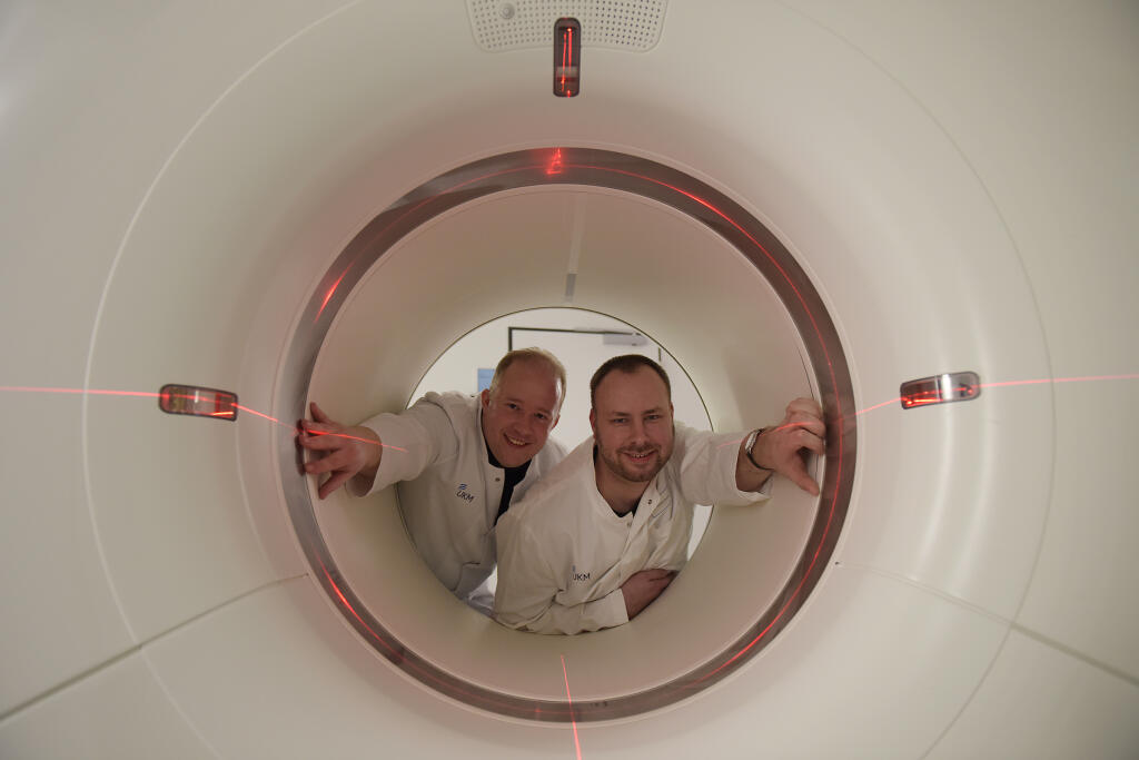
Joining forces physician and physicist can implement new methods like ‘gating’ in the clinic using the PET/CT scanner in the University Hospital Münster.© SFB 656 - Peter Grewer 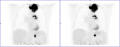
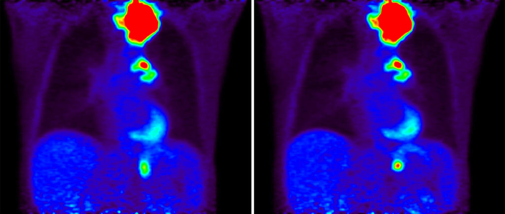
These PET images display the torso of a patient from the shoulders to the navel area, without gating (left) and with gating (right). Particularly in the abdomen there are blur effects on the left image.© UKM - Florian Büther, Thomas Vehren, Michael Schäfers 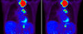
The shining knobs show a pulmonary tumour (top), a lymph node metastasis (middle) and metasteses in the adrenal glands (bottom). The gated image on the right shows size and activity of these tumours more precisely.© UKM - Florian Büther, Thomas Vehren, Michael Schäfers