Nanoanalysis with ToF-SIMS/Laser-SNMS
To understand the important physical and chemical properties of nanostructured systems, it is essential to know their 3D chemical composition. This requires answering three central questions: 1. Which elements, isotopes, functional groups and molecular compounds are present? 2. How are these components distributed in three dimensions? 3. What are the concentrations of these chemical constituents? Time-of-Flight Secondary Ion-Mass Spectrometry (ToF-SIMS) and its sister technique, Laser Secondary Neutral Mass Spectrometry (Laser-SNMS), are important tools for answering these critical questions.
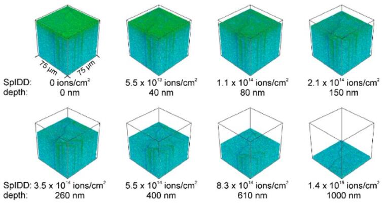
Using state-of-the-art ion gun technology, atomic and molecular species can be imaged in 3 dimensions in both organic and inorganic materials with a lateral resolution of as little as 50 nm and depth resolution of 1 nm. Improvements are however needed to detect many species at low concentrations or in nanoscale volumes. In ToF-SIMS, the vast majority of material sputtered from the sample is desorbed as neutral species that cannot be detected by the mass spectrometer. This limits the useful spatial resolution and detections limits. Improving ionization efficiency is essential for nanoscale analysis of biological samples and nanomaterials.
Methods for improving ionization efficiency for nanoscale imaging
The Arlinghaus research group is exploring two methods for improving ionization efficiency for nanoscale imaging: 1. Matrix Enhanced Secondary Ion Mass Spectrometry (ME-SIMS) and 2. Laser post-ionization Secondary Neutral Mass Spectrometry (Laser-SNMS). In ME-SIMS, samples are coated with an ultrathin layer of an organic compound that increases ionization of target molecules. Although exciting improvements have been observed with ME-SIMS, the fundamentals are poorly understood and results are often highly variable. Our focus is on exploring factors that influence the matrix enhancement effect including optimizing methods of matrix deposition, exploring the influence of temperature and humidity, studying mass transport of analytes in the matrix and varying the chemical structures of the matrix and analyte.
Alternatively, the sputtered neutral species can be ionized using a laser pulse (Laser-SNMS) in order in increase detection efficiency, which is essential to study nanoscale structures and low concentration species. With Laser-SNMS, we have demonstrated enhanced sensitivity up to 5 orders of magnitude over ToF-SIMS. However, detection of large organic ions is challenging due to photofragmentation. Further research is needed to understand the interplay between sputtering conditions, such as primary ion cluster size and energy, laser parameters, including timing, wavelength and power density, and sample parameters such as the matrix and target analyte. Further experiments are underway combining matrix enhancement and laser post-ionization.
In addition to these fundamental ion formation studies, the group is developing multivariate data analysis methods, image fusion techniques and advanced sample preparation techniques. The group has developed a unique combined cryo-ToF-SIMS/Laser-SNMS instrument with integrated high vacuum cryo-preparation/cryo-sectioning for nanoanalytical applications in life sciences. Additionally, application of these methods in materials science, tribology and nanotechnology are being investigated.