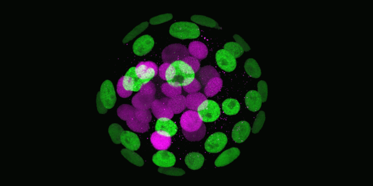
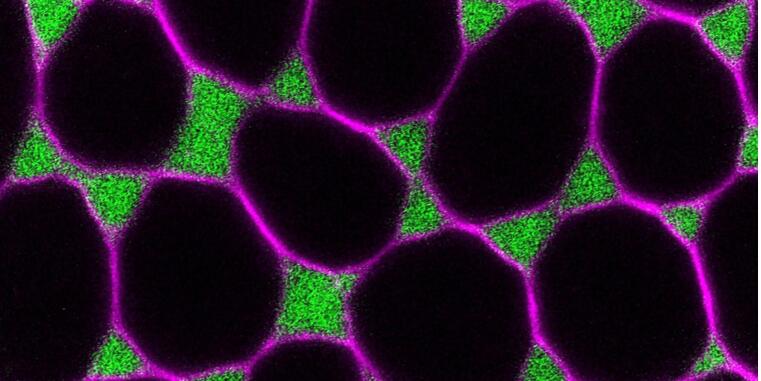
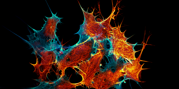

The precise spatial and temporal organization of cellular differentiation, essential for organismal development and physiology, depends on the correct formation and function of cellular interfaces. These interfaces facilitate the efficient transfer of materials and information between cells, through the dynamic assembly of molecular platforms at the plasma membrane. Such platforms integrate external and internal signals and transduce mechanical forces, guiding development and ensuring correct responses to physiological and pathophysiological changes. The Collaborative Research Center (CRC) 1348 focuses on understanding how these dynamic cellular interfaces are formed and how they control decision-making in cells and tissues.
We have developed a comprehensive strategy combining molecular modeling, structural biology, biochemistry, organic synthesis, genetics, cellular and developmental biology. This approach aims to unravel the molecular mechanisms governing cellular interactions and responses in individual cells as well as in multicellular organisms. Leveraging advances in high-resolution imaging, optogenetics, single-cell technologies, synthetic biology, and engineering, CRC 1348 aims to not only understand but also to precisely manipulate dynamic processes at the plasma membrane. Our goal is to provide a deep understanding of the molecular principles that govern cellular interface formation and function, setting the foundation for future efforts to recreate biological complexity from the bottom up.
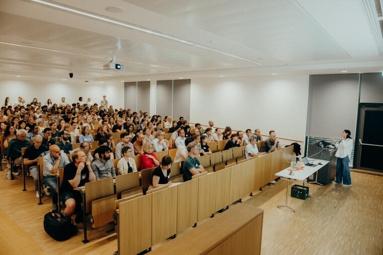
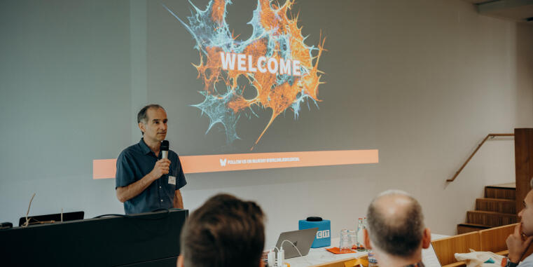
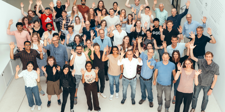
This year’s SFB 1348 meeting has come to a close. 3 days of inspiring science, stimulating discussions, and valuable exchange made it a truly rewarding event. Many thanks to all participants - we’re already looking forward to the next one!
See photo gallery here.
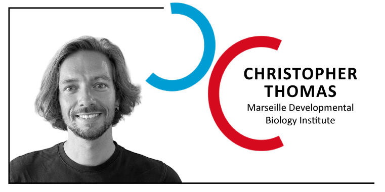
Exciting seminar with Christopher Thomas about "Revealing the Secrets of Ovulation".
Click here for more information.
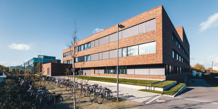
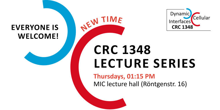
The CRC 1348 office is located in the Multiscale Imaging Center (MIC). Our seminars take place in the MIC auditorium.
From Münster central station (Münster Westf Hbf) you can reach the MIC by bus in around 30 minutes.
Bus stops:
(Status as of November 2023)
(Status as of November 2023)