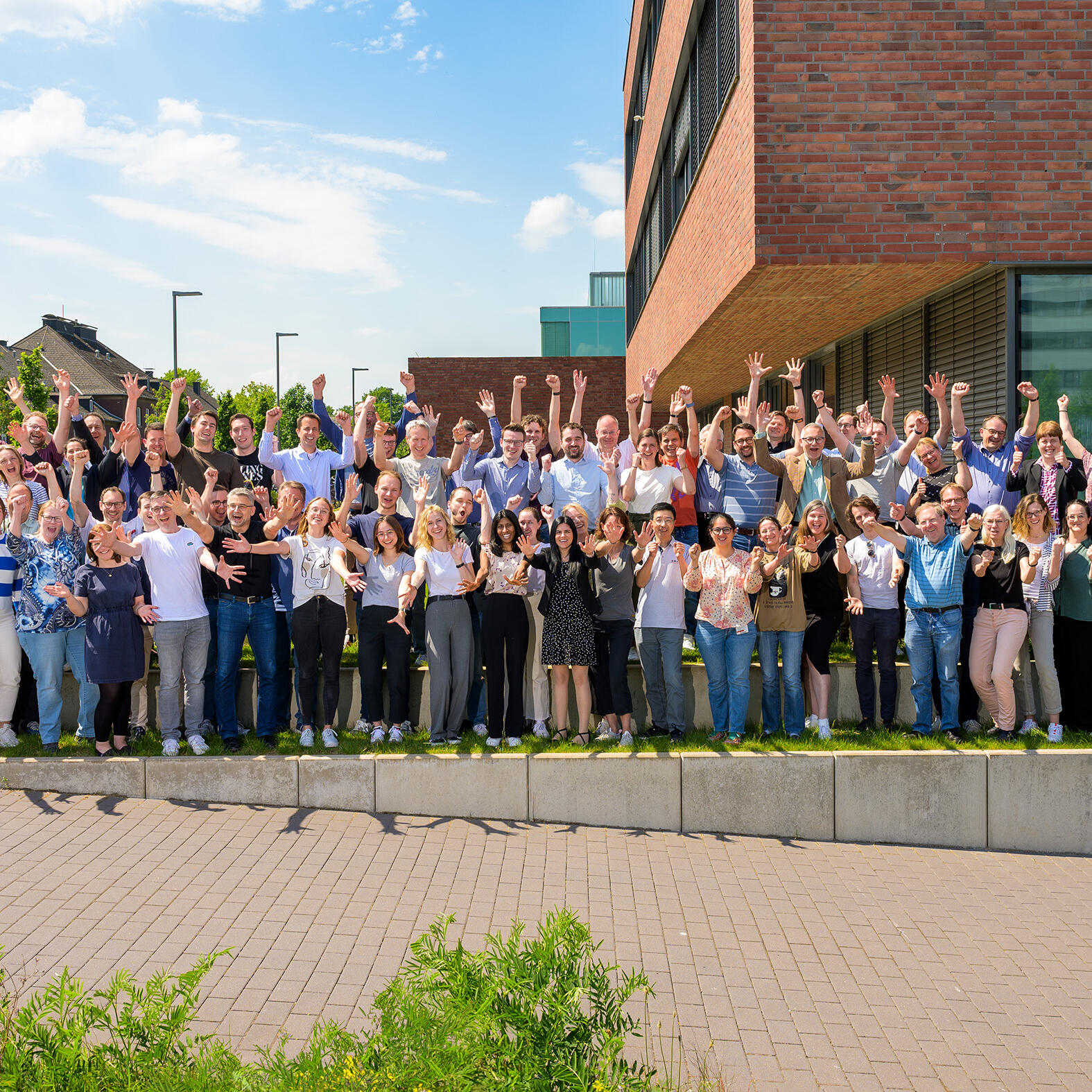
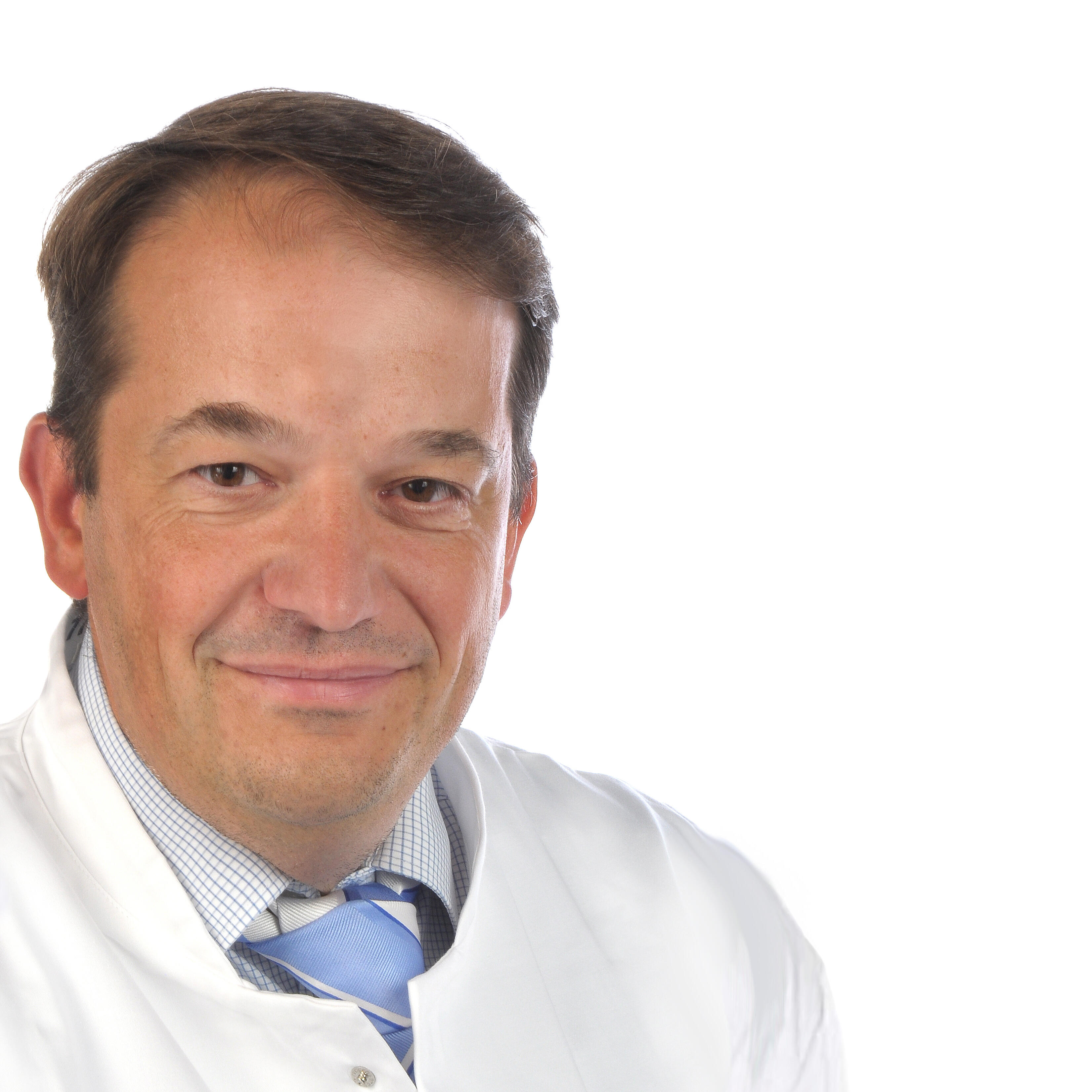
Translational research takes place worldwide in several sequential phases. Nuclear medicine specialist Professor Philipp Backhaus is primarily involved in the early phases of translation, specialising in the imaging and therapy of tumours and inflammation. For several months, his team has been contributing to the development of the drug “OncoACP3”, which aims to improve the examination of prostate cancer.
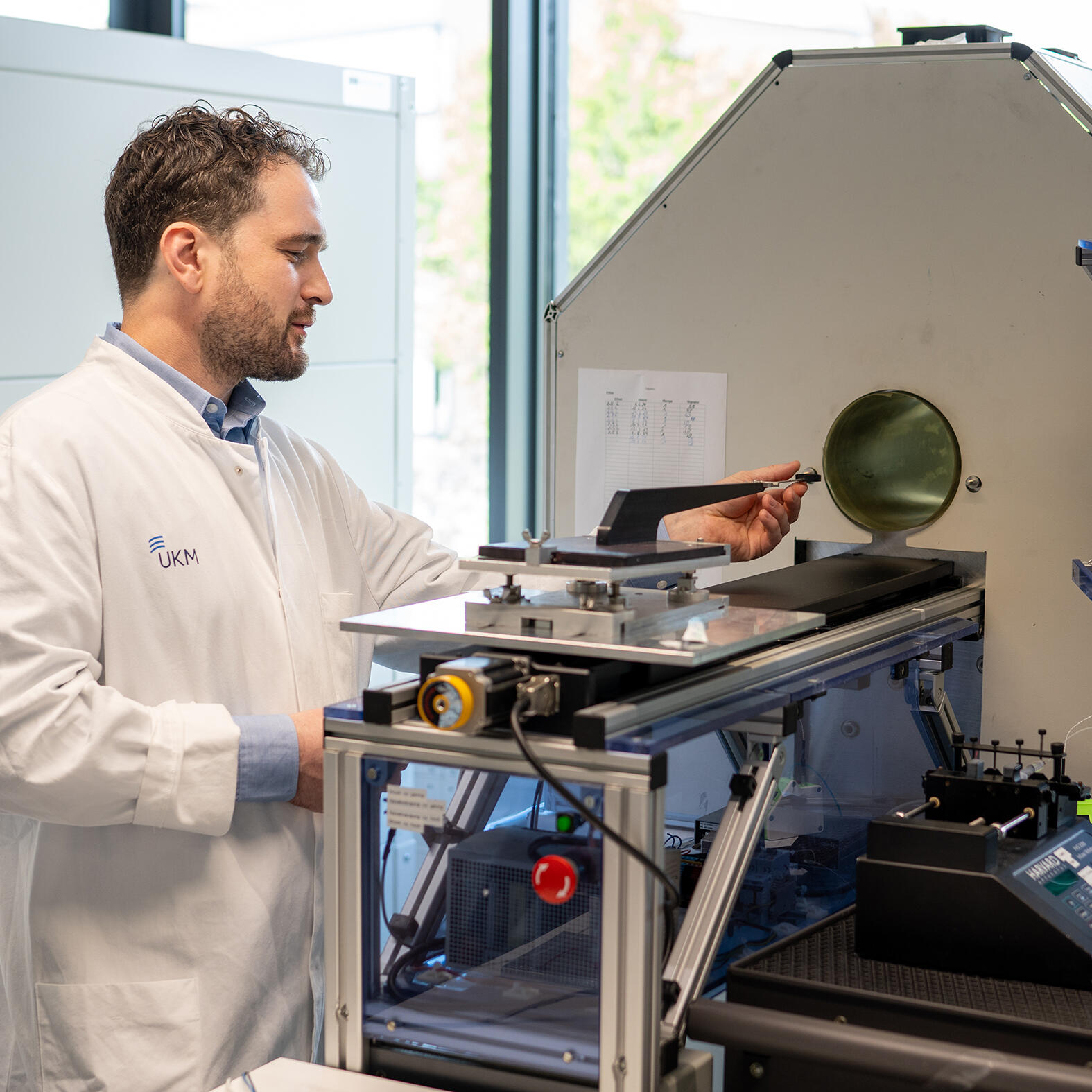
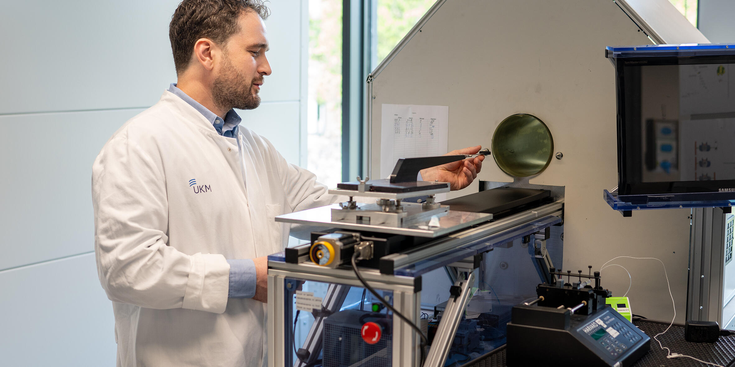
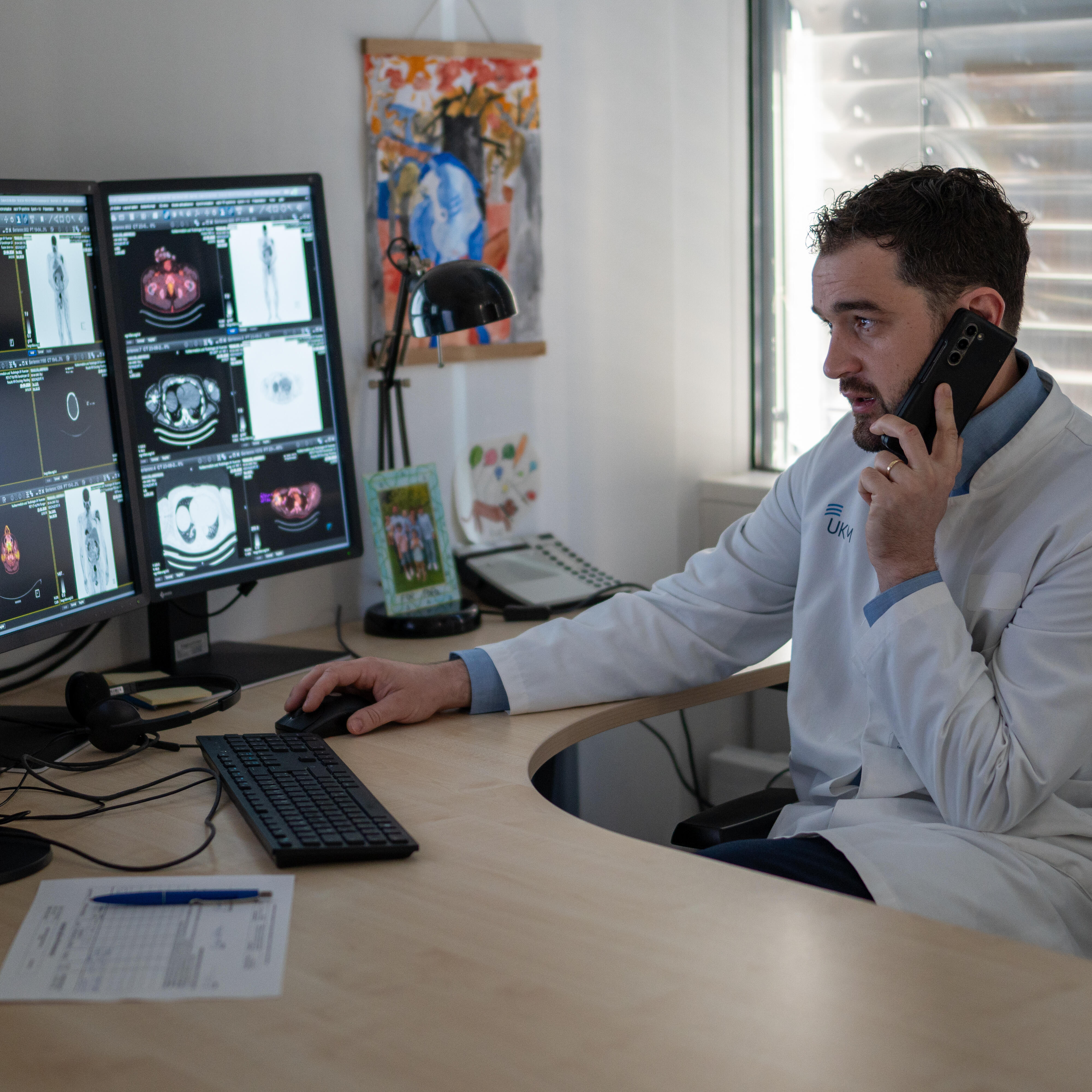
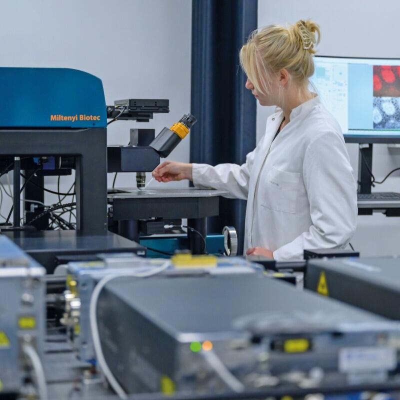
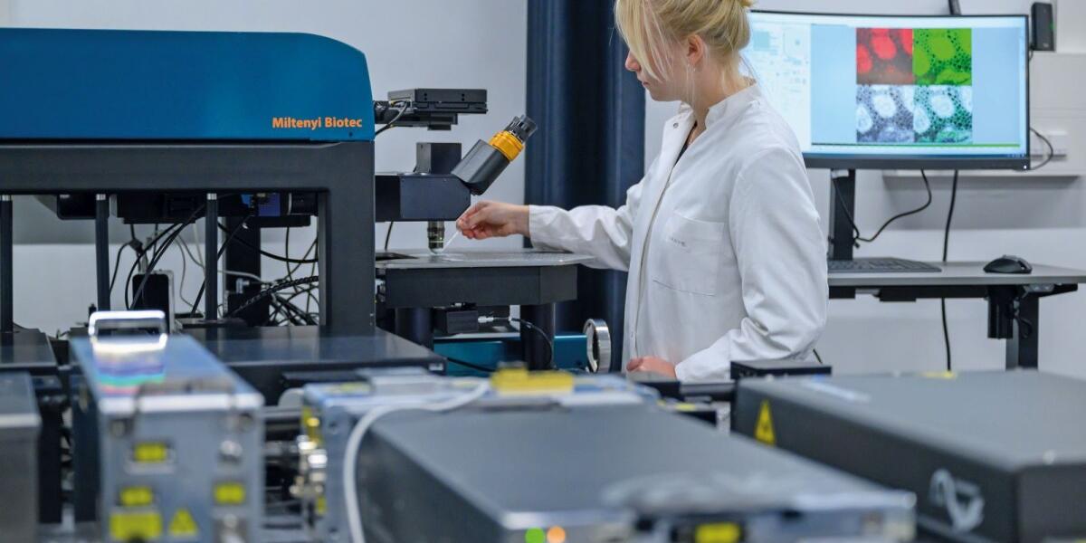
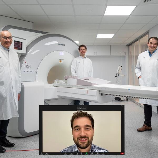
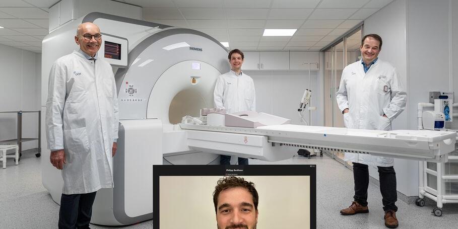
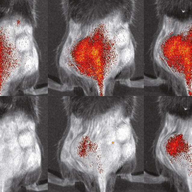
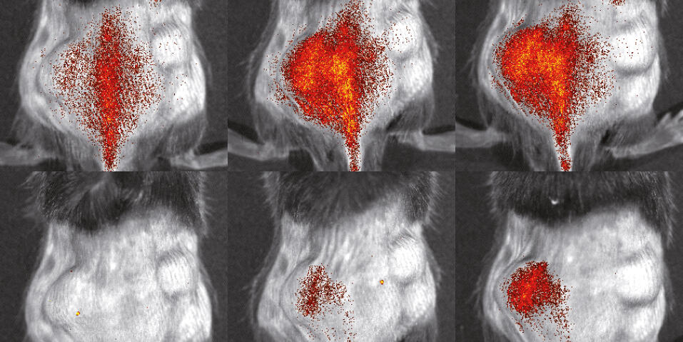
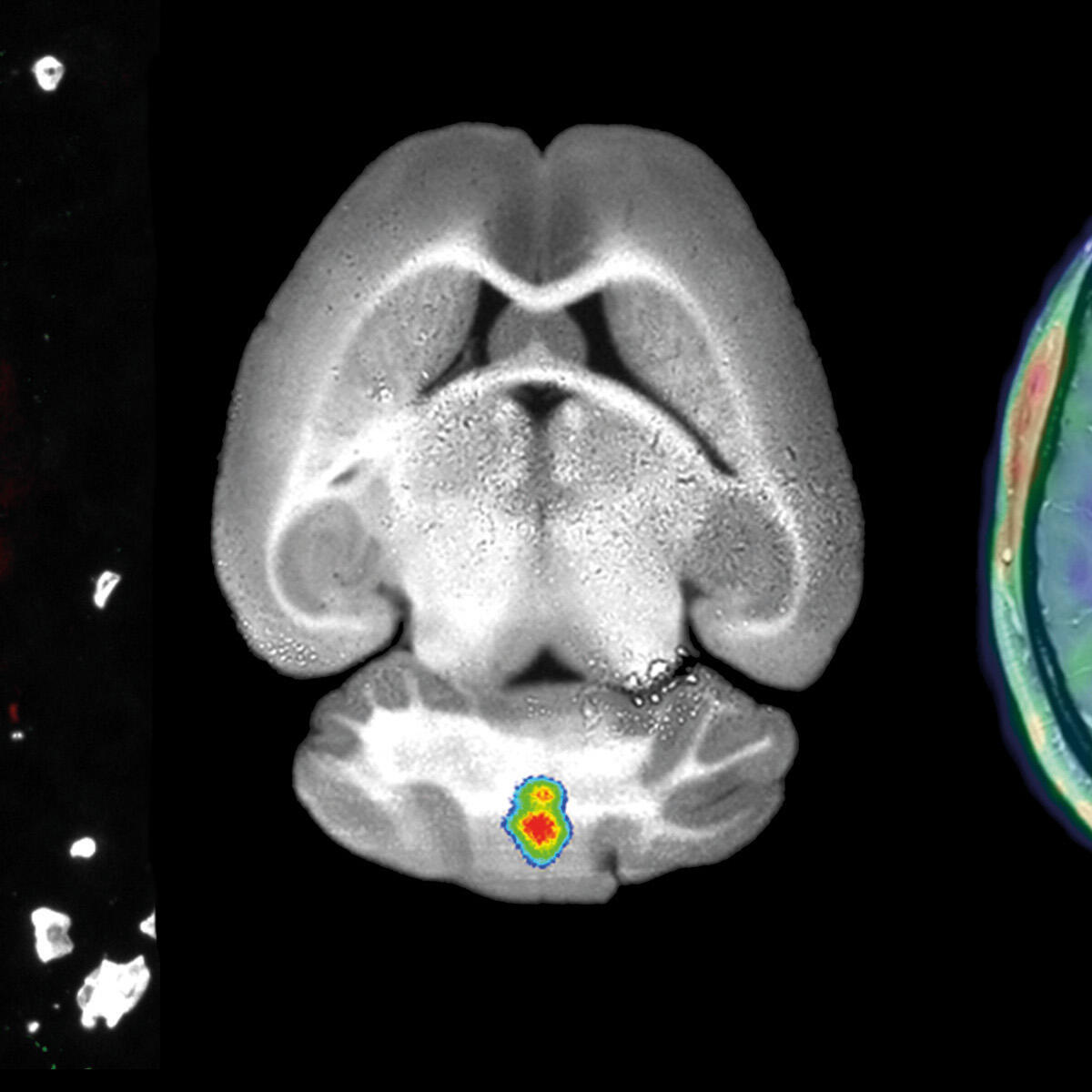
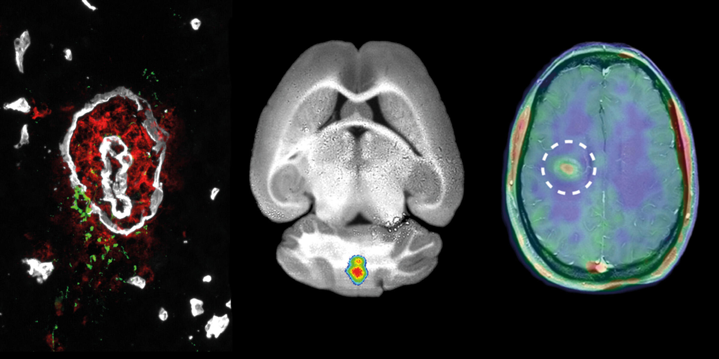
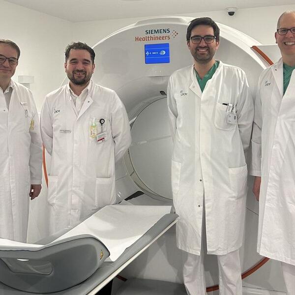
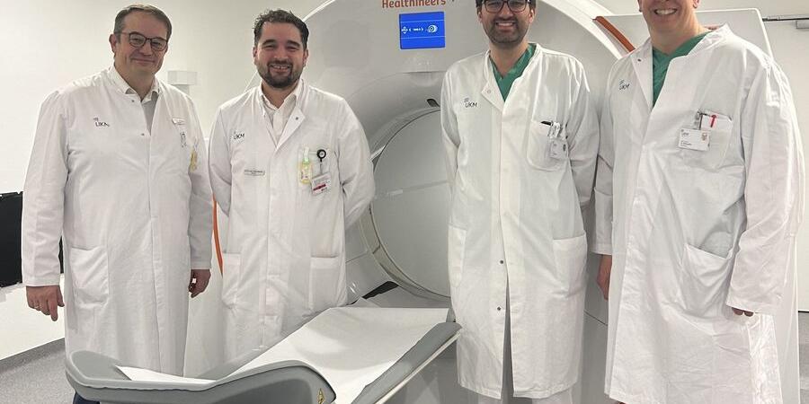
Nuclear medicine specialist Prof Philipp Backhaus and his research partners have evaluated a new radiotracer for PET imaging in prostate cancer that targets the molecule ACP3. Compared to an established PSMA-targeted tracer, it showed superior visualization of disease extent in many cases and led to a change in treatment in a relevant number of patients. The study was awarded “Paper of the Month” by the Medical Faculty.
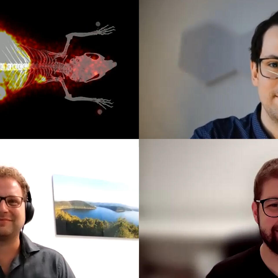
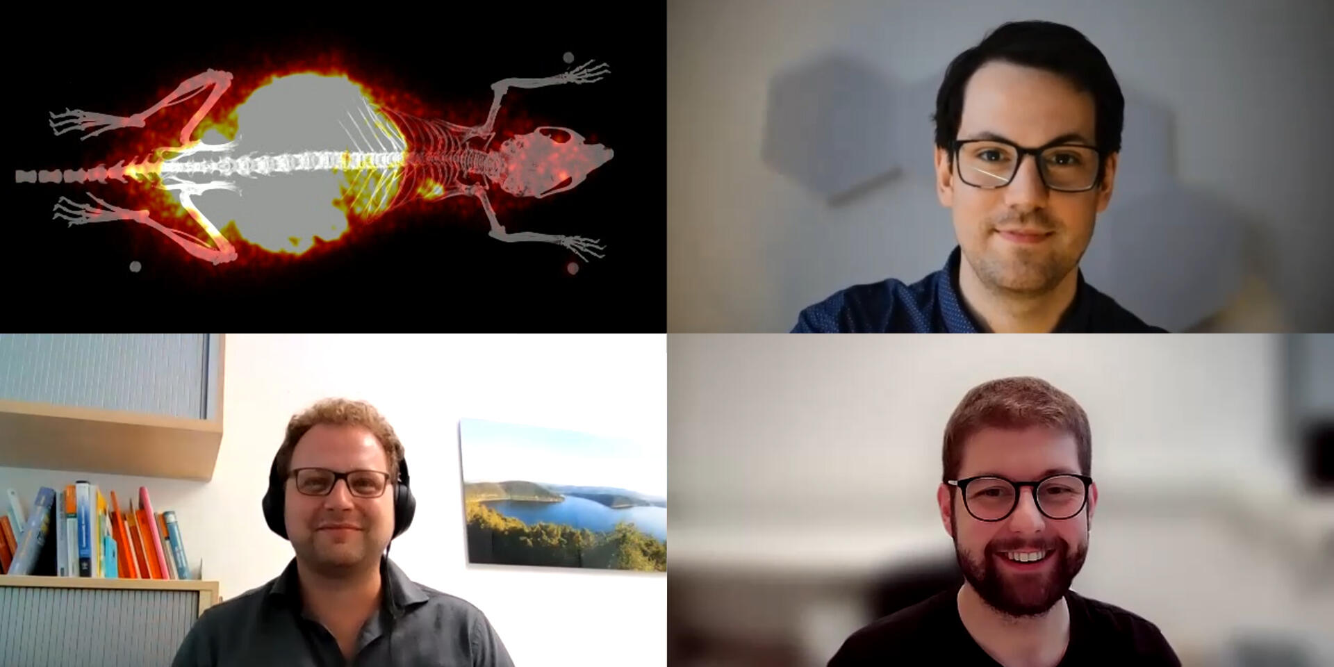
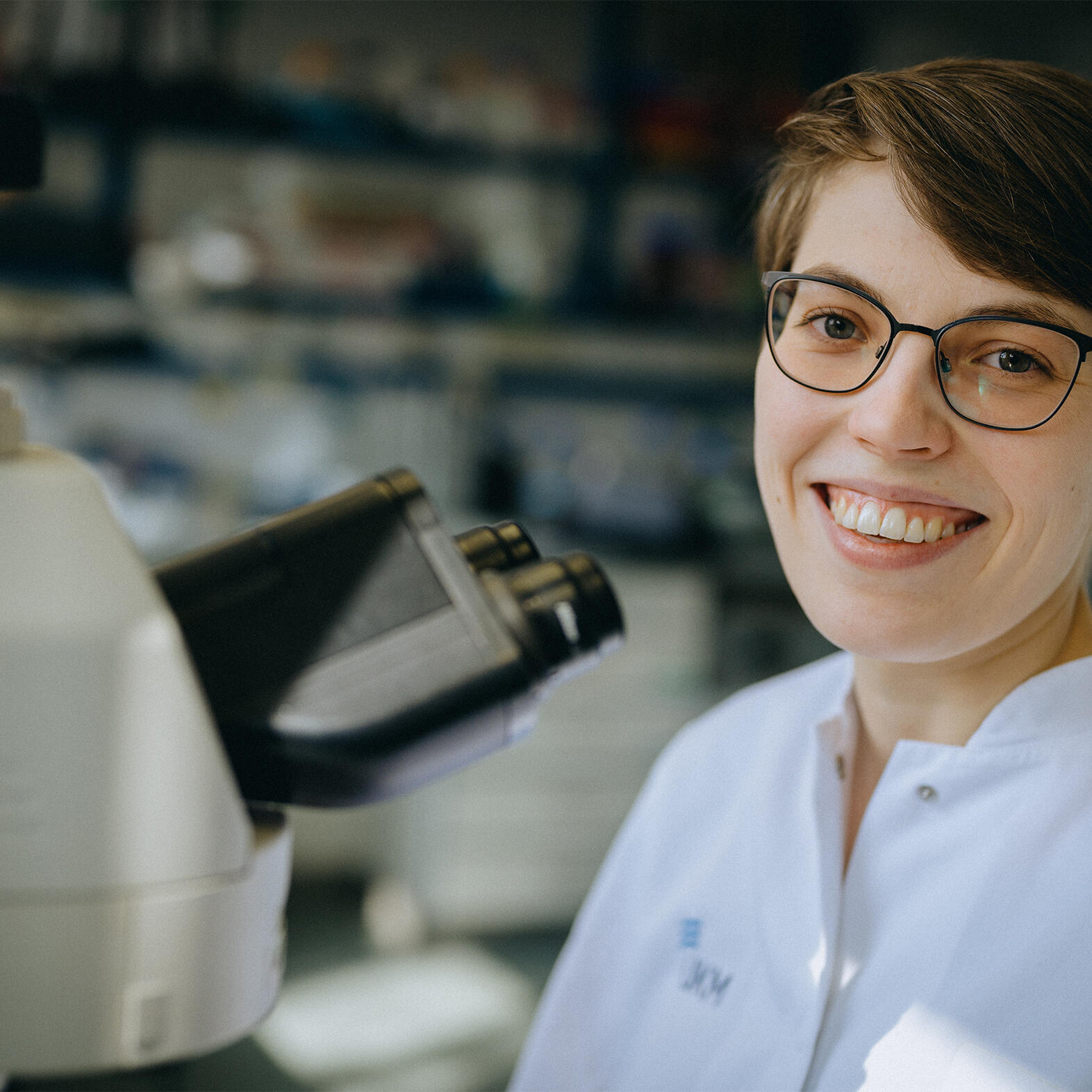
Nils Marquardt, a doctoral researcher in Medical Science, investigates how individual, moving cells in the body, for example, immune cells, can be visualized and tracked using positron emission tomography. In November, he attended the IEEE Medical Imaging Conference in Japan. In addition to the scientific program, there was also time to experience Japanese culture. He shares his impressions in this guest contribution.
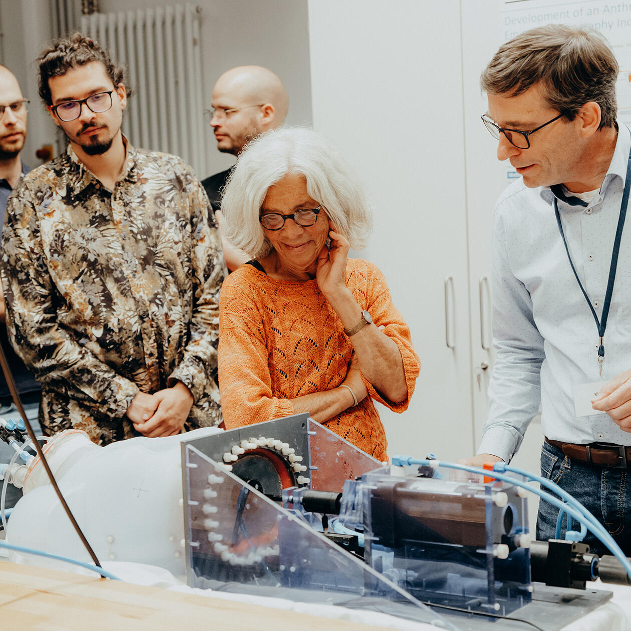
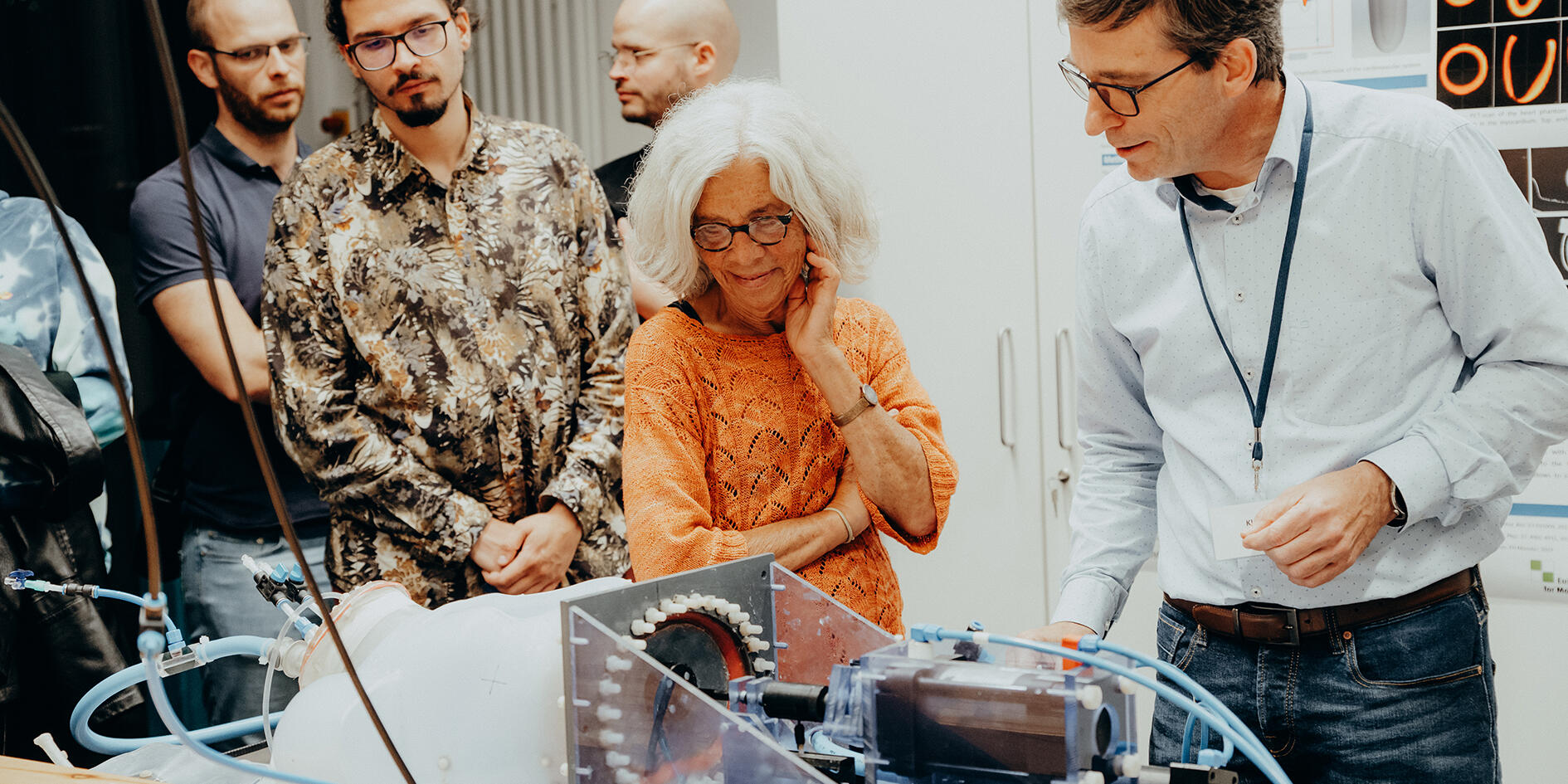
On September 12, 2025, the University and University Hospital Münster opened their doors to the public during the “Lange Nacht der Universitätsmedizin”. At the Multiscale Imaging Centre, many visitors took the opportunity to gain live insights into various areas of research.
The Collaborative Research Centre 1450 “inSight – Multiscale imaging of organ-specific inflammation” at the University of Münster will receive approximately 13 million euros from the German Research Foundation for a second funding period of four years. In this project, researchers are investigating how the body regulates inflammation in different organs and are, to this end, developing a specific multiscale imaging methodology.
Emmy Noether junior research group leader Dr Maria Florencia Sánchez has been working at the European Institute for Molecular Imaging for several months. Dr Sánchez, who was born in Argentina, and her team are investigating how cells communicate with each other and how they perceive and react to their environment. She also supports students and young scientists at the University of Münster.
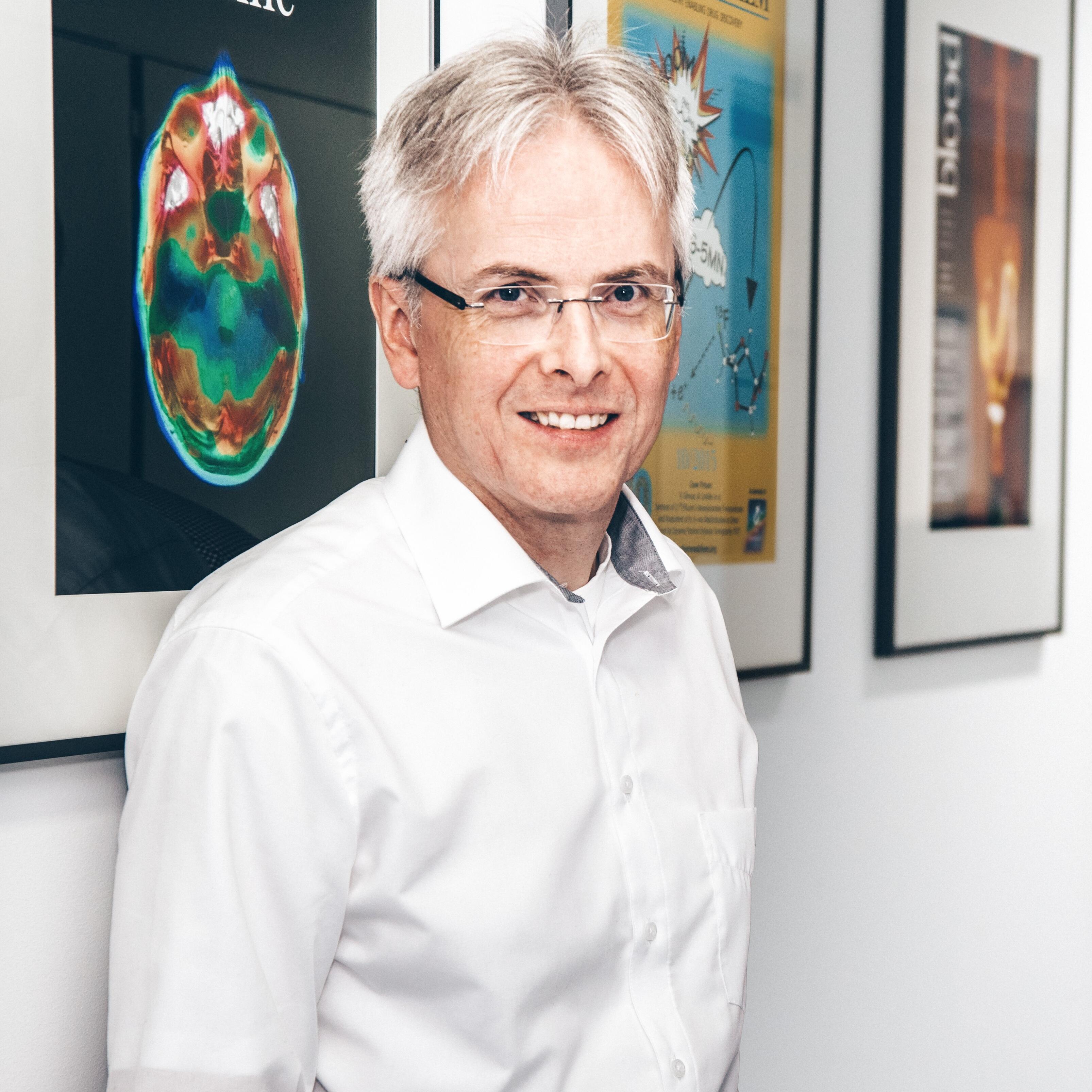
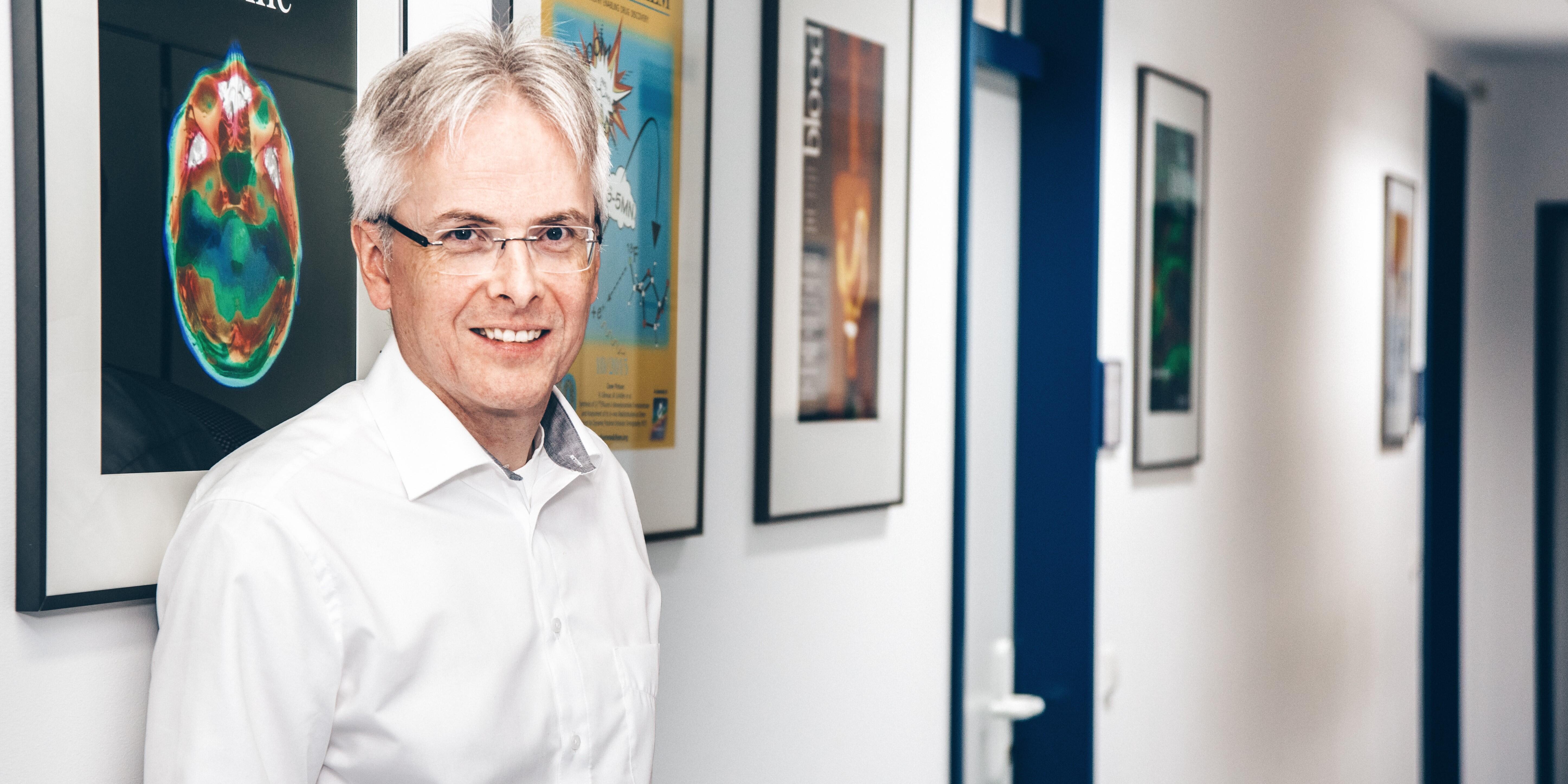
As a clinician scientist, junior professor Dr Philipp Backhaus is good at juggling the demands of research and hospital work. A specialist in nuclear medicine with six children, he hopes his research will help bring about concrete improvements in medical care. This is a dual role that requires good organisational abilities and the support of those around him.
Microscopes make it possible to take ever deeper and more precise looks at the smallest of details, and in ever higher resolutions. This article looks at some of the techniques used by researchers at the University of Münster including, among others, high-performance cryogenic electron microscopy available at Prof. Christos Gatsogiannis’ lab, three-photon microscopy used by Prof. Friedemann Kiefer’s group, and insights into confocal laser scanning microscopy provided by Prof. Stefan Luschnig’s group.
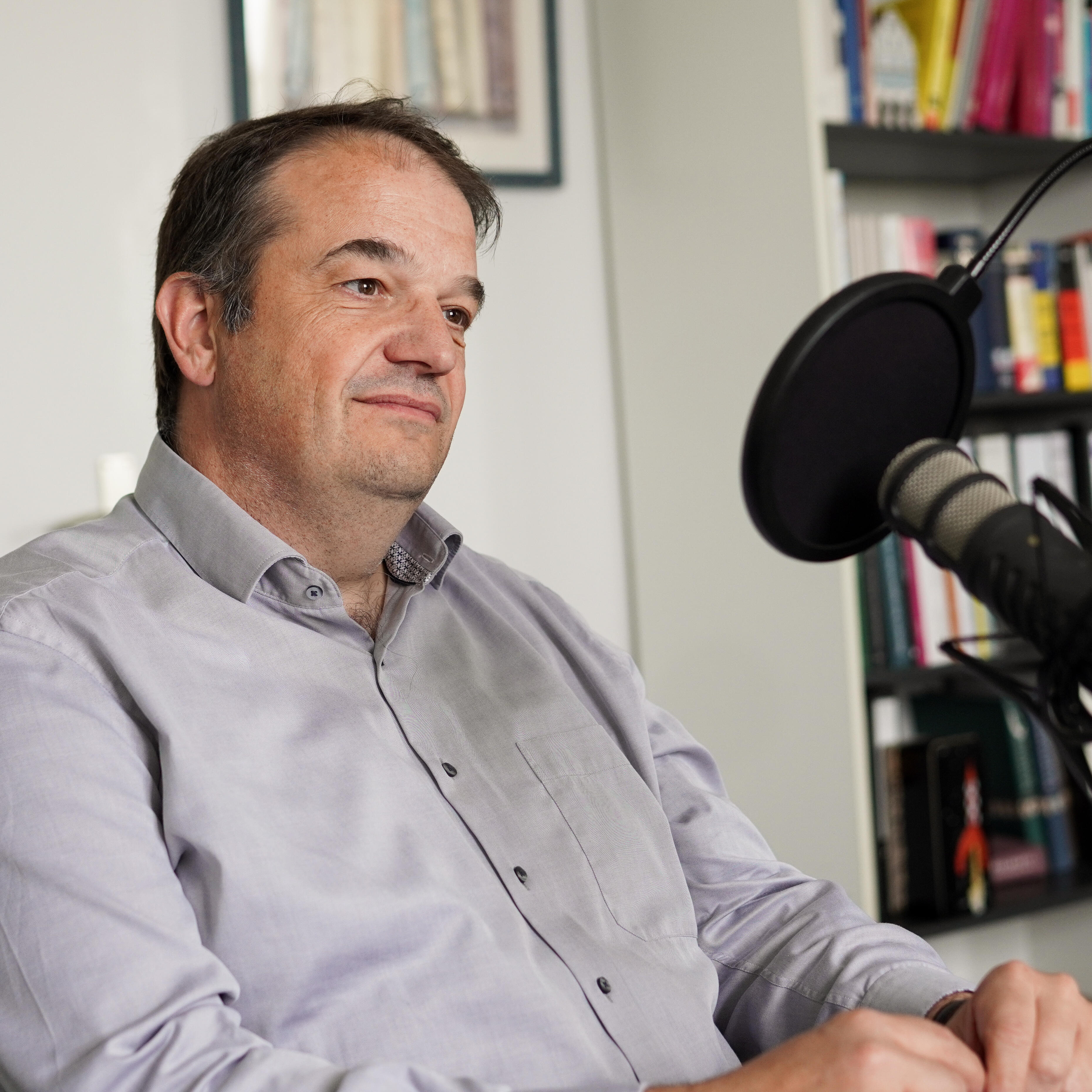
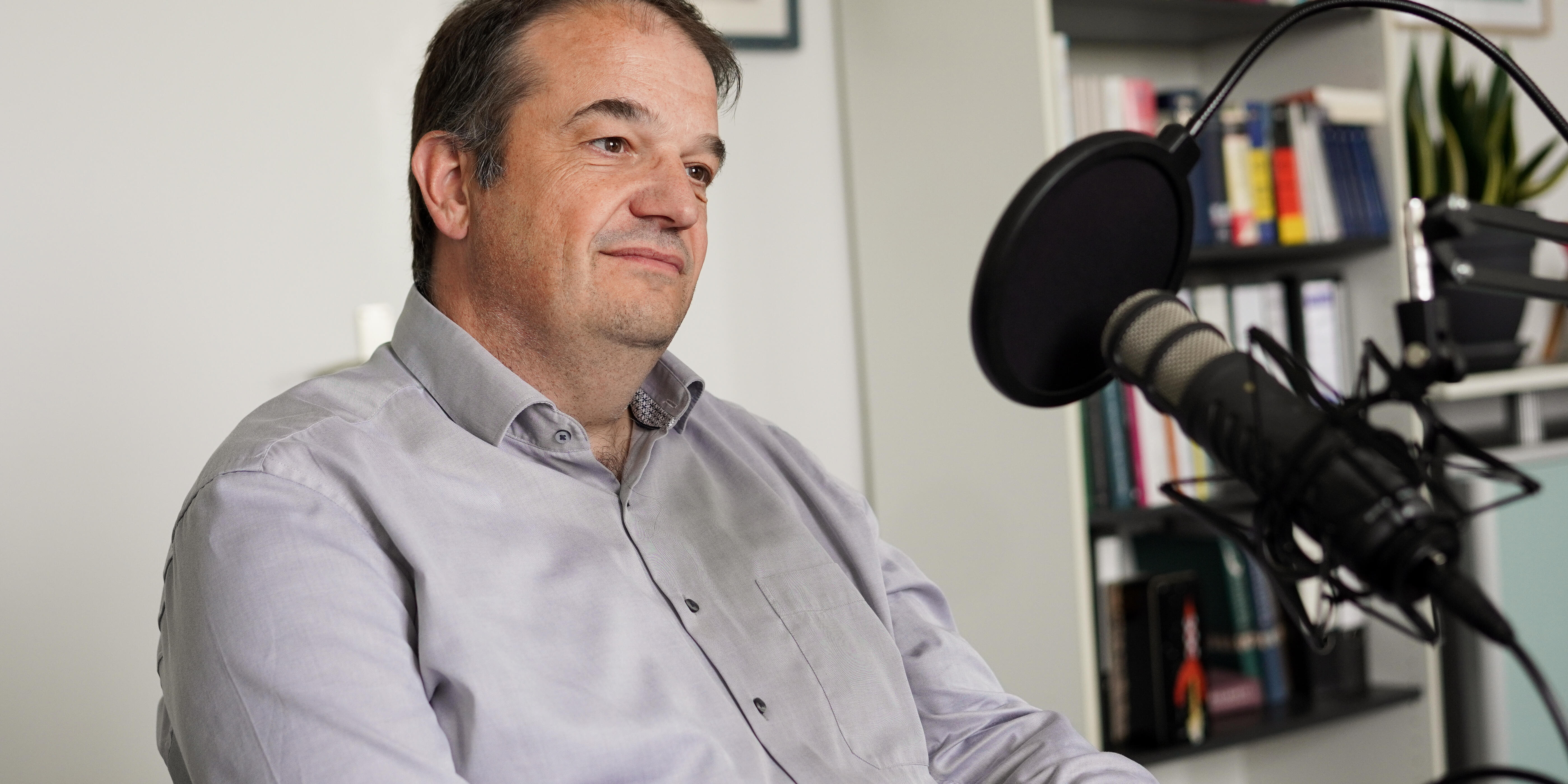
Science needs specialised researchers. For many research questions, however, cooperation with colleagues from other disciplines is just as important. Using the example of the Collaborative Research Centre “inSight” Prof Michael Schäfers, a specialist in nuclear medicine, provides insight into research practice in the field of inflammation and imaging. He also talks about “network life” and explains, for example, how a grant application for a research network is created and what role junior scientists play. The podcast is available in German.
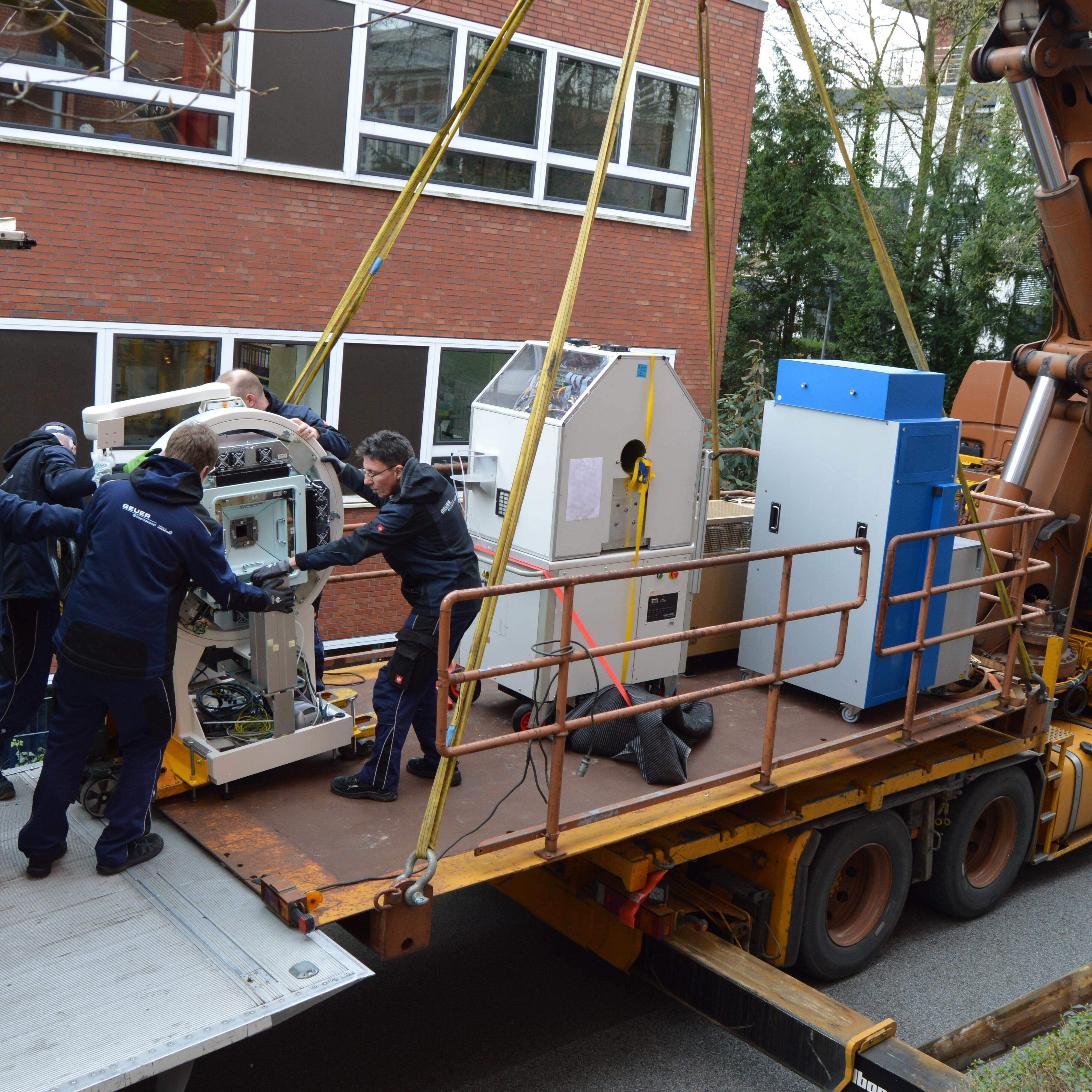
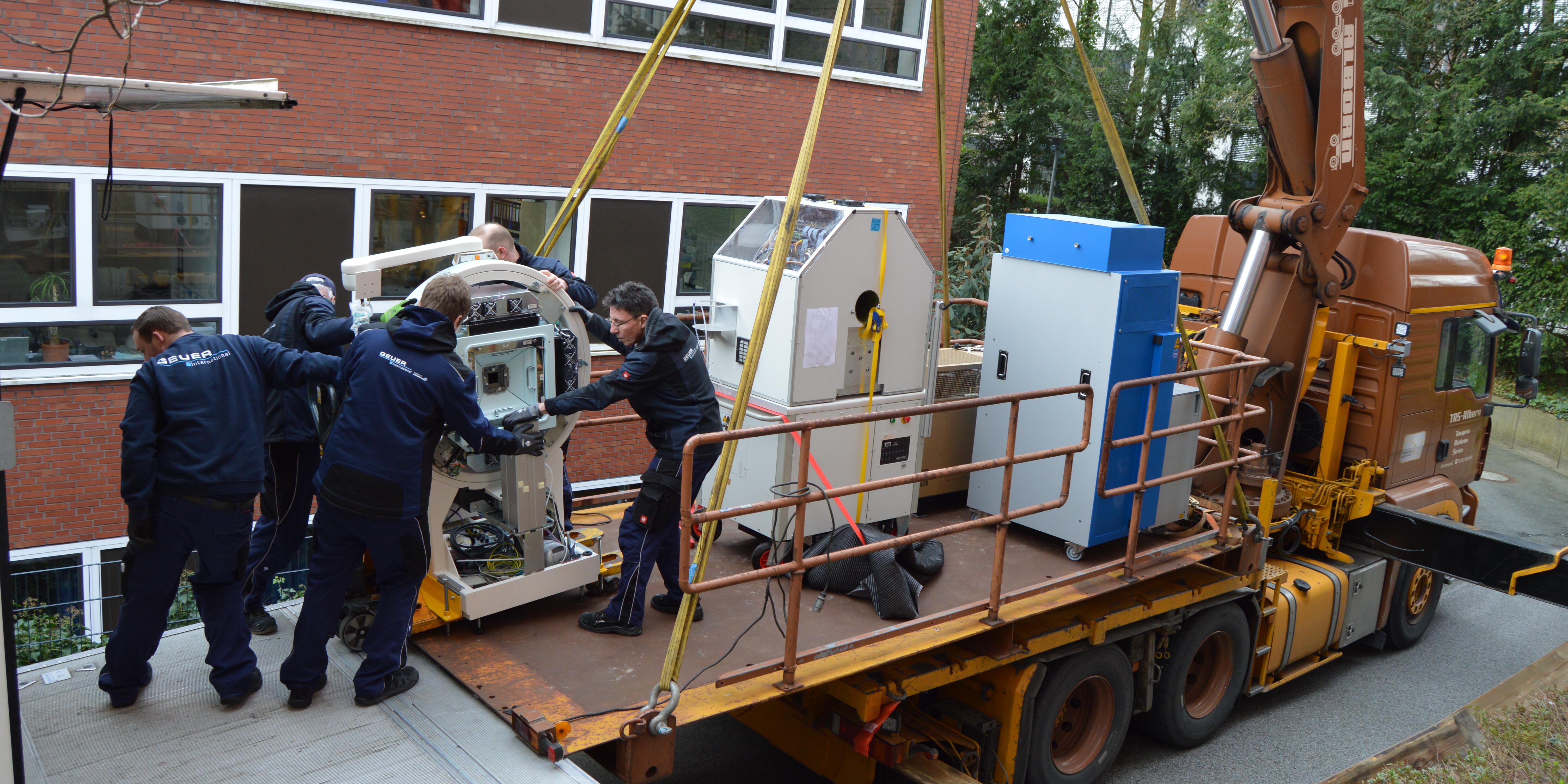
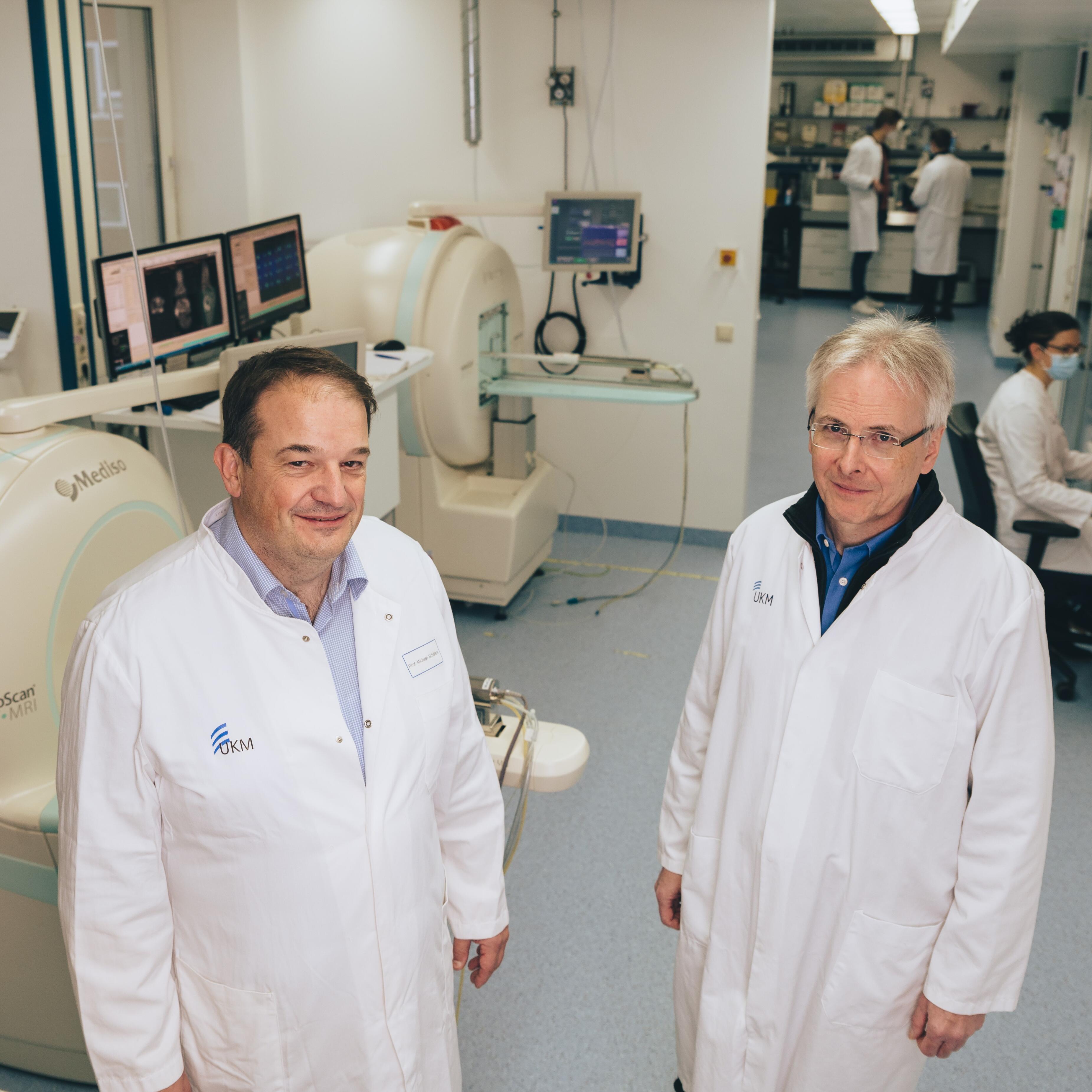
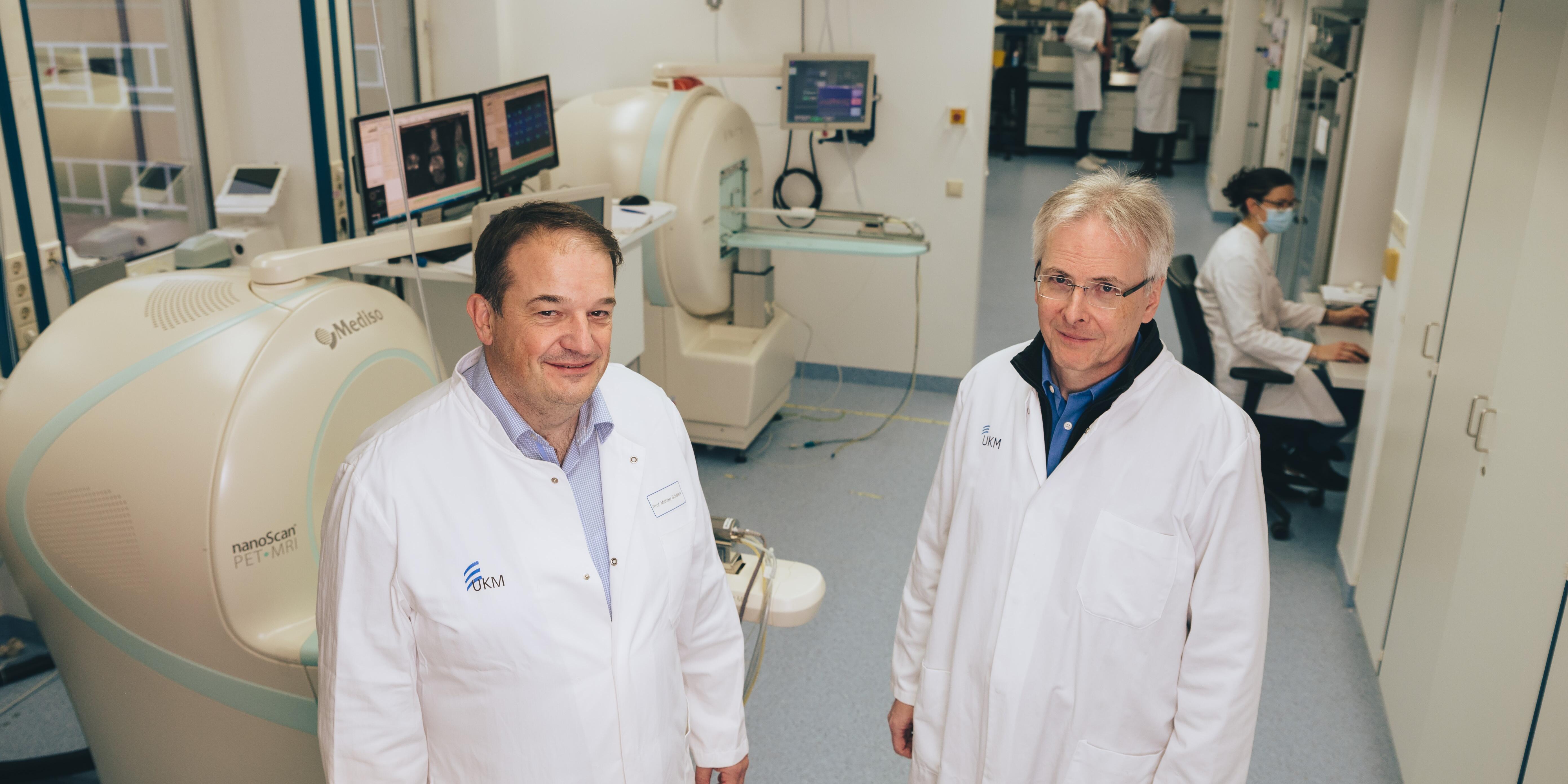
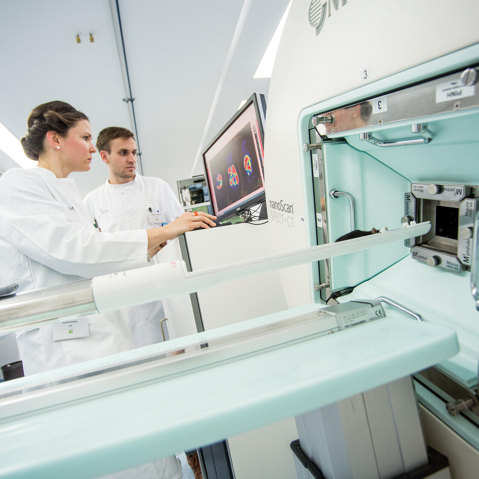
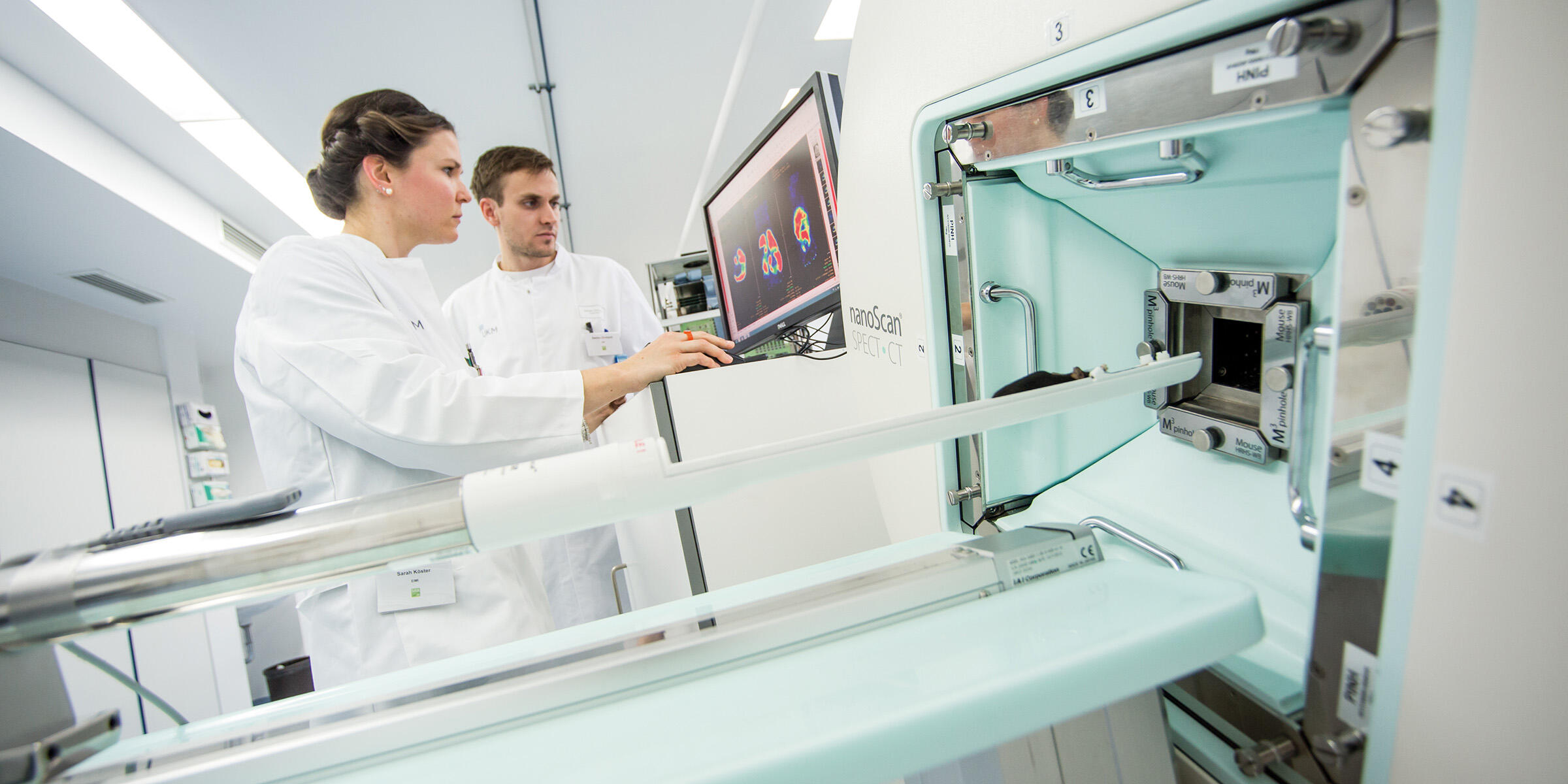
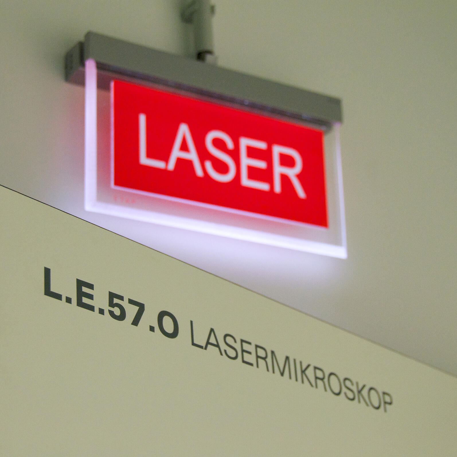
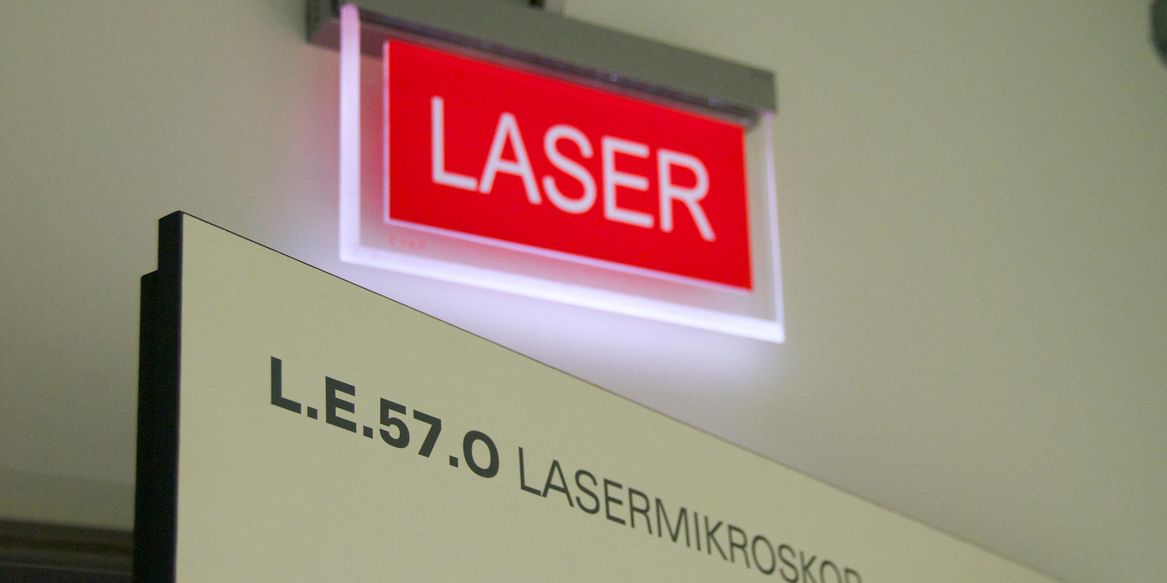
As of March 2023, the European Institute for Molecular Imaging (EIMI) is located in the Multiscale Imaging Centre (MIC) on Röntgenstraße 16 in Münster. In this research building, working groups from our university bring together a wide range of state-of-the-art imaging techniques to investigate biomedical questions. EIMI director Prof. Michael Schäfers is the spokesperson for MIC, and our team is happy about their new "home"! Our photo gallery shows how our imaging devices were transported to our new labs.
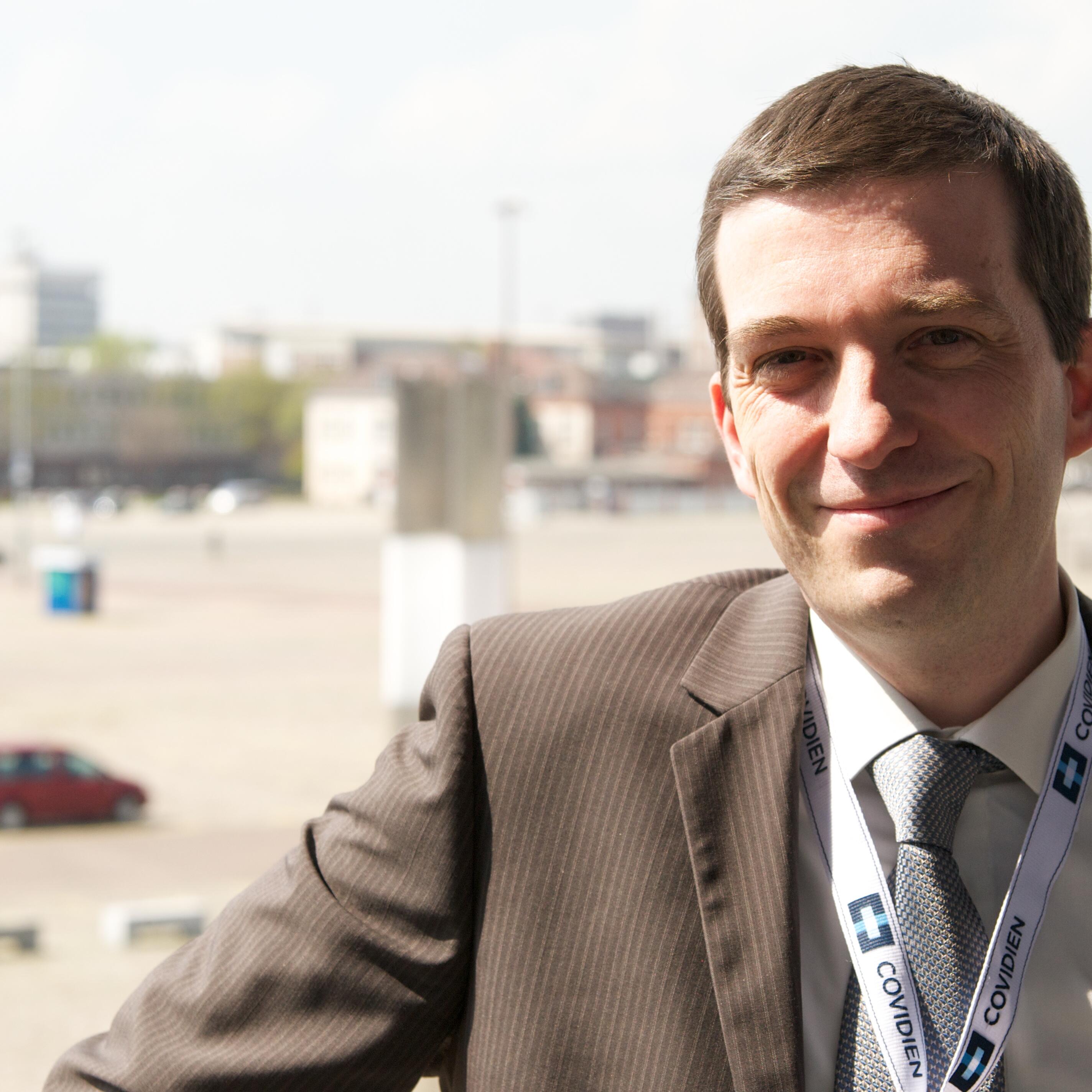
Nuclear medicine specialist Dr. Philipp Backhaus and his research partners examined breast cancer patients, for the first time systematically using a radiotracer that binds to the fibroblast activation protein. In particular small cancer lesions could be newly detected, and in combination with MRI, the new PET imaging method influenced further treatment in three of 19 patients. The study, published in “Radiology”, was awarded “Paper of the Month” by the Medical Faculty.
Medical professional Nadine Heiden is training to become a specialist physician while actively pursuing research. “I always wanted to do both,” she says – and a close connection between research and patient care can only be beneficial. Although the dual qualification is challenging, Nadine Heiden provides insight into how it is working out for her.
A research team led by biochemist Prof Andrea Rentmeister and nuclear medicine specialist Prof Michael Schäfers has, for the first time, utilised so-called SNAP-tag technology to radioactively label cells in living organisms. The method opens up the prospect of examining cells with different imaging techniques and at different temporal stages. The study was published in “Chemical Communications”.
Stefanie Bobe wants to dive deeper into medicine looking at the biomedical basis for improved diagnostic and therapeutic options. Therefore, she did an additional, science-oriented Master's degree in experimental medicine parallel to her medical studies and, in the working group led by biochemist Prof Friedemann Kiefer, became part of an interdisciplinary research team.
The new Collaborative Research Center "inSight" at Münster University receives funding from the German Research Foundation amounting to approximately ten million euros. The researchers aim to gain a comprehensive understanding of how the body regulates inflammation in different organs and, to this end, develop a specific imaging methodology that brings together information from single cells to entire organisms.
In a new video series the University of Münster introduces junior researchers. The first one is Cristina Barca, a PhD student at the European Institute for Molecular Imaging: Using biomedical imaging, she is investigating how well certain drugs work in treating strokes. In the video she provides an insight into her everyday working life and explains what is so special about being a scientist.
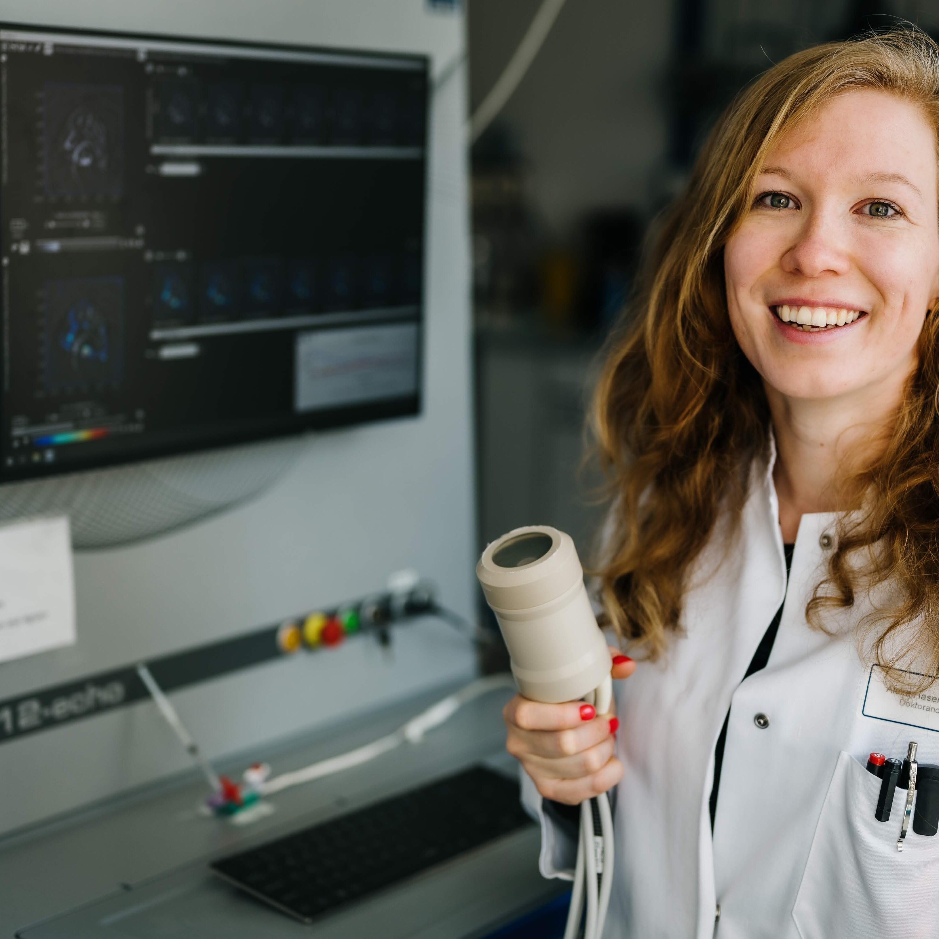
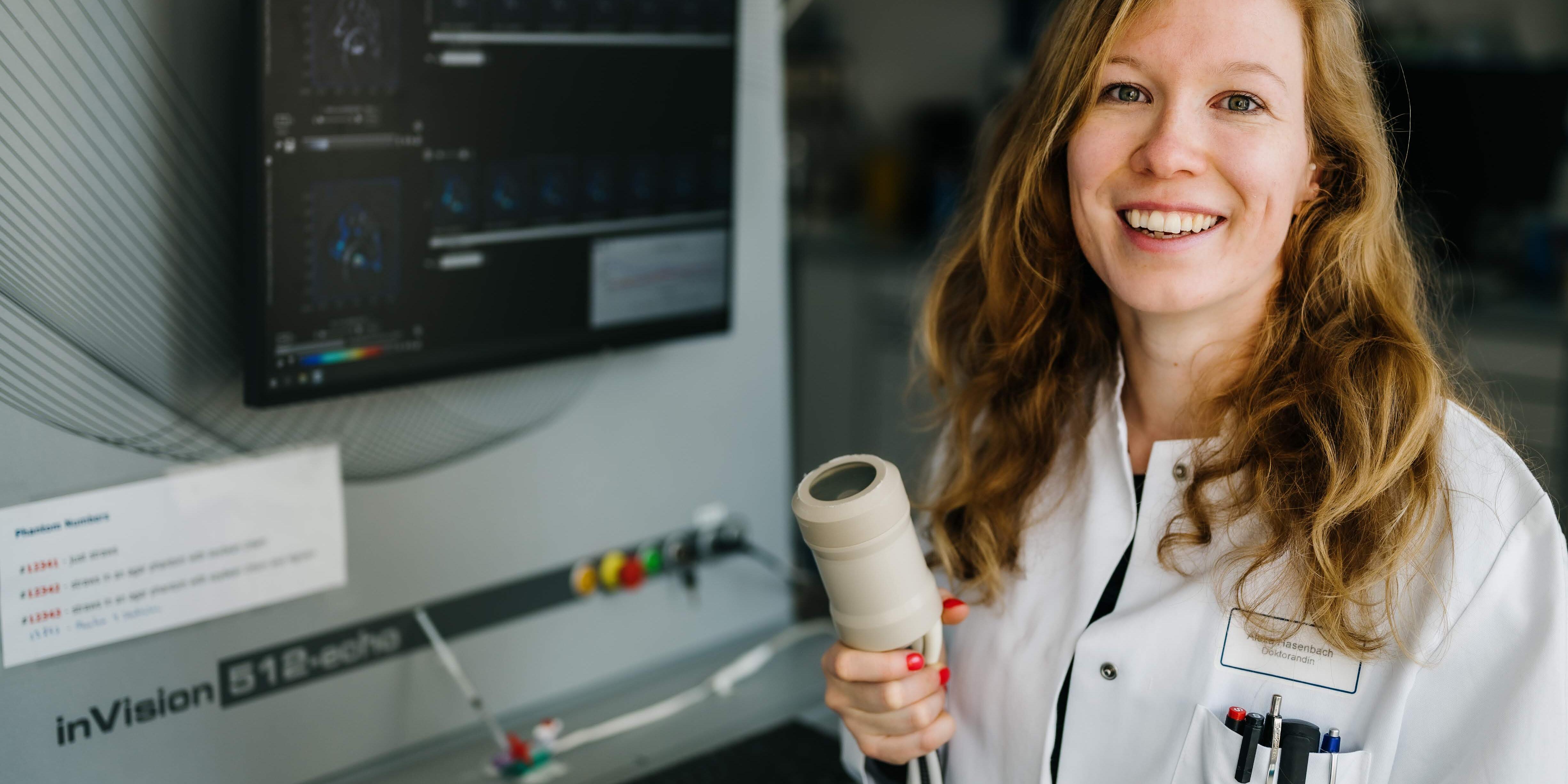
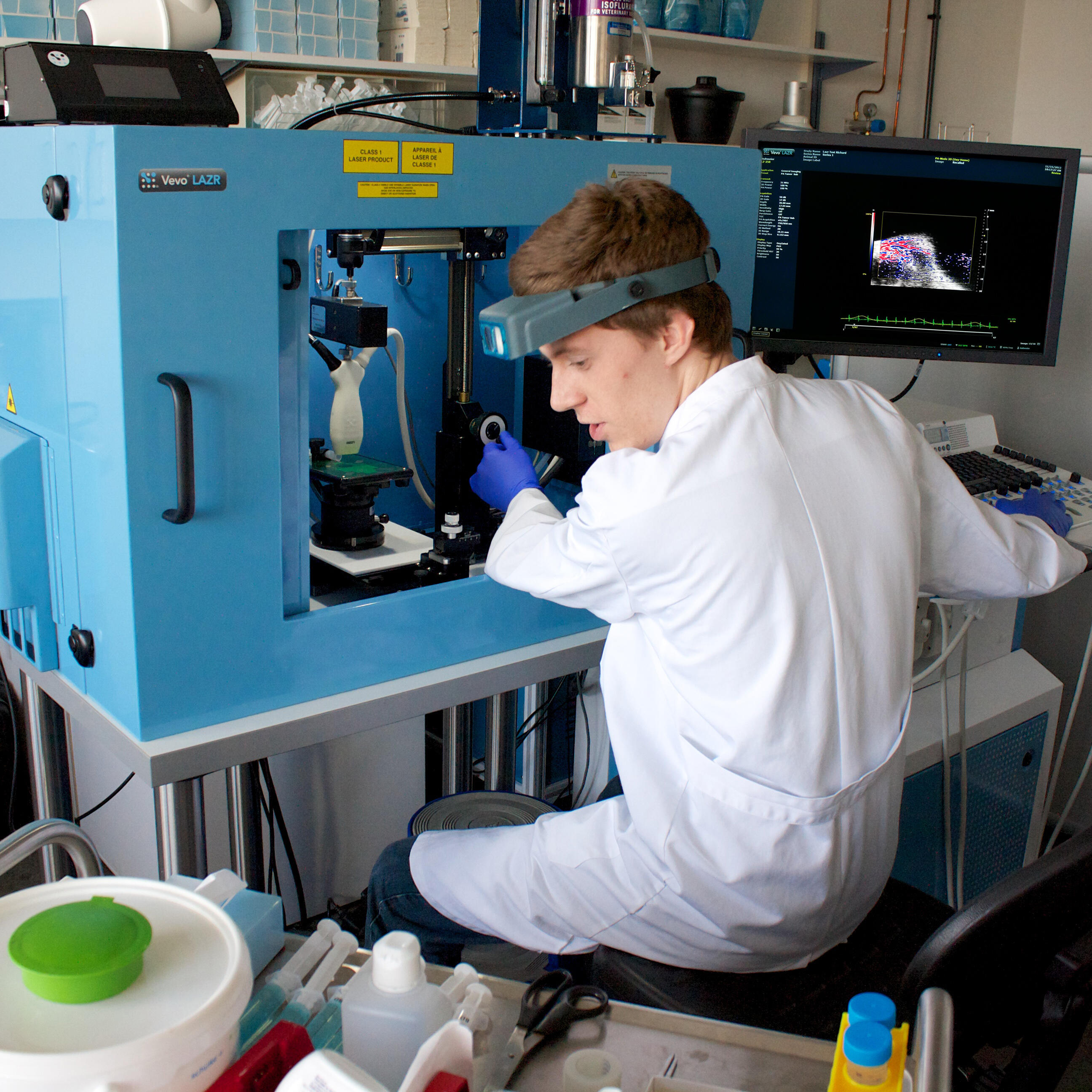
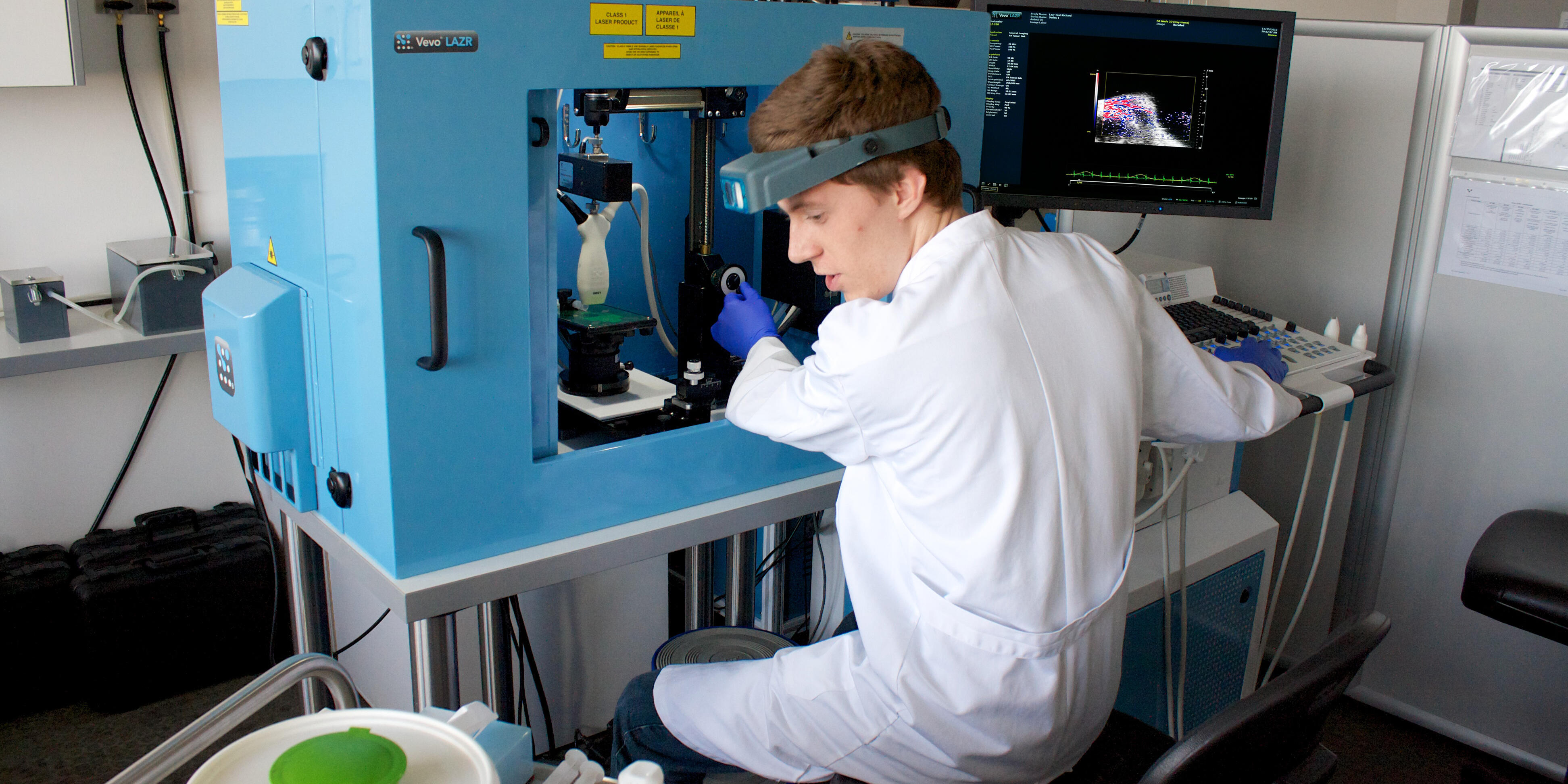
Laser light that cannot be seen, and sounds that cannot be heard: this combination produces something that is all the more visible – images from inside the body. Photoacoustics is the name of this method, whose purpose is to acoustically record the sounds of molecules. During her PhD thesis, biologist Alexa Hasenbach investigated inflammatory processes.
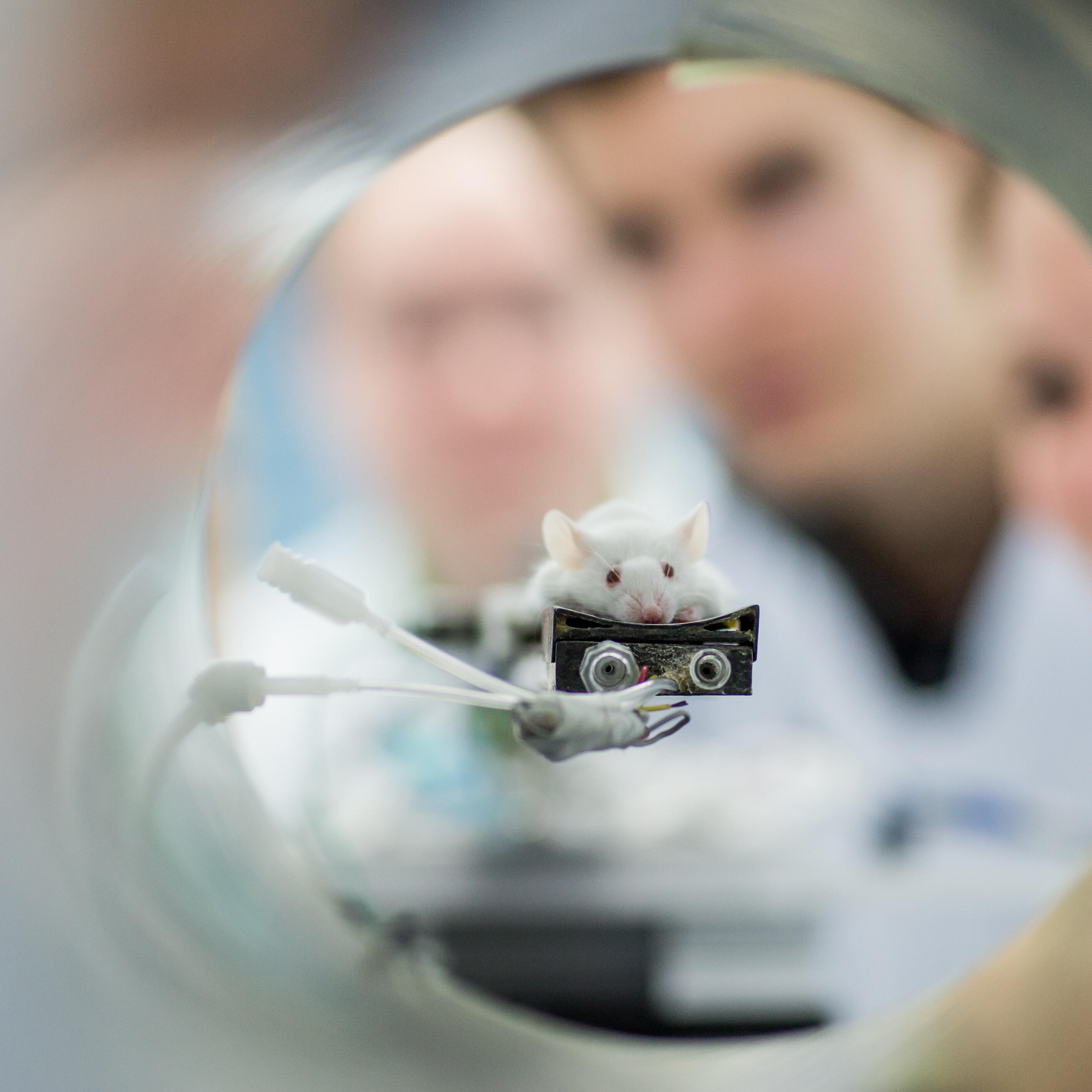
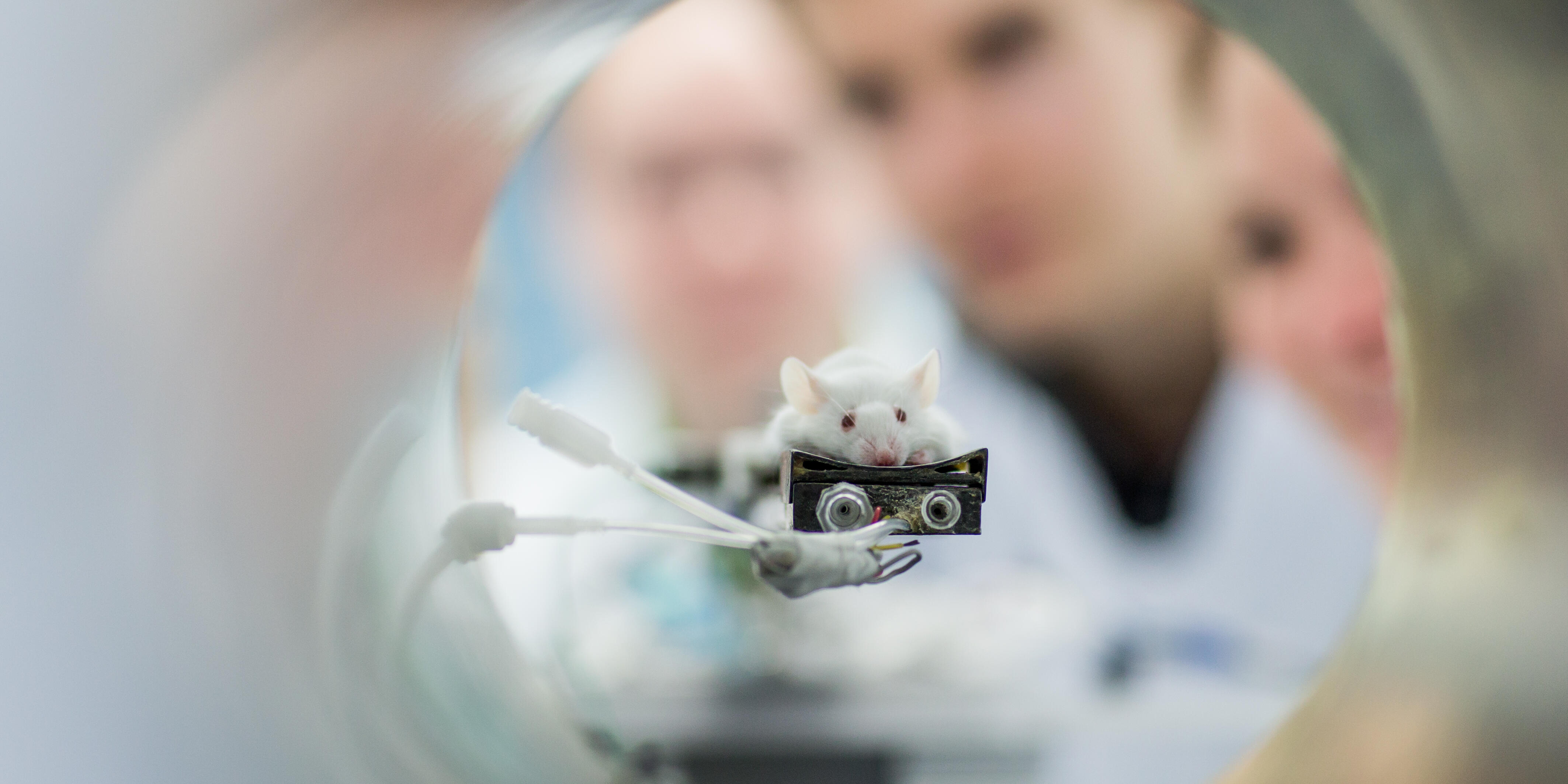
Scientists at the University of Münster use a broad spectrum of imaging techniques to investigate structures and processes in the body. Last week, they shared their knowledge with international junior researchers: The participants of the tenth annual Mouse Imaging Academy spent five days training on different methods for examining mice.
How do immune cells behave in the body? What happens during immunotherapy? To answer these questions, the European Union brings together leading experts from research and the pharmaceutical industry. The Europe-wide research project "Immune-Image", which is funded with 30 million euros over five years and in which scientists from Münster University are involved, started on 1 October.
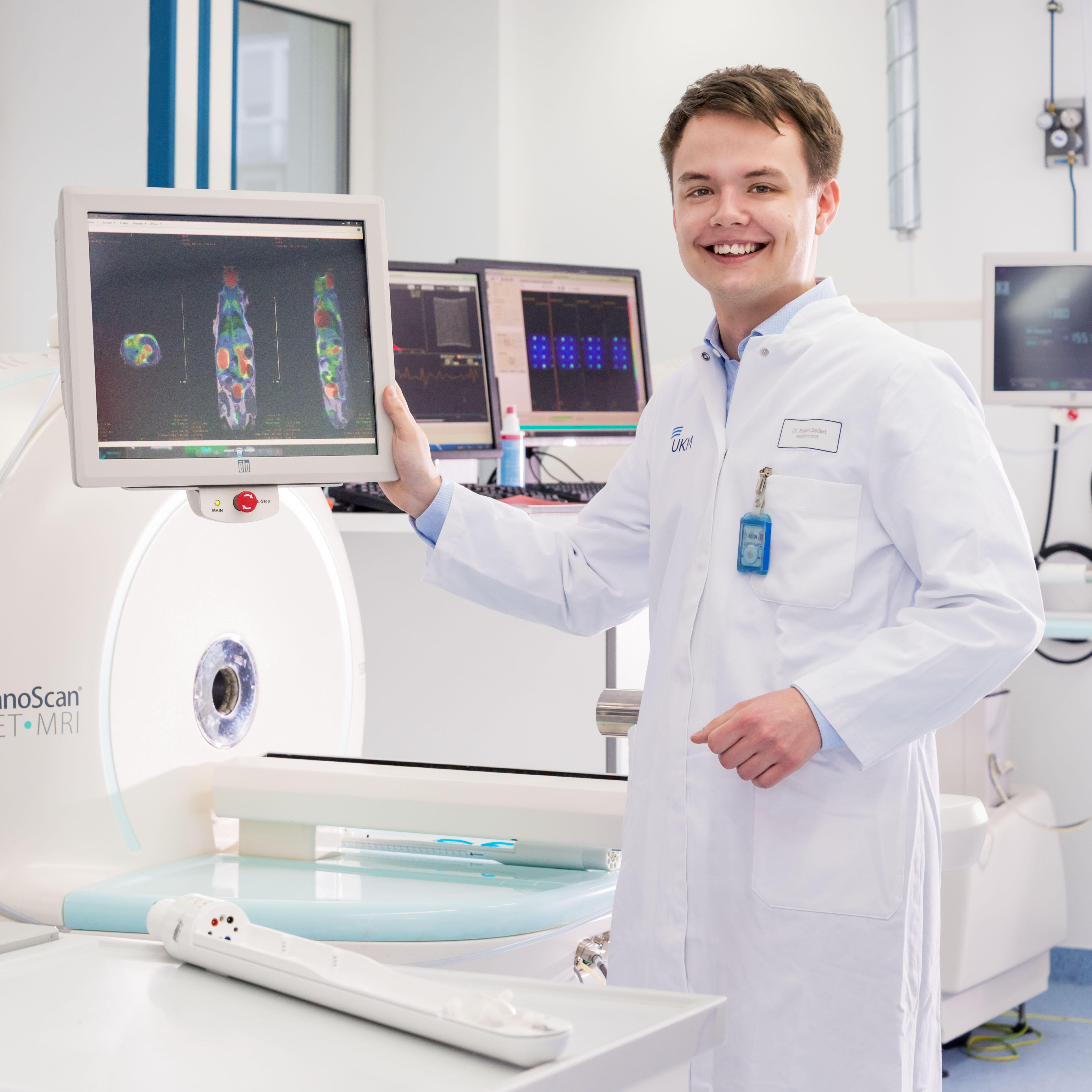
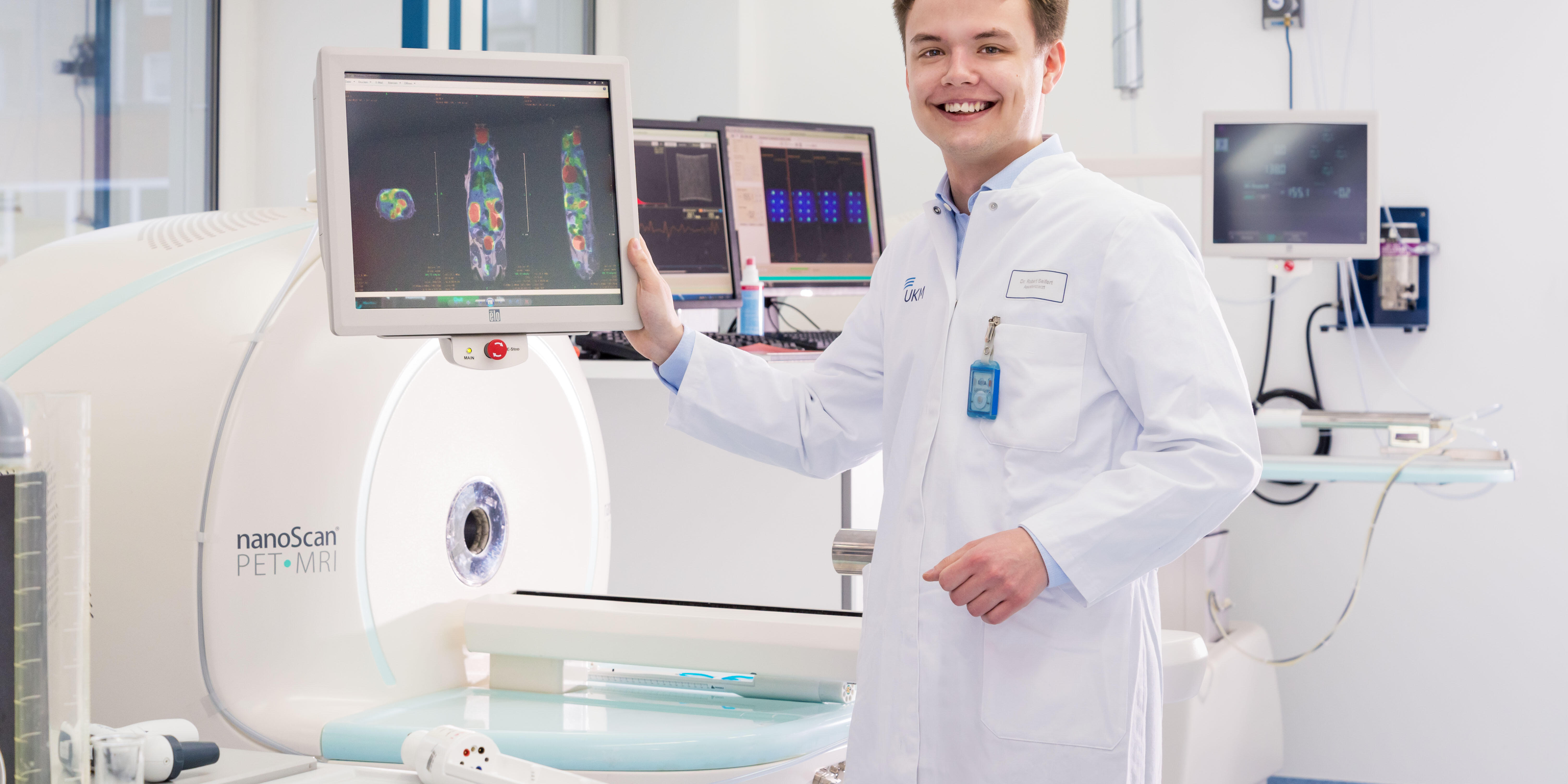
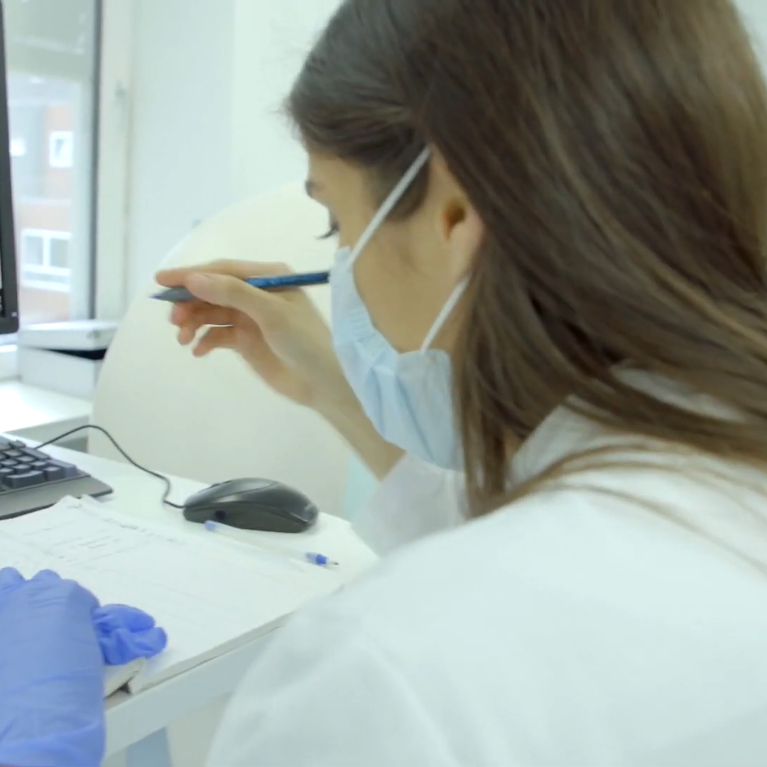
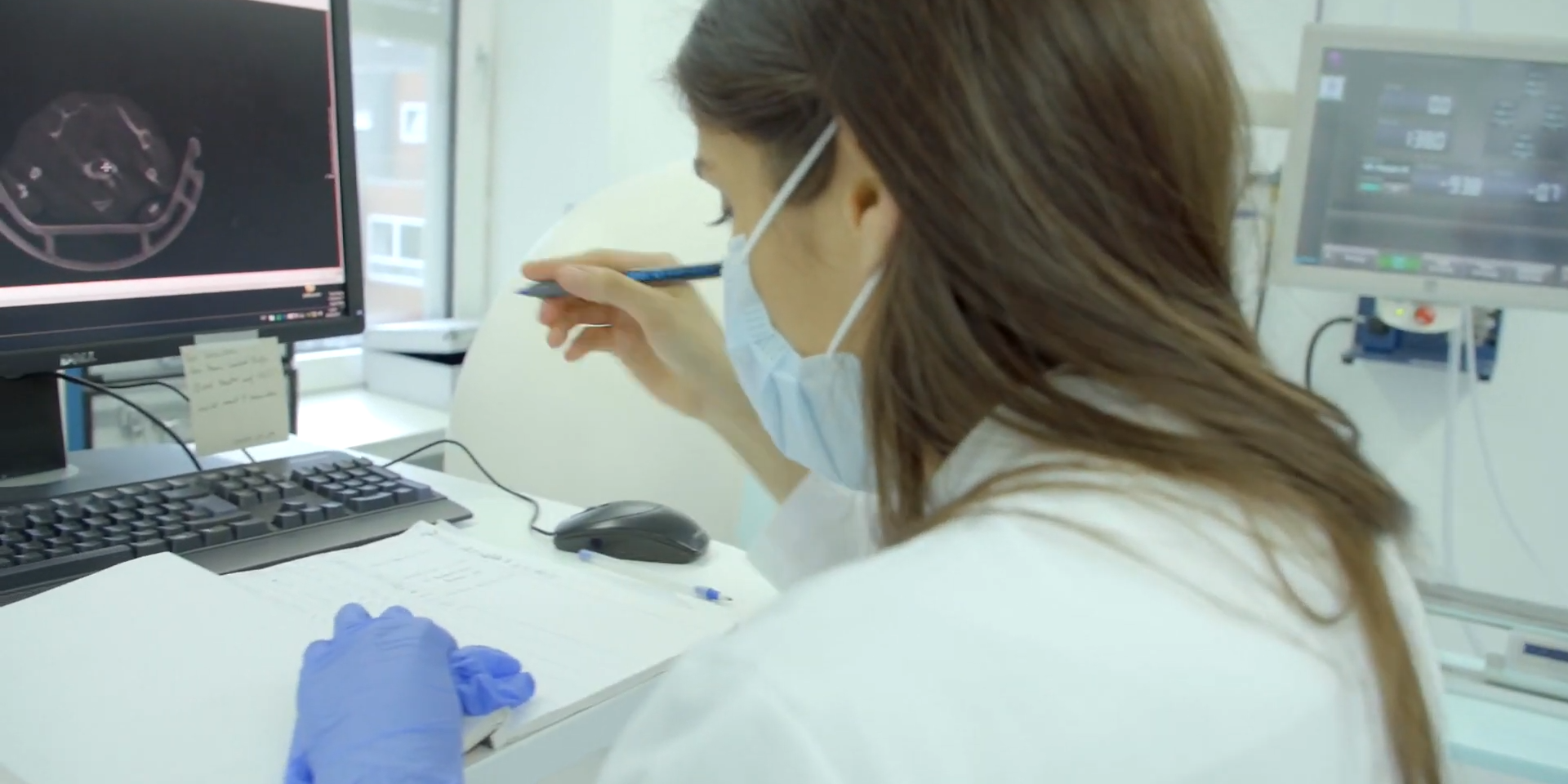
The MD thesis of Dr. Robert Seifert, a physician, is based on an interdisciplinary cooperation, supported by the Cells-in-Motion Cluster of Excellence. He and his colleagues developed an algorithm for the precise analysis of image data. A WWU dissertation prize was awarded for this.
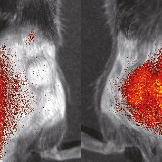
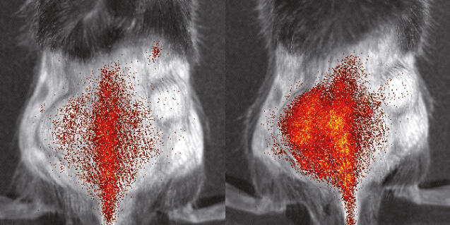
Immunologists and imaging specialists at the Cells-in-Motion Cluster of Excellence have jointly developed a method enabling them to better evaluate and study the activity of inflammatory cells in mice. The study has been published in the “Theranostics” journal.
In November, CiM researchers Prof. Michael Schäfers and Prof. Stefan Schlatt demonstrated experiments with mice to journalists and gave insights into how different animals are kept at Münster University. The University has compiled an experience report and a collection of links to press articles.
Prof. Friedemann Kiefer performs research at the Cells-in-Motion Cluster of Excellence, investigating how lymphatic vessels are formed and preserved. In his work, though, he constantly looks beyond the boundaries of his own field to develop new ideas. His overall aim is to make contributions of lasting value.
One part of the foundation of the Cells-in-Motion Cluster of Excellence celebrates its tenth anniversary: the European Institute for Molecular Imaging. How has the institute developed over the past ten years? What were special moments? In an interview, the EIMI Directors look back.
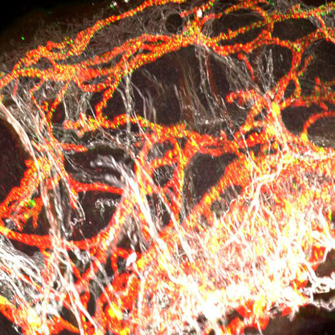
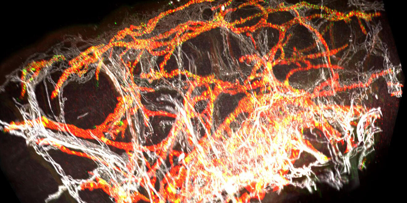
Researchers at the Cells-in-Motion Cluster of Excellence have developed a new method for producing digital 3D reconstructions of blood and lymphatic vessels from tissue samples and then creating images of them for analysis. The study has been published in the “JCI Insight” journal.
Prof. Michael Schäfers is fascinated by the possibilities offered by positron emission tomography, which enables to visualize molecular processes inside the body. With the help of this technology he would like to show inflammatory processes, e.g. to predict future heart attacks.
How can processes in the body that are normally hidden from the human eye, such as inflammation or disease, be made visible? To do this, scientists at the Cells-in-Motion Cluster of Excellence use a broad range of imaging technologies and work on developing innovative imaging strategies.
Researchers at the Cells-in-Motion Cluster of Excellence have succeeded in visualizing, for the first time, ongoing inflammation in the brain in patients suffering from multiple sclerosis. The study has been published in the prestigious journal "Science Translational Medicine".
Bacterial infections can have serious consequences – for example, when they colonize an artificial heart valve. There is especially problematic when the bacteria are resistant to several antibiotics. Researchers are looking for new methods of treatment as well as for ways to find centres of infection in the body, for example by means of special sugar molecules. Chemists, physicists, biologists and physicians were all involved in the study.
Without oxygen, cells cannot survive. Until now, scientists could not observe the effect that a reduced oxygen supply can have on individual cells. This was technically not feasible before. Scientists from Münster have now developed a reporter, which allows them to see an acute lack of oxygen of cells using light microscopy.
For Prof. Klaus Schäfers everything revolves around motion correction. He and his team aim to make clinical images even more exact. And they have no lack of ideas for improving high-tech equipment.
How can you take more sharply defined pictures from the body’s interior – even though motion comes in? Many of our scientists deal with this question in the labs of the Cells-in-Motion Cluster of Excellence. A newly developed programme now enables physicians to do away with the so-called respiratory belt in positron emission tomography (PET). This belt often has only limited uses in the case of people who are seriously ill.
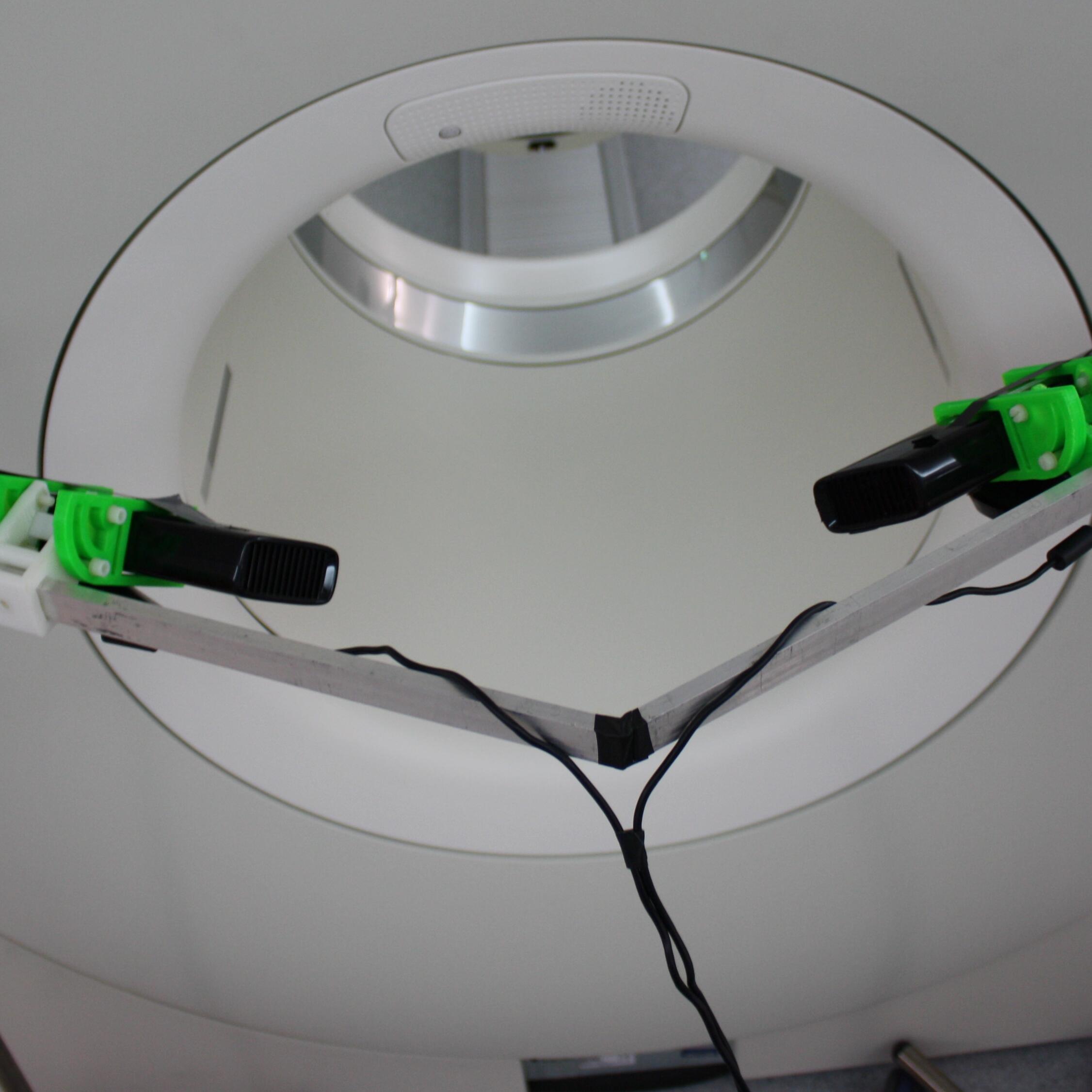
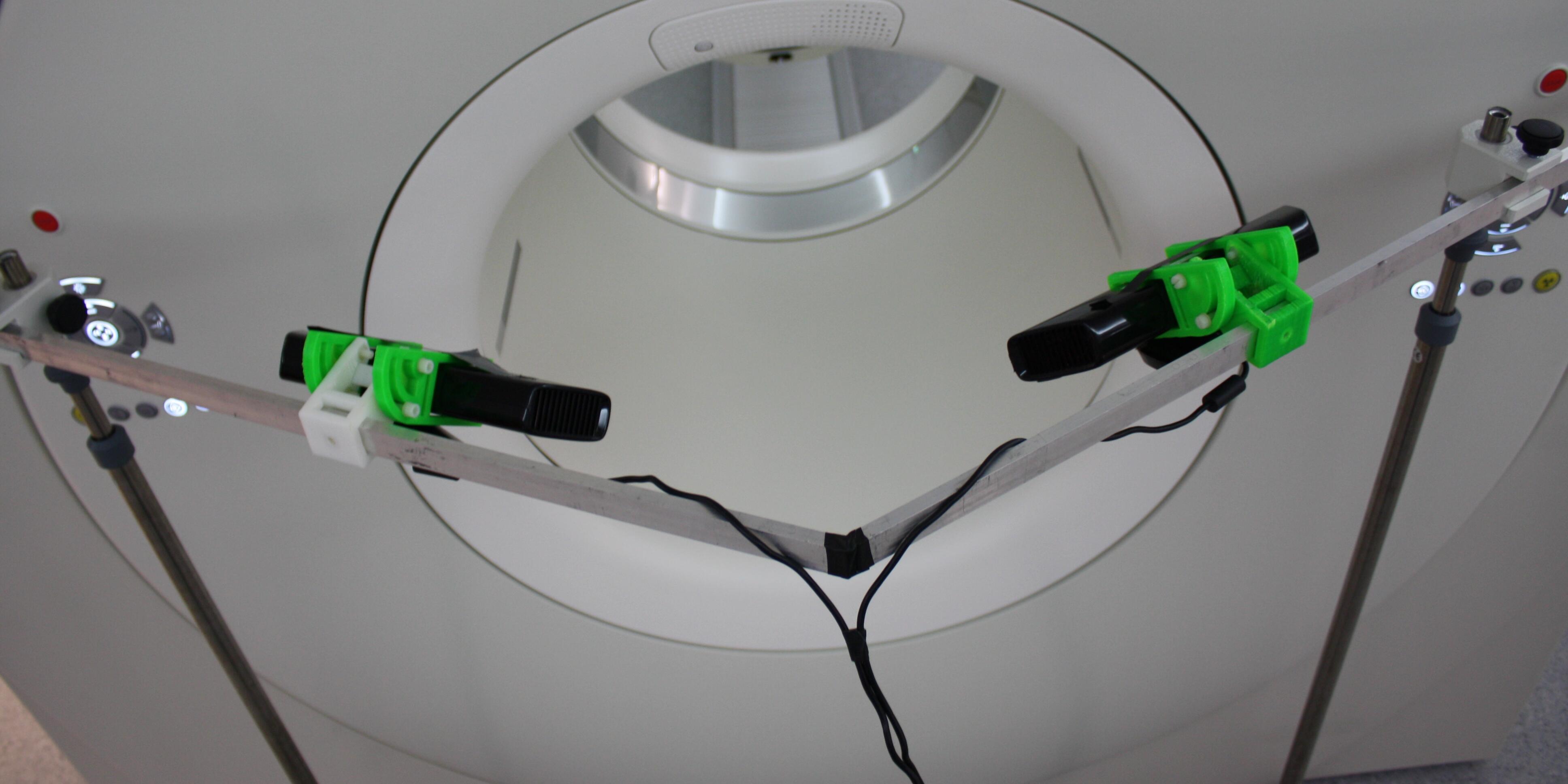
It does not always need to be the most expensive high tech product to optimize medical technology. A scientist from a research group in the Cells-in-Motion Cluster of Excellence used the Xbox technology for an experiment. With the help of this affordable technology Mirco Heß shows scientists a way to better understand clinical images of the inside from the outside.
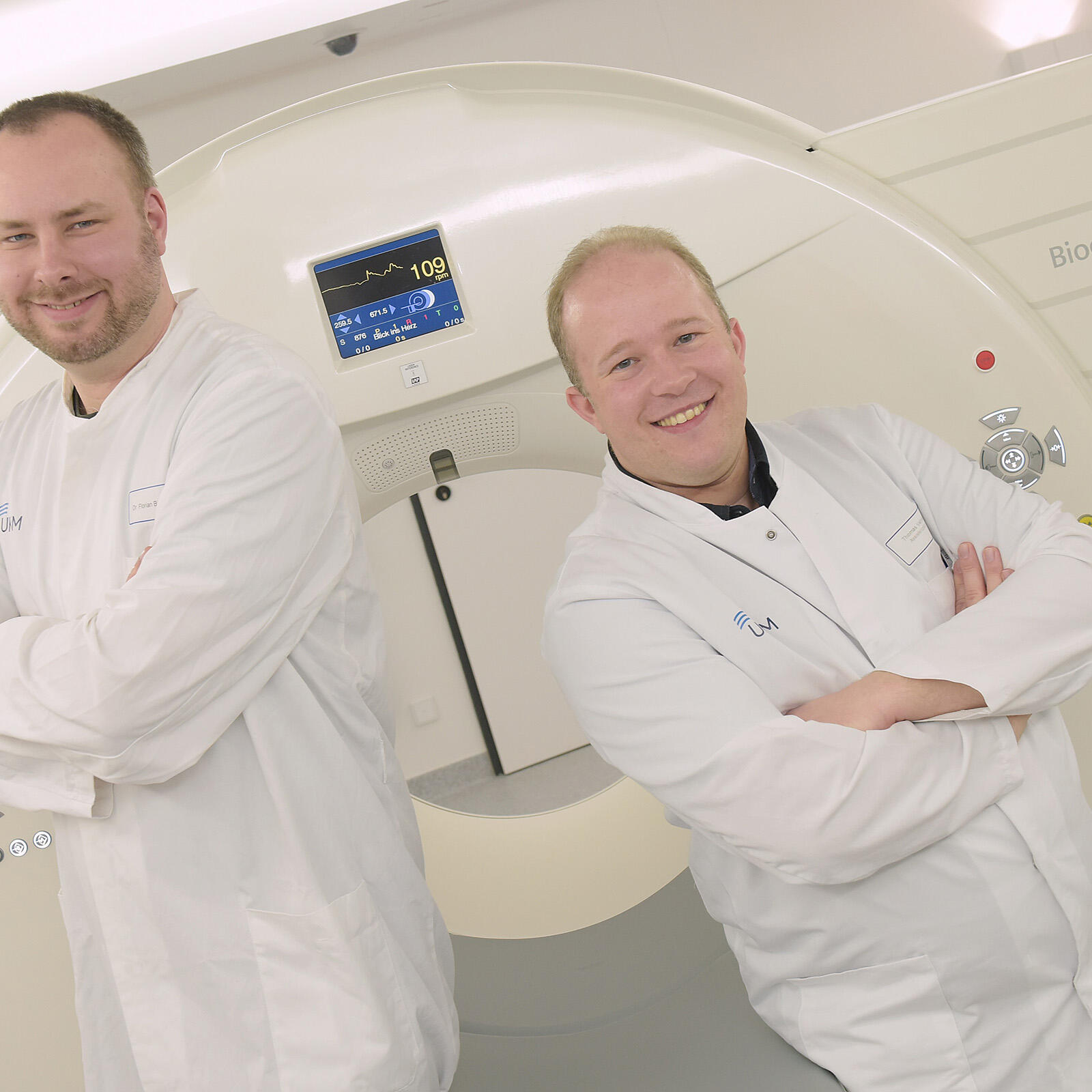
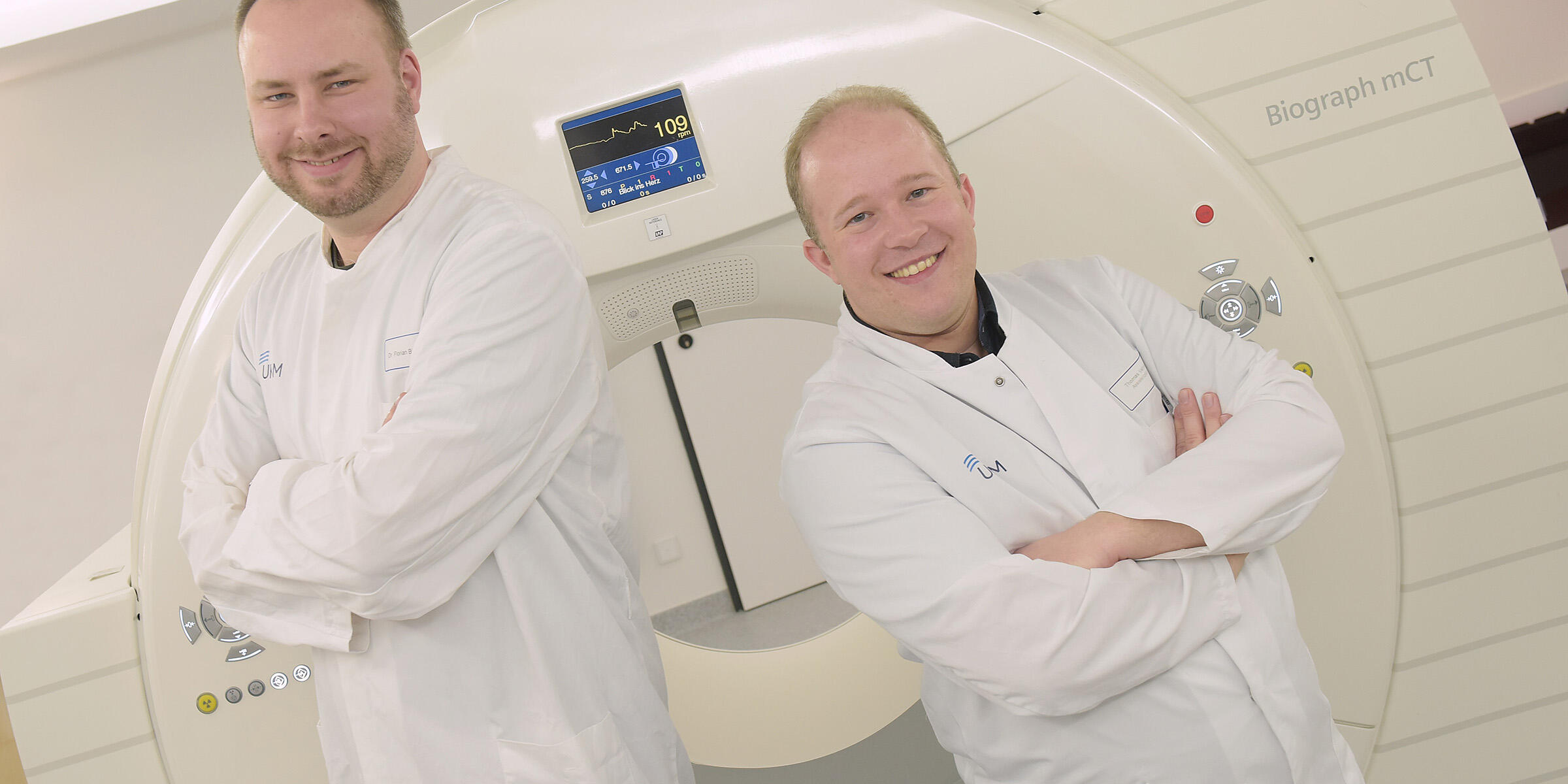
If you move during an examination with a whole body scanner the resulting images from inside the body will be blurred. A patient’s breathing is already enough to make tomographic images less precise. That is why medical physicist Dr. Florian Büther and nuclear medicine specialist Dr. Thomas Vehren develop new methods which shall improve the quality of medical images.
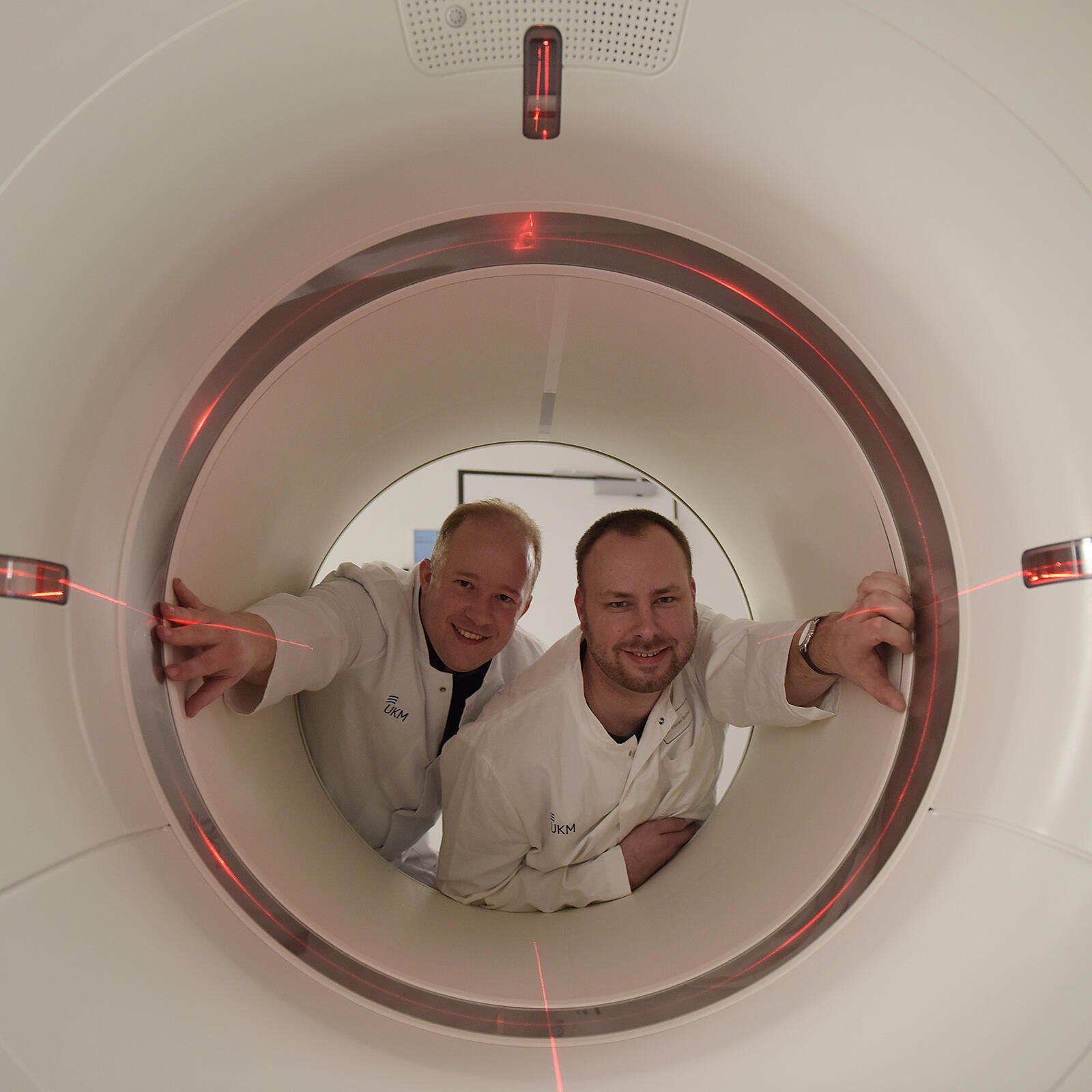
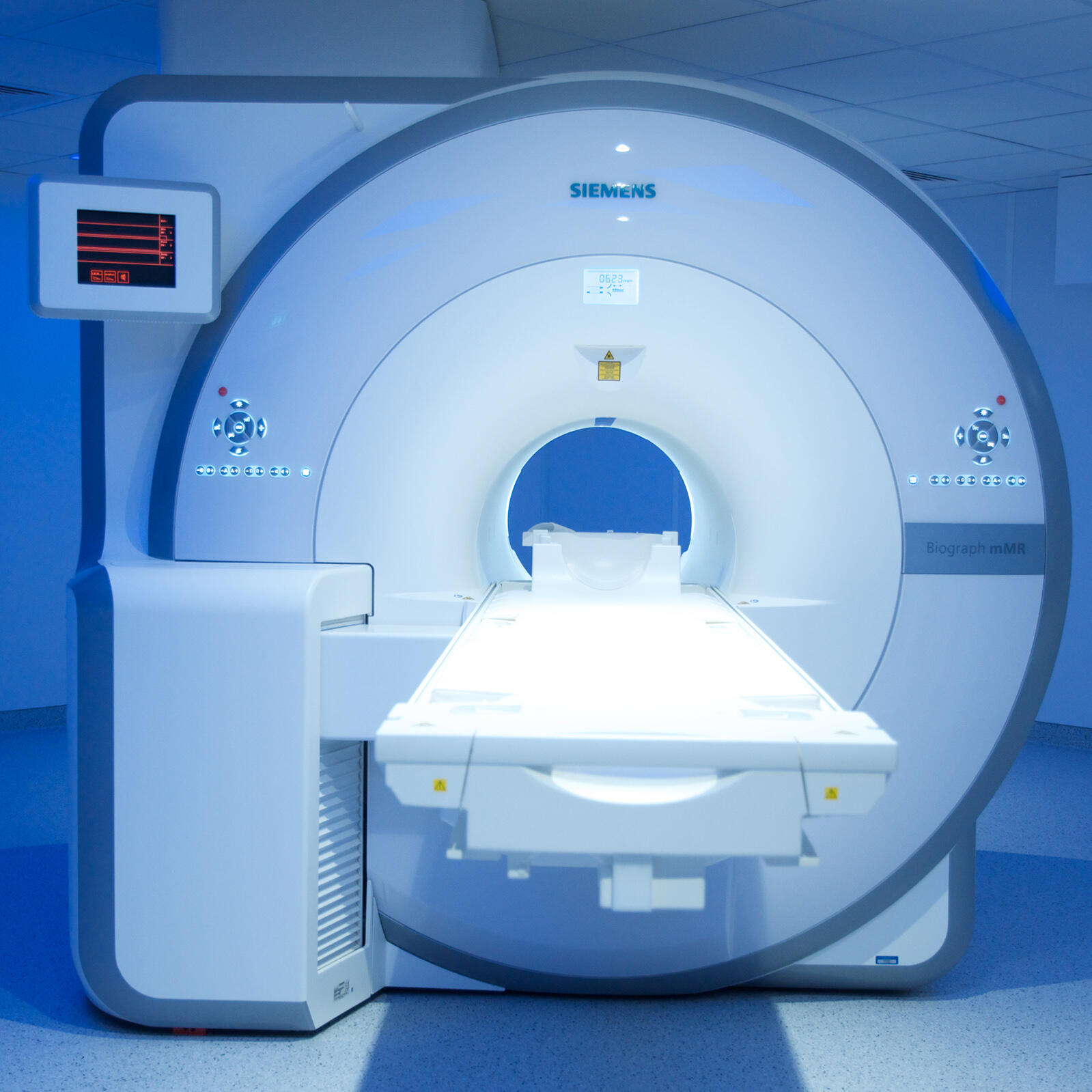
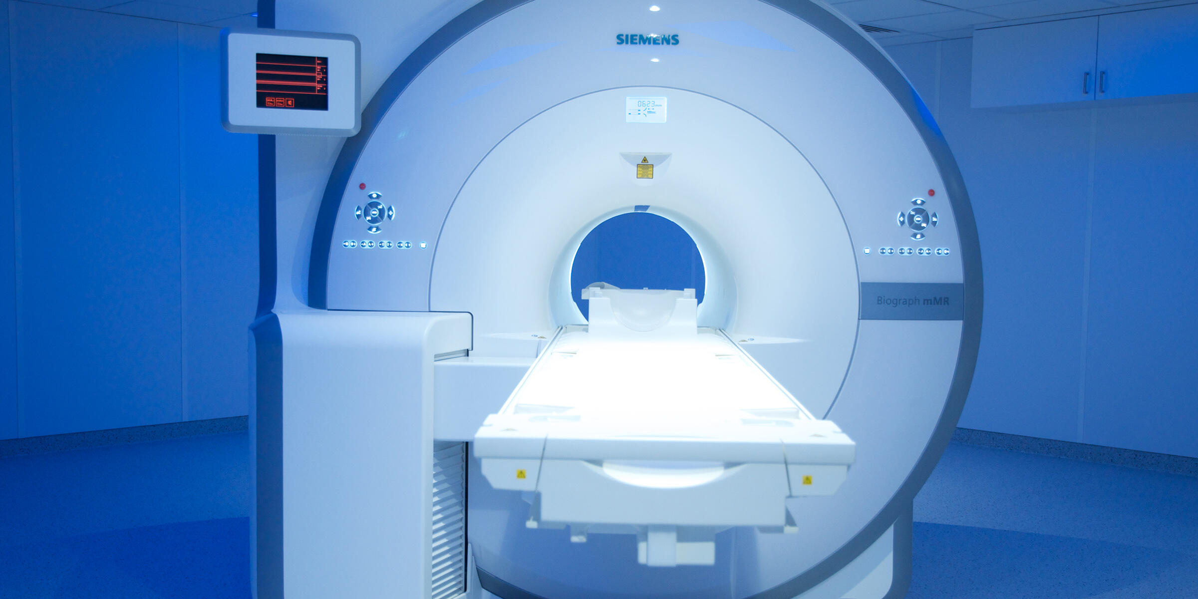
Scanning before dissection – to look inside the human body physicians nowadays use a set of imaging techniques. Just recently, the University Hospital Münster has installed an innovative hybrid system which combines PET and MRI. The high end scanner generates both unique images and challenges for the physicians.
When coronary arteries become inflamed, the vessel walls thicken and may rupture – leading to an acute heart attack. Dr. Thomas Vogl, Dr. Andreas Faust und Dr. Sven Hermann develop substances to visualise inflamed vessel walls with molecular imaging and to assess the individual risk.
Cellular structures of different dimensions can be studied specifically by light microscopy – ranging from processes in the whole organism to the smallest components of individual cells. In the last thirty years, microscopes have benefitted from fluorescence and 3D-techniques and are extremely powerful, explain Prof. Jürgen Klingauf and Prof. Friedemann Kiefer.
Just recently the Cells-in-Motion Cluster of Excellence (CiM) obtained a new device for 'photoacoustic imaging'. Prof. Michael Schäfers from the team of CiM coordinators explains in an interview with Christina Heimken why this prototype is especially important.