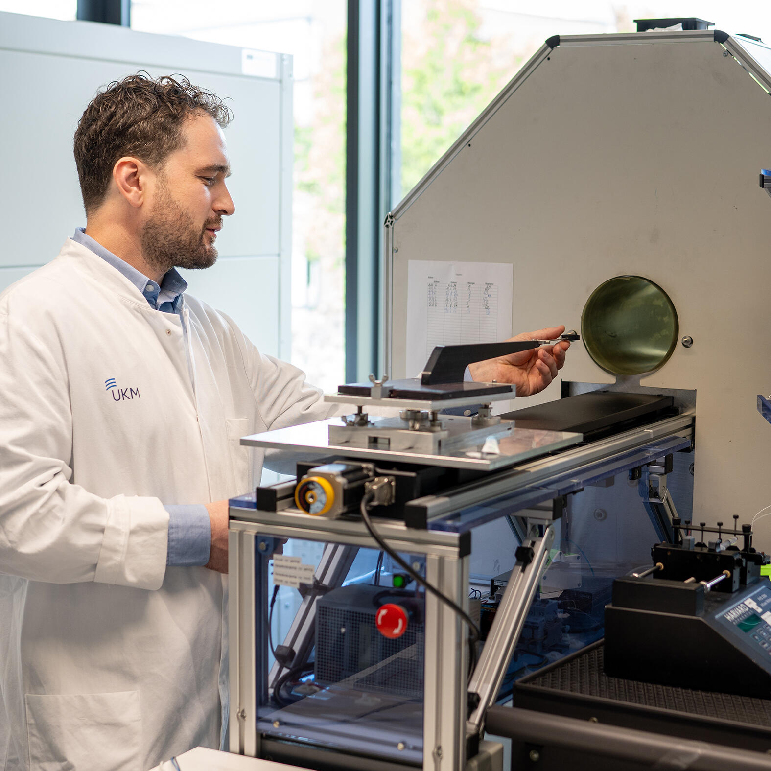
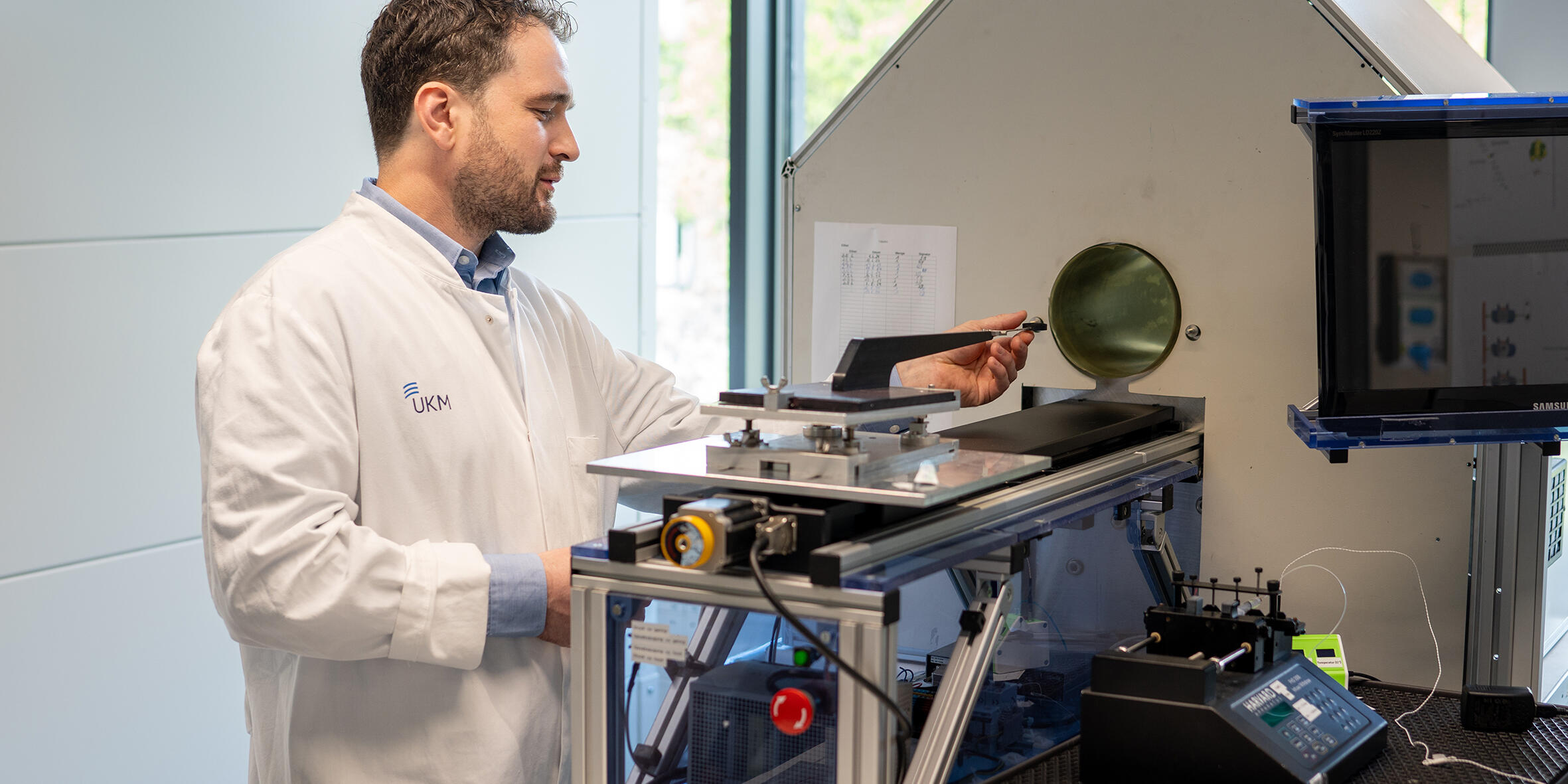
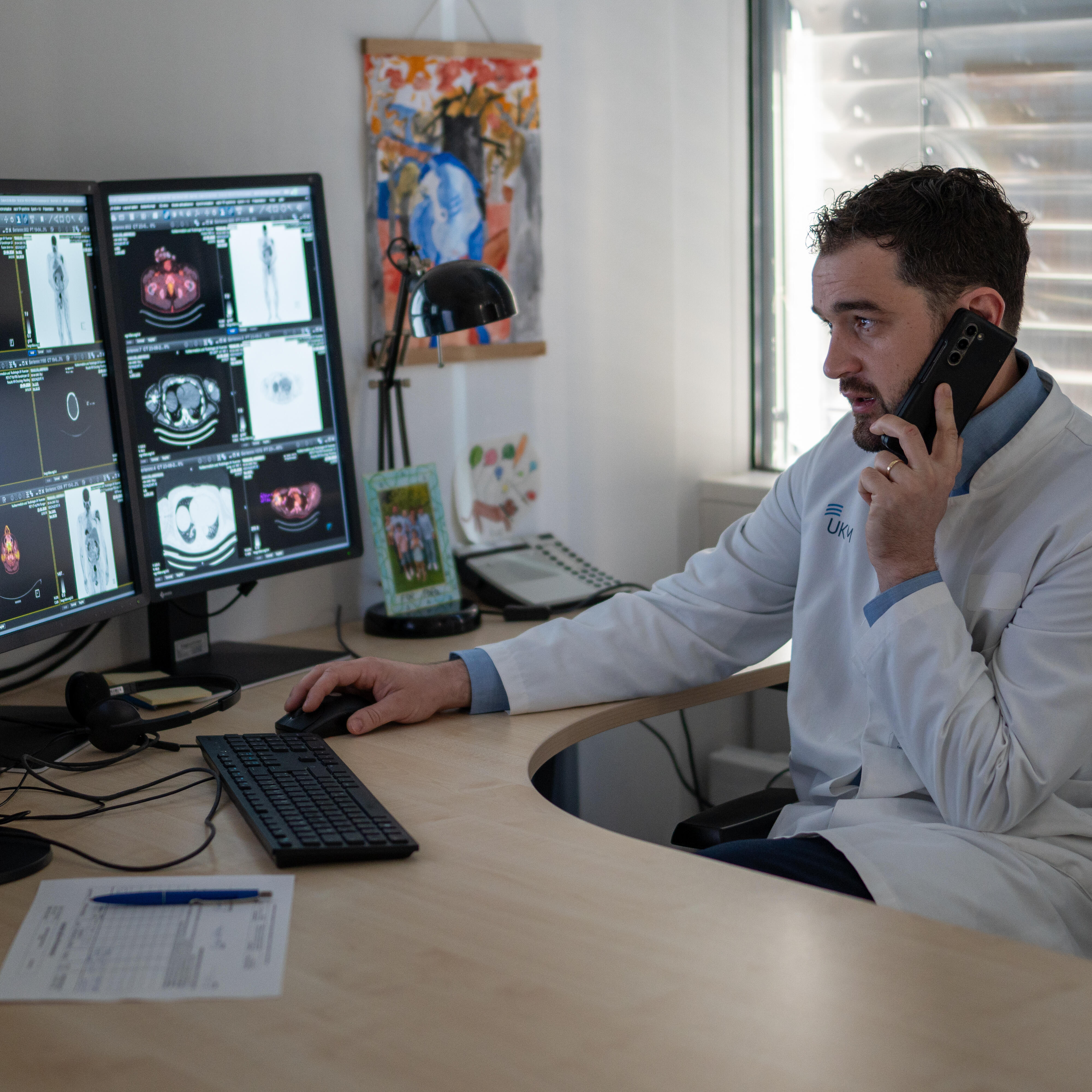
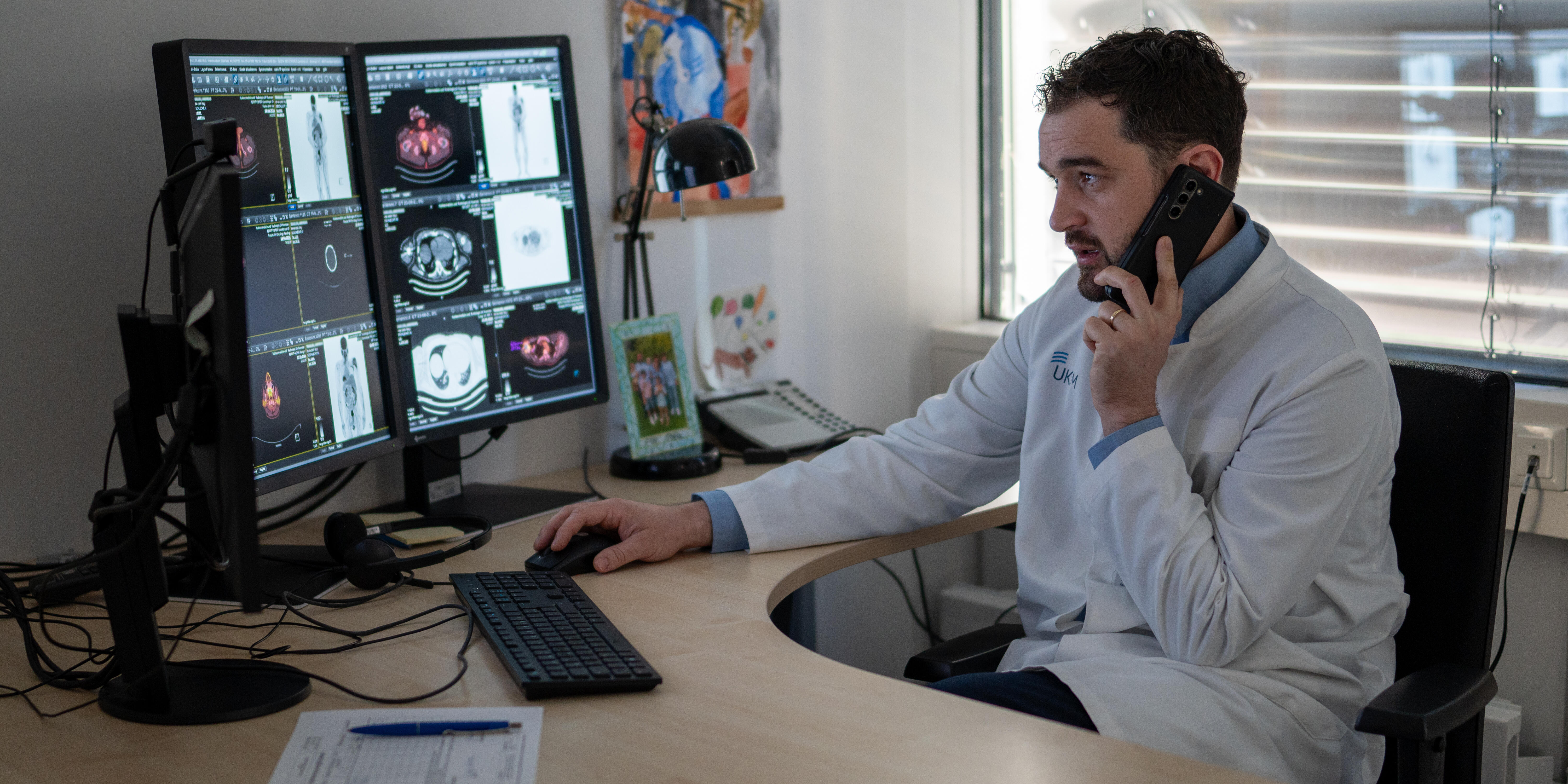
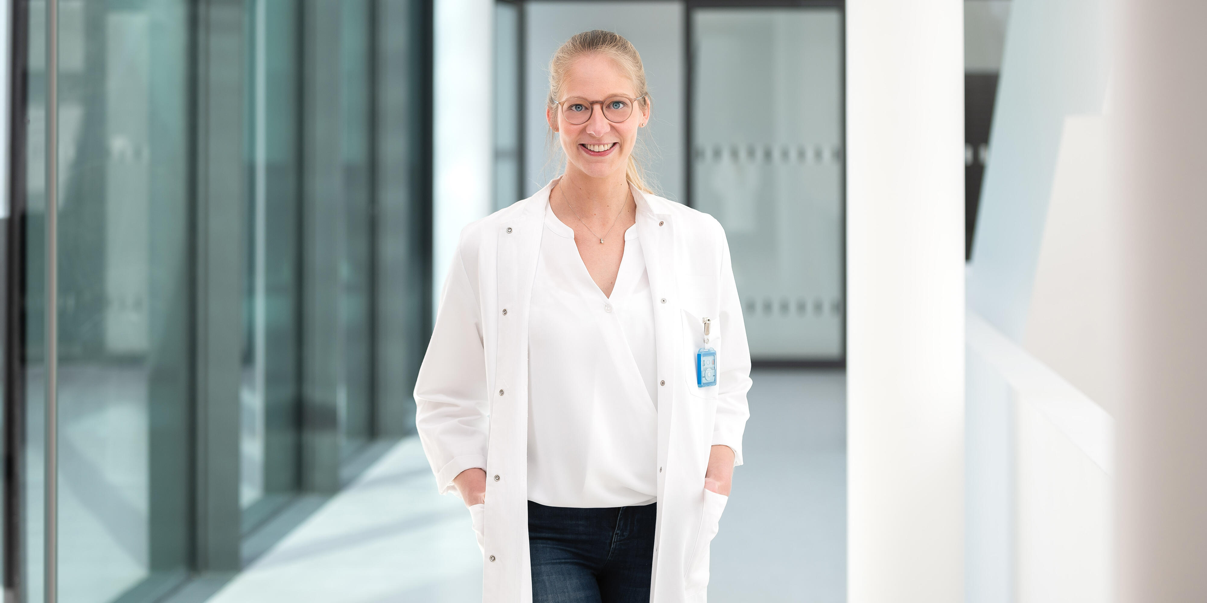
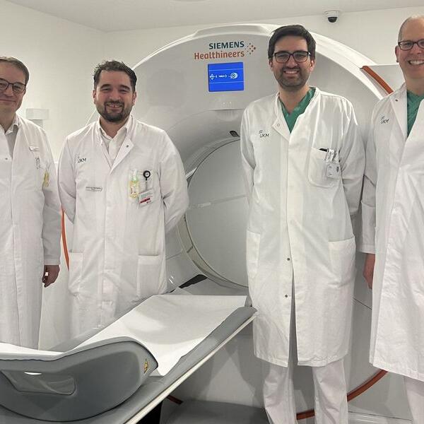
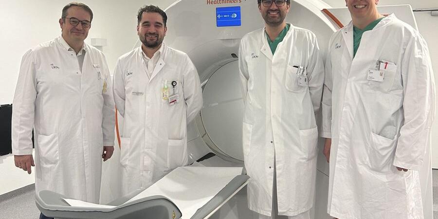
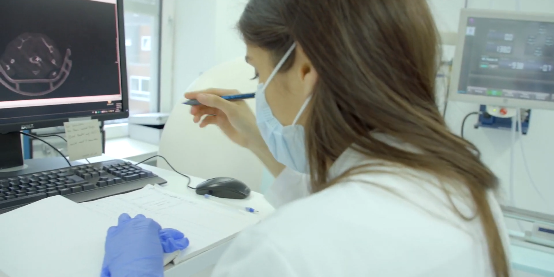
Translational research takes place worldwide in several sequential phases. Nuclear medicine specialist Professor Philipp Backhaus is primarily involved in the early phases of translation, specialising in the imaging and therapy of tumours and inflammation. For several months, his team has been contributing to the development of the drug “OncoACP3”, which aims to improve the examination of prostate cancer.
Nuclear medicine specialist Prof Philipp Backhaus and his research partners have evaluated a new radiotracer for PET imaging in prostate cancer that targets the molecule ACP3. Compared to an established PSMA-targeted tracer, it showed superior visualization of disease extent in many cases and led to a change in treatment in a relevant number of patients. The study was awarded “Paper of the Month” by the Medical Faculty.
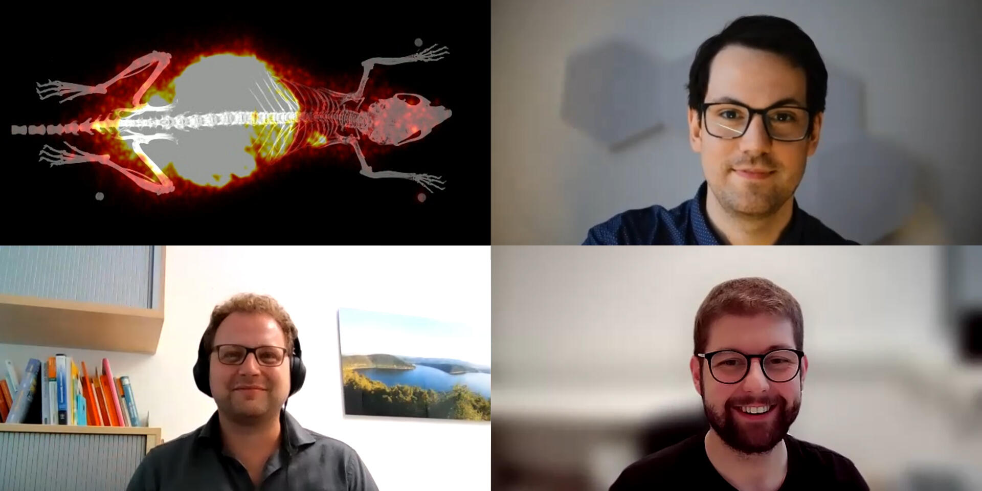
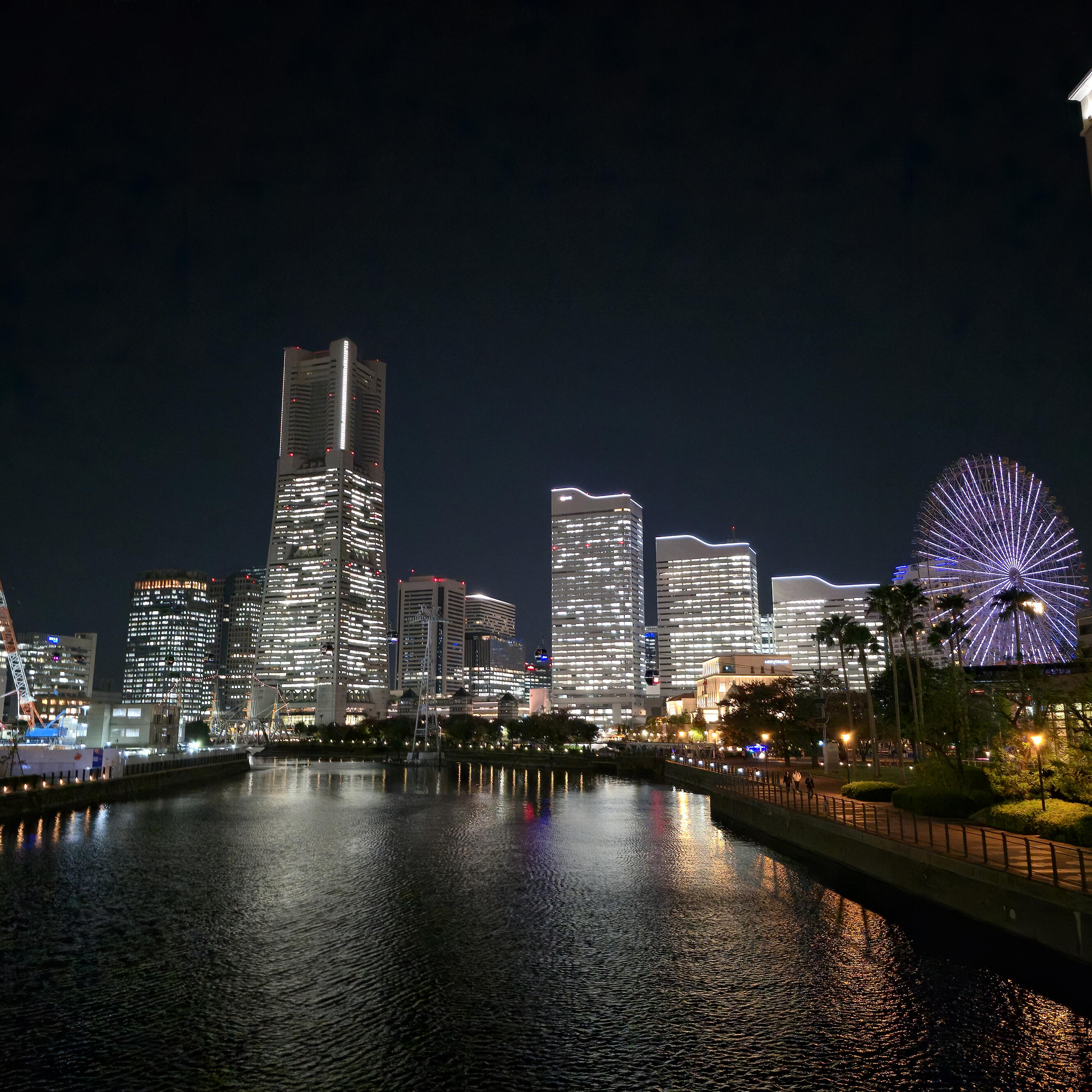
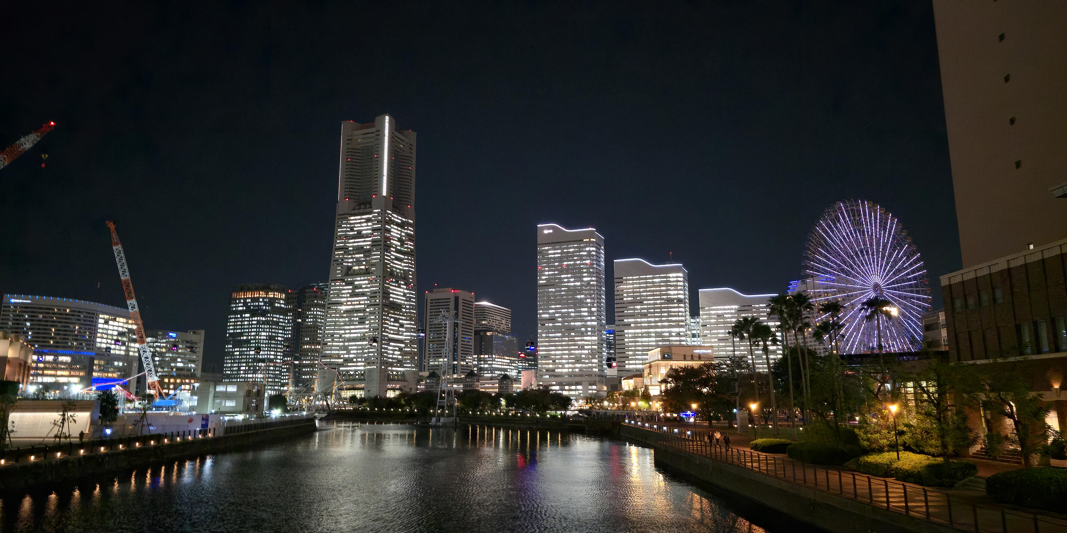
Nils Marquardt, a doctoral researcher in Medical Science, investigates how individual, moving cells in the body, for example, immune cells, can be visualized and tracked using positron emission tomography. In November, he attended the IEEE Medical Imaging Conference in Japan. In addition to the scientific program, there was also time to experience Japanese culture. He shares his impressions in this guest contribution.
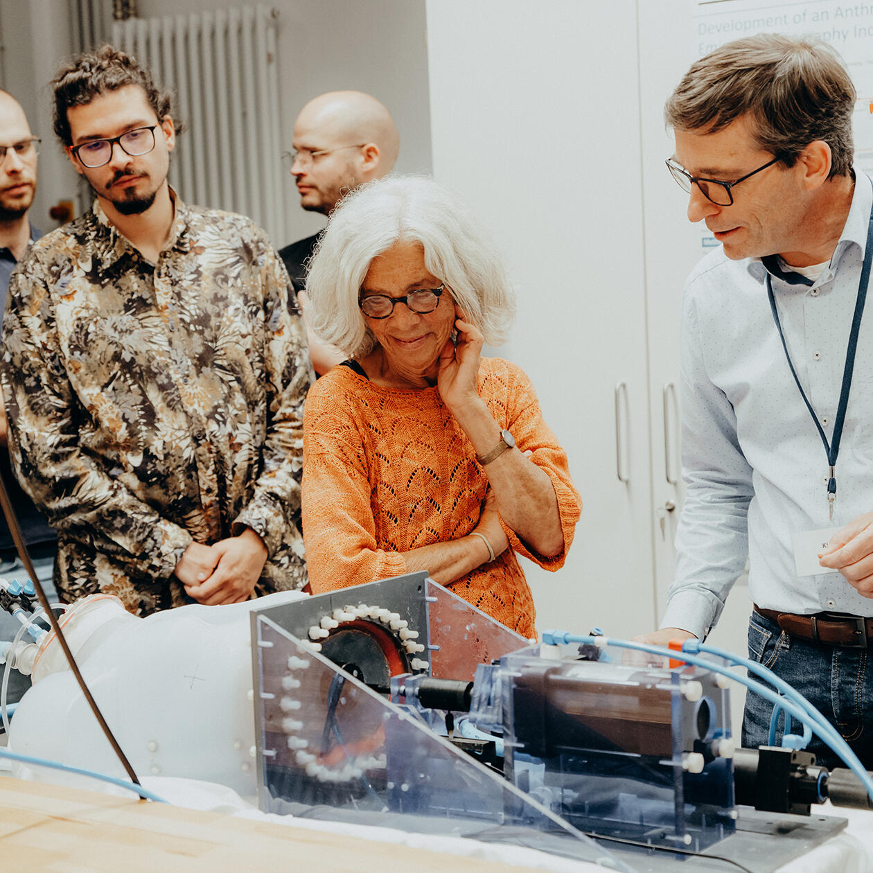
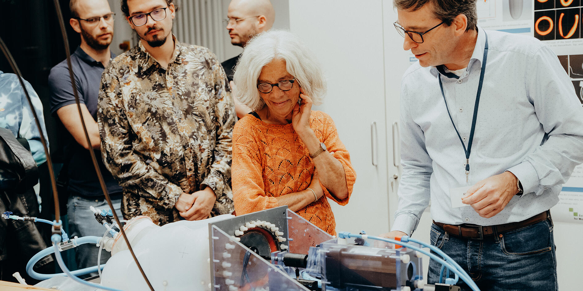
On September 12, 2025, the University and University Hospital Münster opened their doors to the public during the “Lange Nacht der Universitätsmedizin”. At the Multiscale Imaging Centre, many visitors took the opportunity to gain live insights into various areas of research.
The Collaborative Research Centre 1450 “inSight – Multiscale imaging of organ-specific inflammation” at the University of Münster will receive approximately 13 million euros from the German Research Foundation for a second funding period of four years. In this project, researchers are investigating how the body regulates inflammation in different organs and are, to this end, developing a specific multiscale imaging methodology.
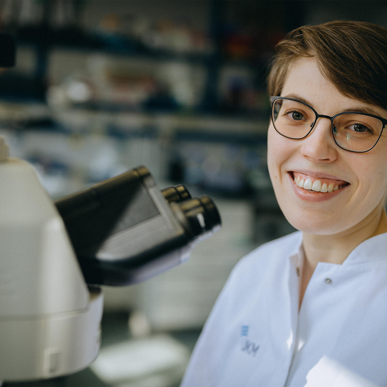
Emmy Noether junior research group leader Dr Maria Florencia Sánchez has been working at the European Institute for Molecular Imaging for several months. Dr Sánchez, who was born in Argentina, and her team are investigating how cells communicate with each other and how they perceive and react to their environment. She also supports students and young scientists at the University of Münster.
As a clinician scientist, junior professor Dr Philipp Backhaus is good at juggling the demands of research and hospital work. A specialist in nuclear medicine with six children, he hopes his research will help bring about concrete improvements in medical care. This is a dual role that requires good organisational abilities and the support of those around him.
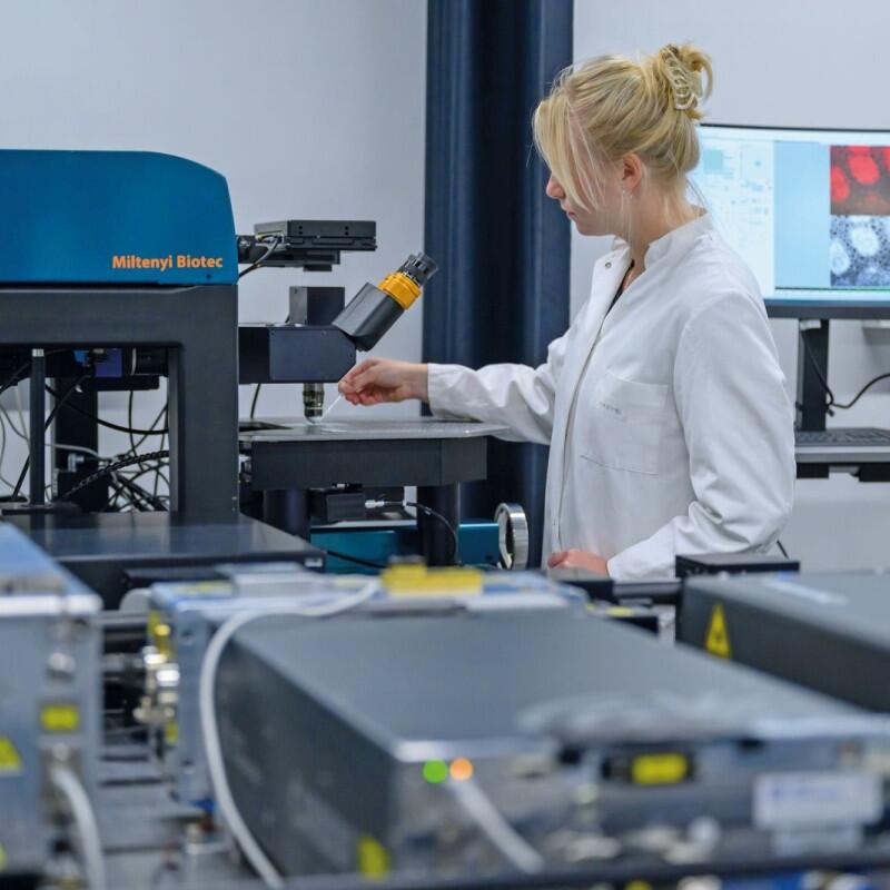
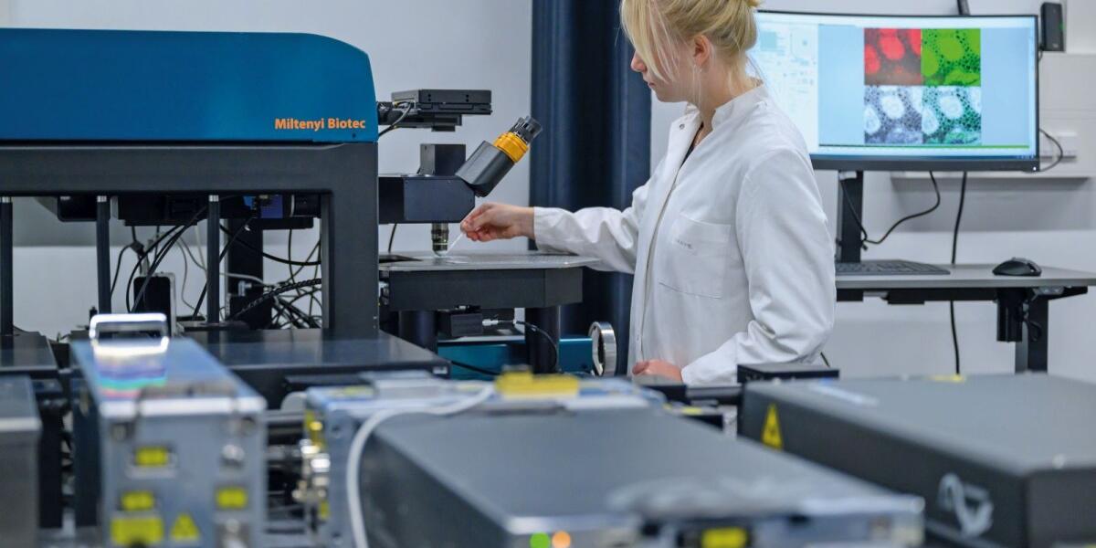
Microscopes make it possible to take ever deeper and more precise looks at the smallest of details, and in ever higher resolutions. This article looks at some of the techniques used by researchers at the University of Münster including, among others, high-performance cryogenic electron microscopy available at Prof. Christos Gatsogiannis’ lab, three-photon microscopy used by Prof. Friedemann Kiefer’s group, and insights into confocal laser scanning microscopy provided by Prof. Stefan Luschnig’s group.
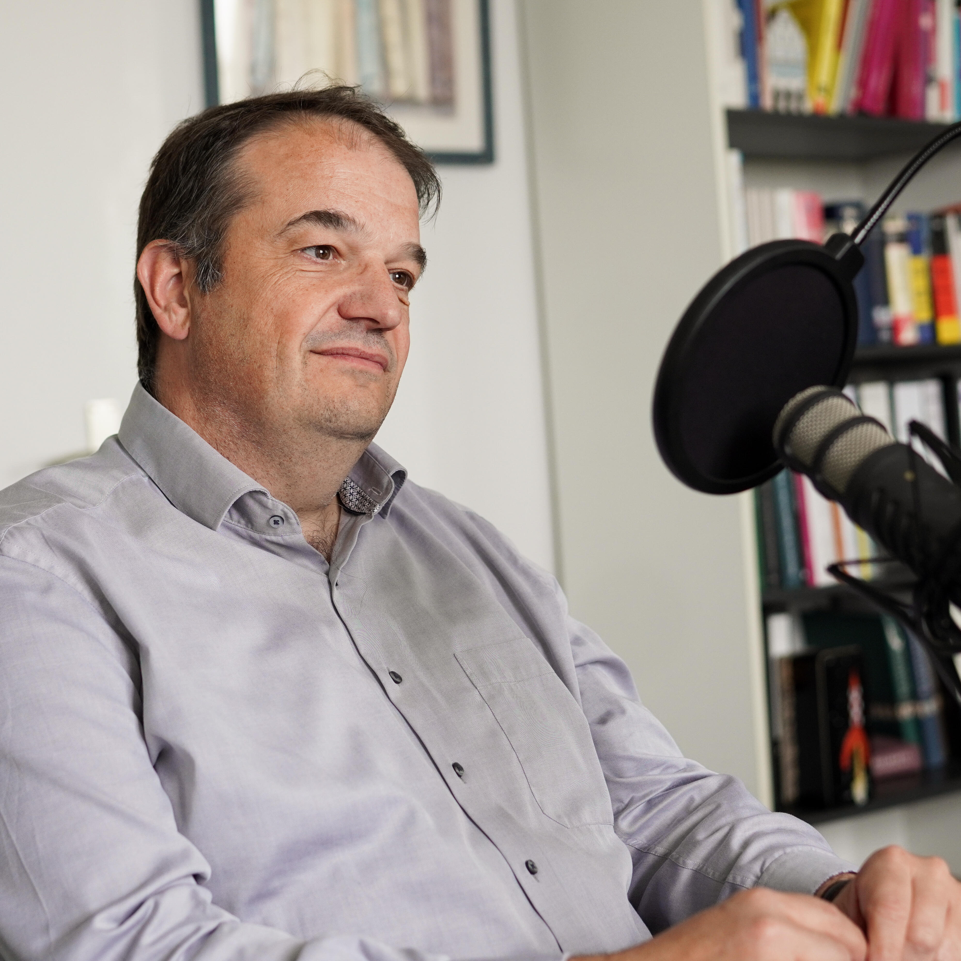
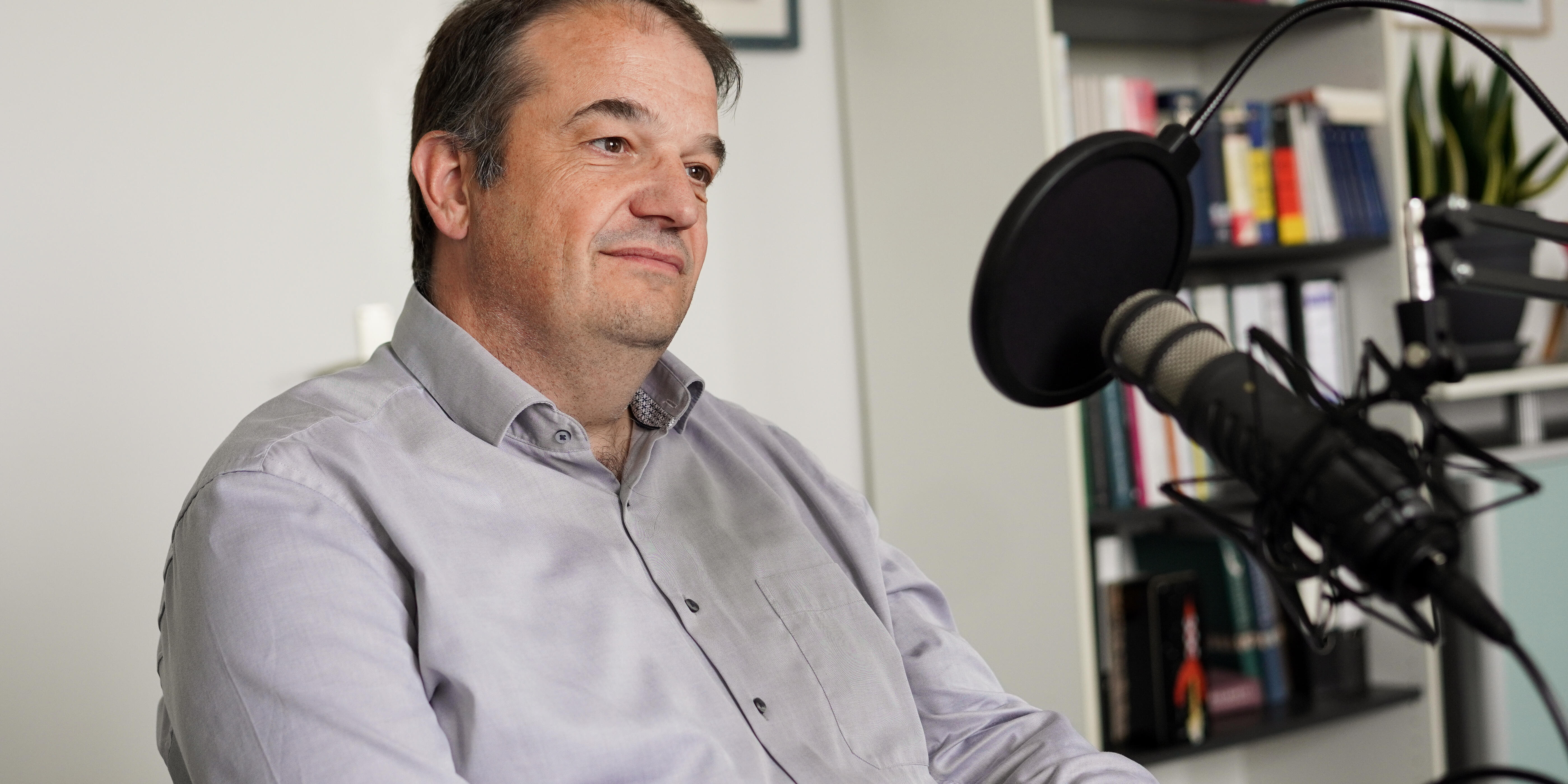
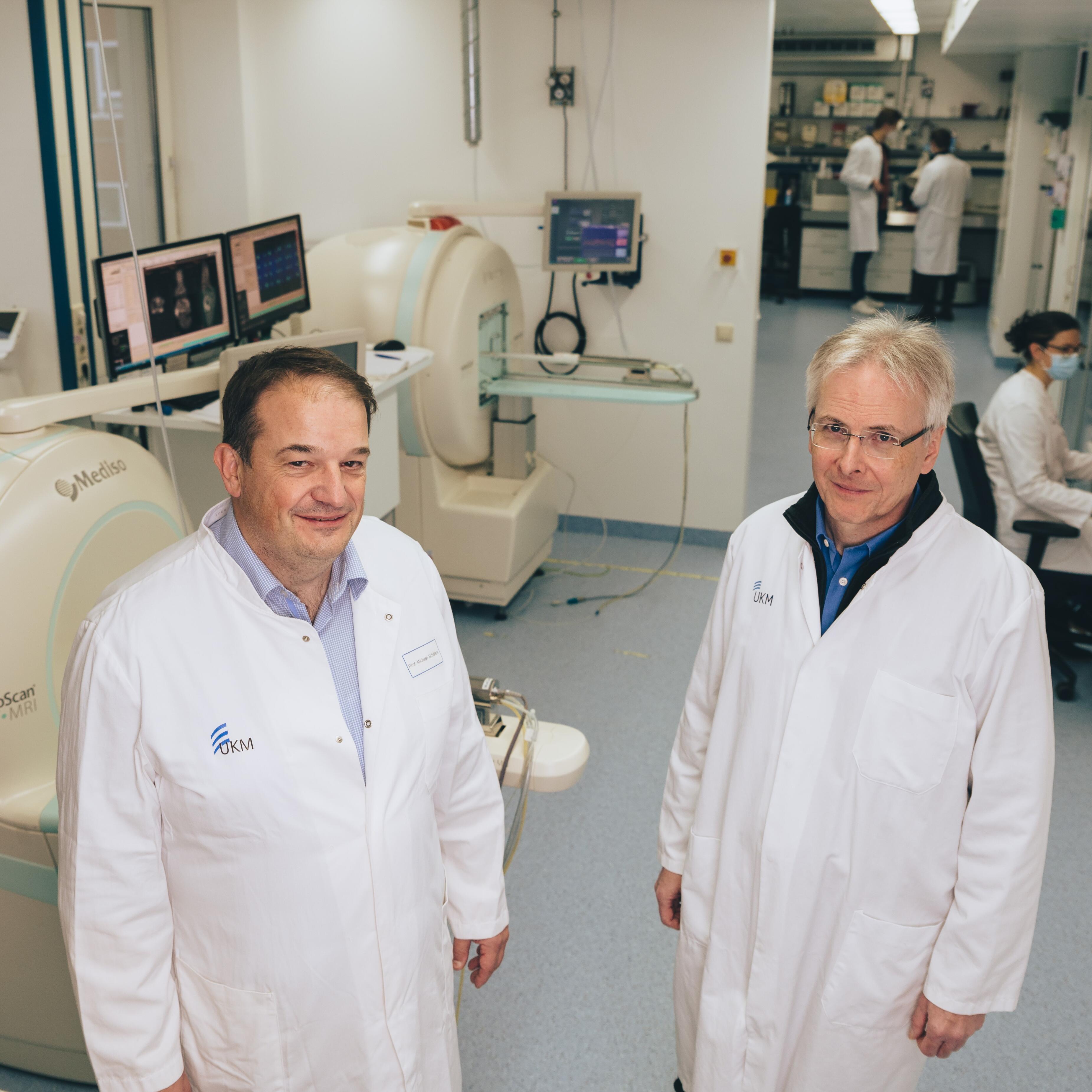
Science needs specialised researchers. For many research questions, however, cooperation with colleagues from other disciplines is just as important. Using the example of the Collaborative Research Centre “inSight” Prof Michael Schäfers, a specialist in nuclear medicine, provides insight into research practice in the field of inflammation and imaging. He also talks about “network life” and explains, for example, how a grant application for a research network is created and what role junior scientists play. The podcast is available in German.
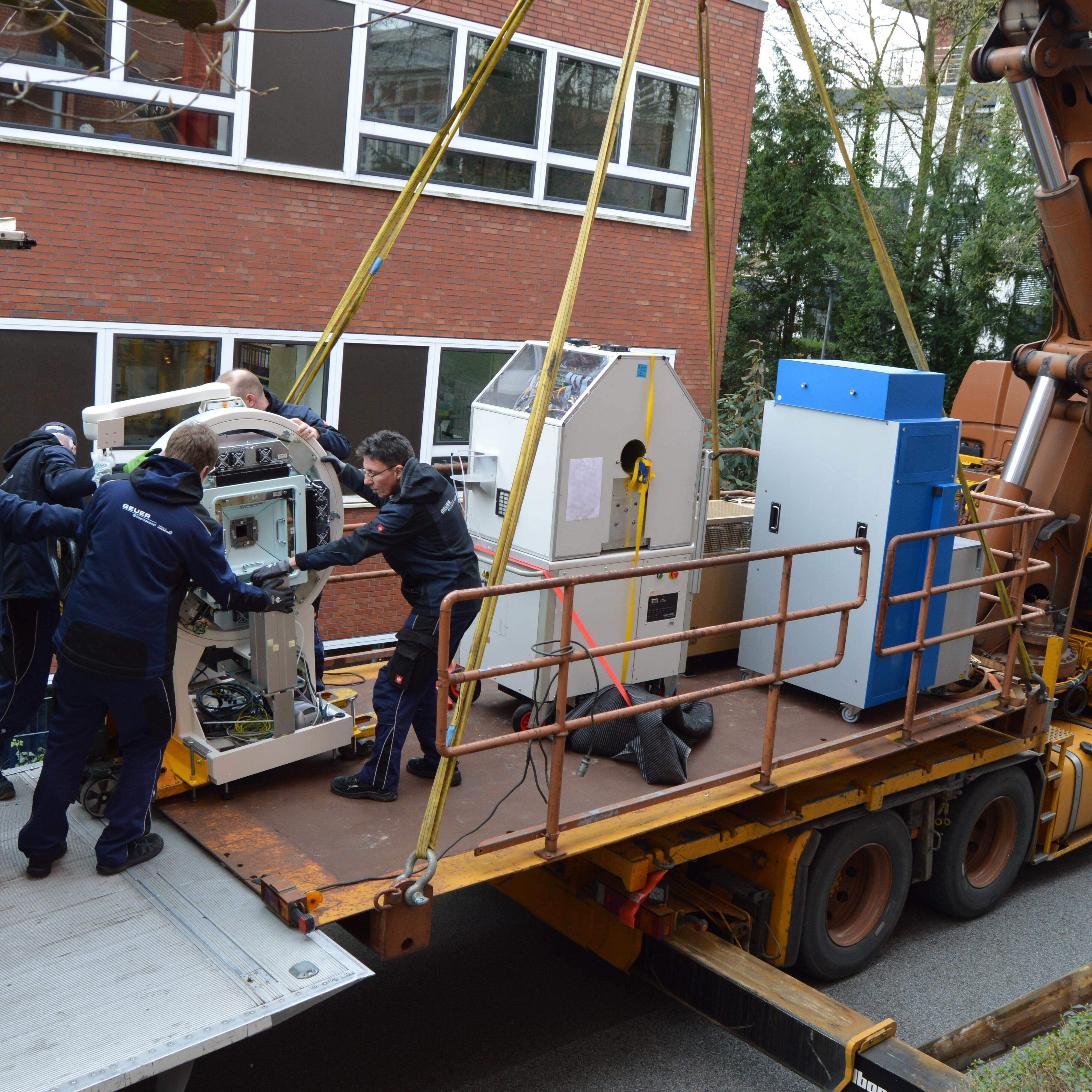
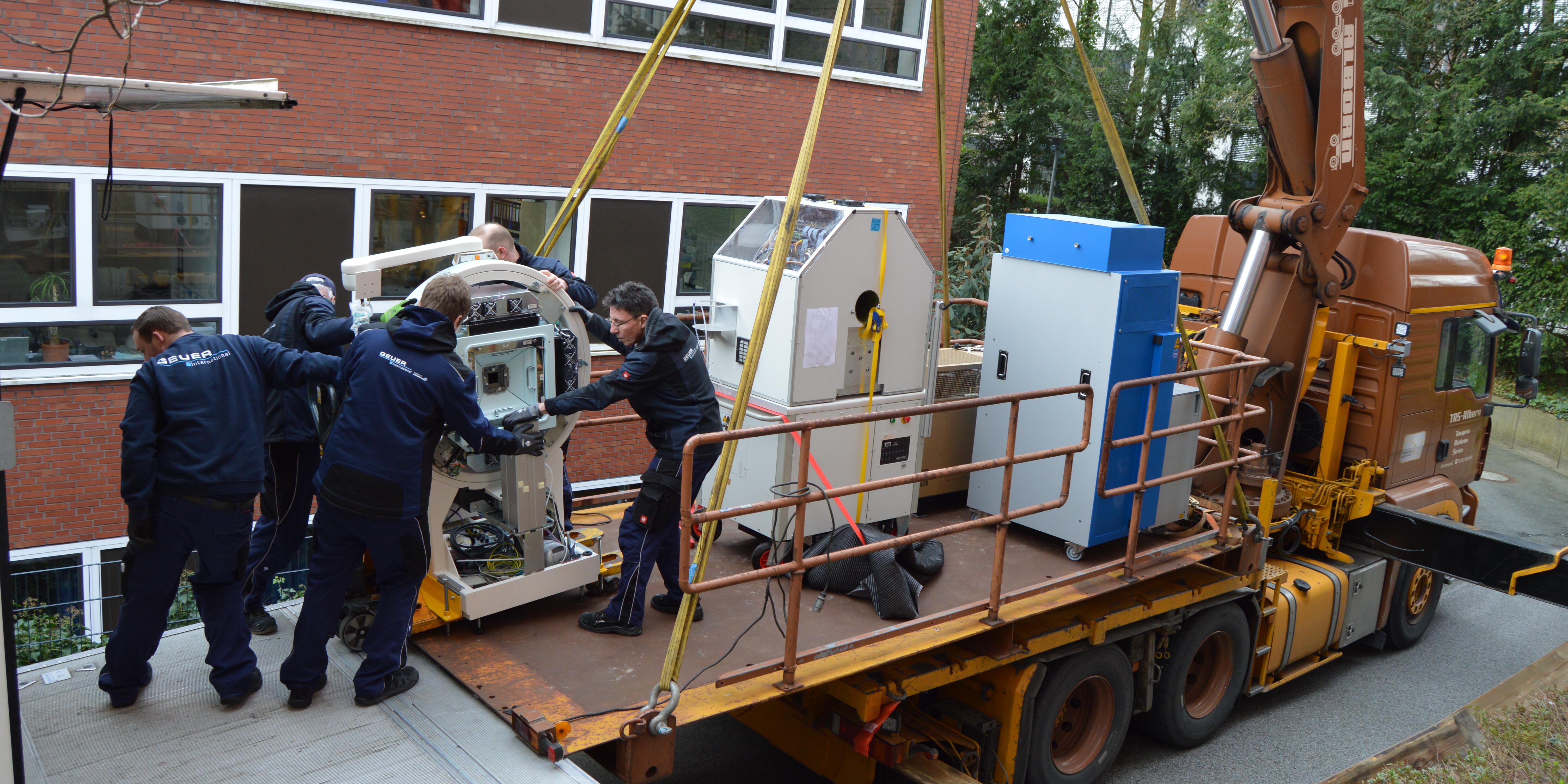
As of March 2023, the European Institute for Molecular Imaging (EIMI) is located in the Multiscale Imaging Centre (MIC) on Röntgenstraße 16 in Münster. In this research building, working groups from our university bring together a wide range of state-of-the-art imaging techniques to investigate biomedical questions. EIMI director Prof. Michael Schäfers is the spokesperson for MIC, and our team is happy about their new "home"! Our photo gallery shows how our imaging devices were transported to our new labs.
Nuclear medicine specialist Dr. Philipp Backhaus and his research partners examined breast cancer patients, for the first time systematically using a radiotracer that binds to the fibroblast activation protein. In particular small cancer lesions could be newly detected, and in combination with MRI, the new PET imaging method influenced further treatment in three of 19 patients. The study, published in “Radiology”, was awarded “Paper of the Month” by the Medical Faculty.
Medical professional Nadine Heiden is training to become a specialist physician while actively pursuing research. “I always wanted to do both,” she says – and a close connection between research and patient care can only be beneficial. Although the dual qualification is challenging, Nadine Heiden provides insight into how it is working out for her.
A research team led by biochemist Prof Andrea Rentmeister and nuclear medicine specialist Prof Michael Schäfers has, for the first time, utilised so-called SNAP-tag technology to radioactively label cells in living organisms. The method opens up the prospect of examining cells with different imaging techniques and at different temporal stages. The study was published in “Chemical Communications”.
Stefanie Bobe wants to dive deeper into medicine looking at the biomedical basis for improved diagnostic and therapeutic options. Therefore, she did an additional, science-oriented Master's degree in experimental medicine parallel to her medical studies and, in the working group led by biochemist Prof Friedemann Kiefer, became part of an interdisciplinary research team.
The new Collaborative Research Center "inSight" at Münster University receives funding from the German Research Foundation amounting to approximately ten million euros. The researchers aim to gain a comprehensive understanding of how the body regulates inflammation in different organs and, to this end, develop a specific imaging methodology that brings together information from single cells to entire organisms.
In a new video series the University of Münster introduces junior researchers. The first one is Cristina Barca, a PhD student at the European Institute for Molecular Imaging: Using biomedical imaging, she is investigating how well certain drugs work in treating strokes. In the video she provides an insight into her everyday working life and explains what is so special about being a scientist.
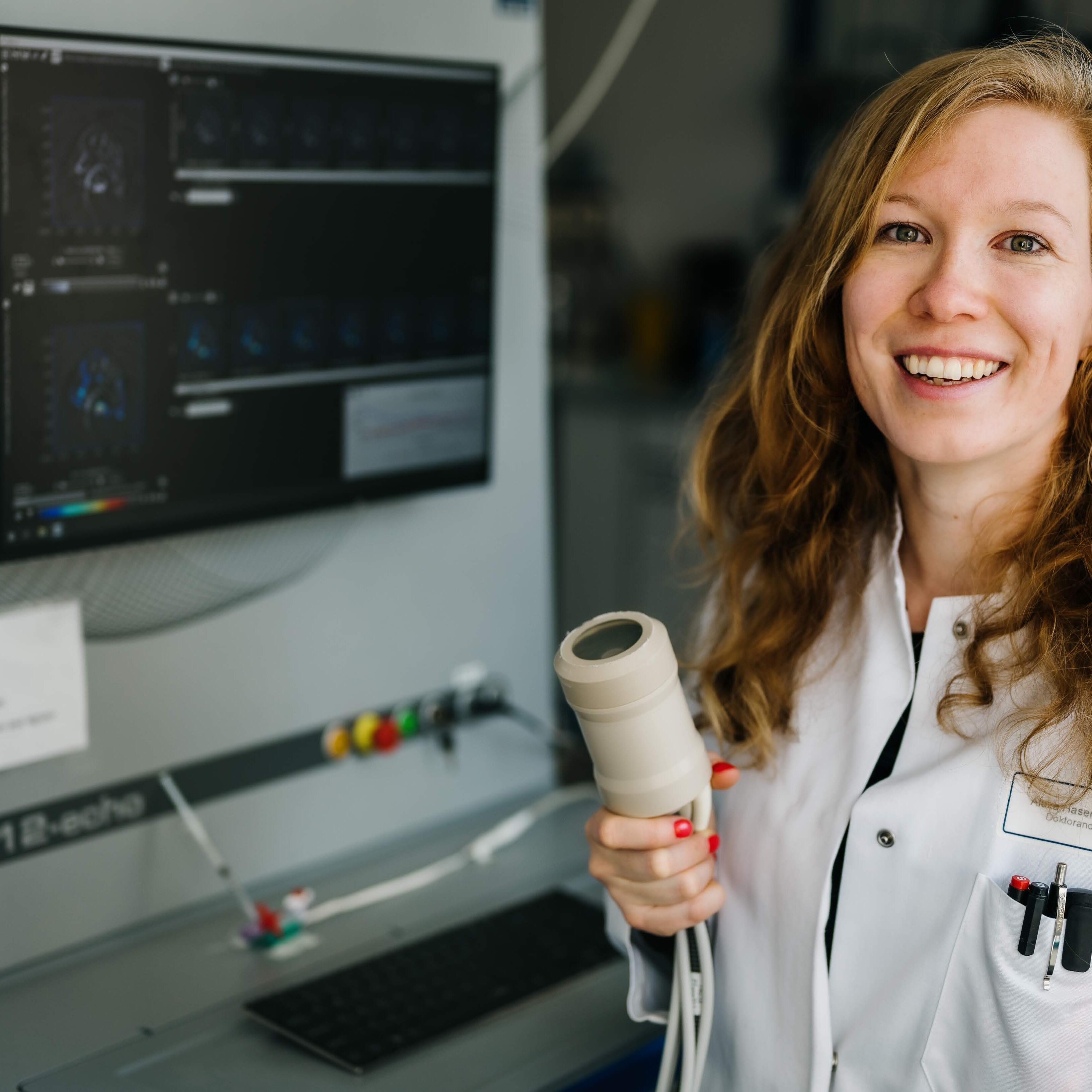
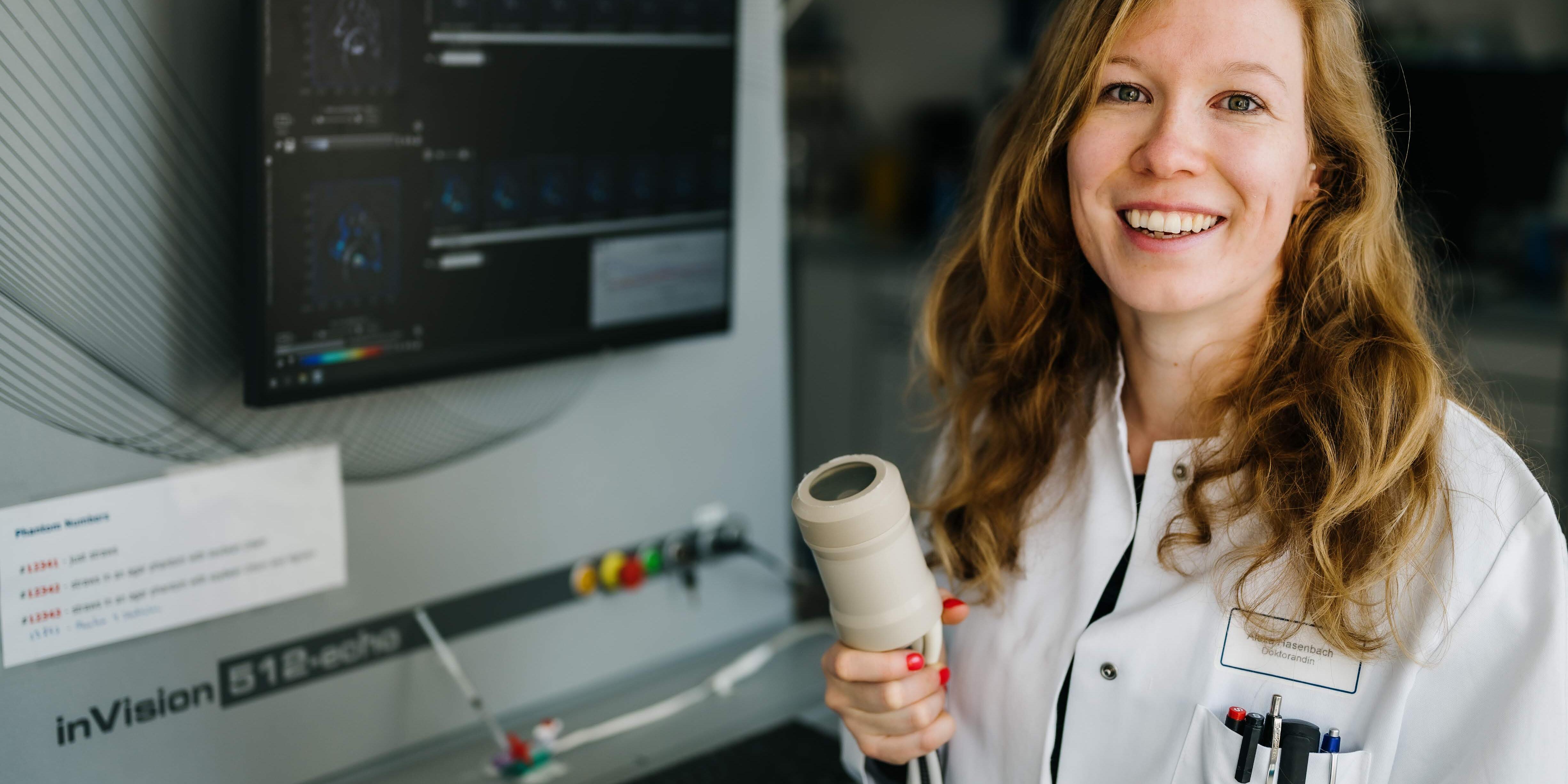
Laser light that cannot be seen, and sounds that cannot be heard: this combination produces something that is all the more visible – images from inside the body. Photoacoustics is the name of this method, whose purpose is to acoustically record the sounds of molecules. During her PhD thesis, biologist Alexa Hasenbach investigated inflammatory processes.