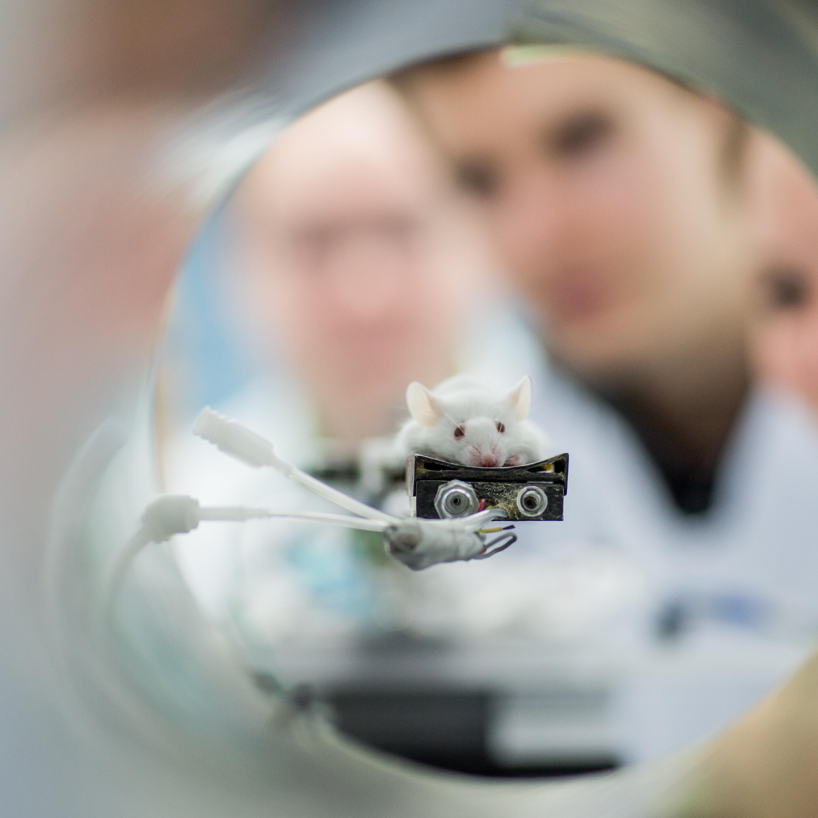
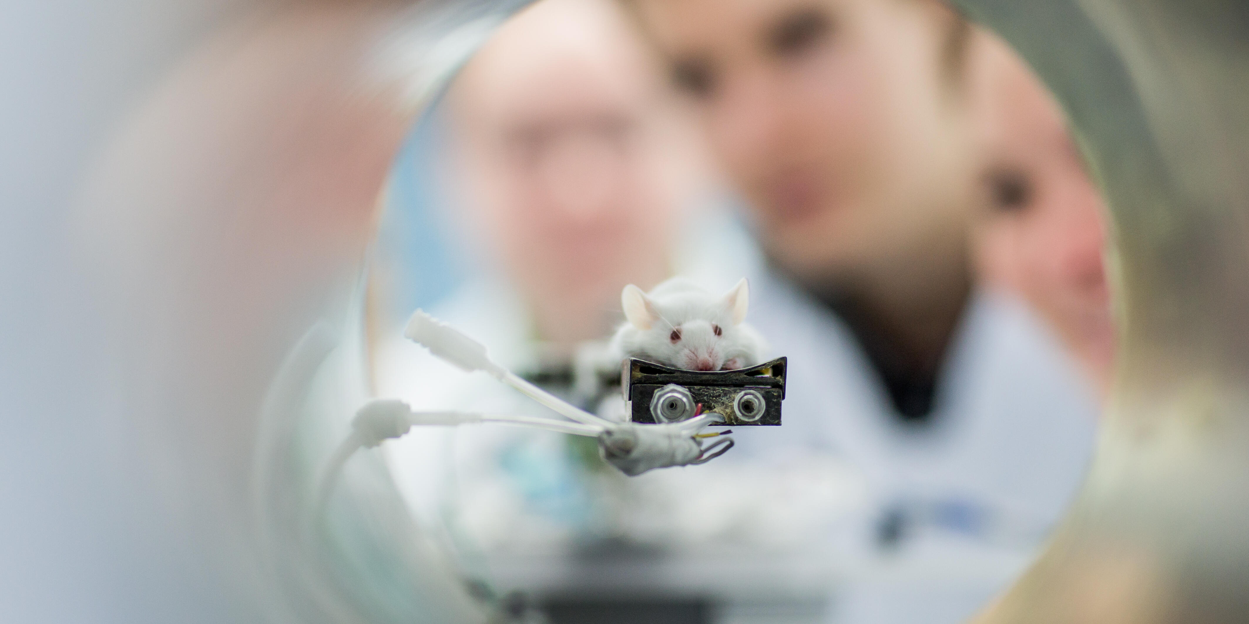
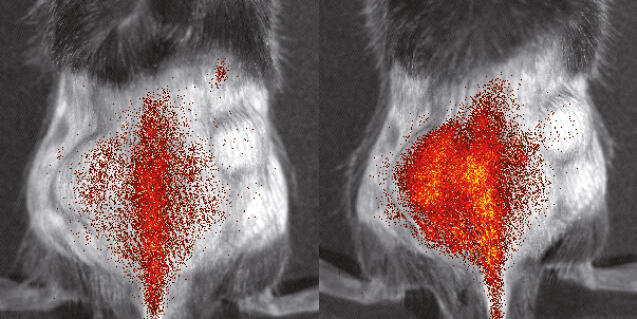
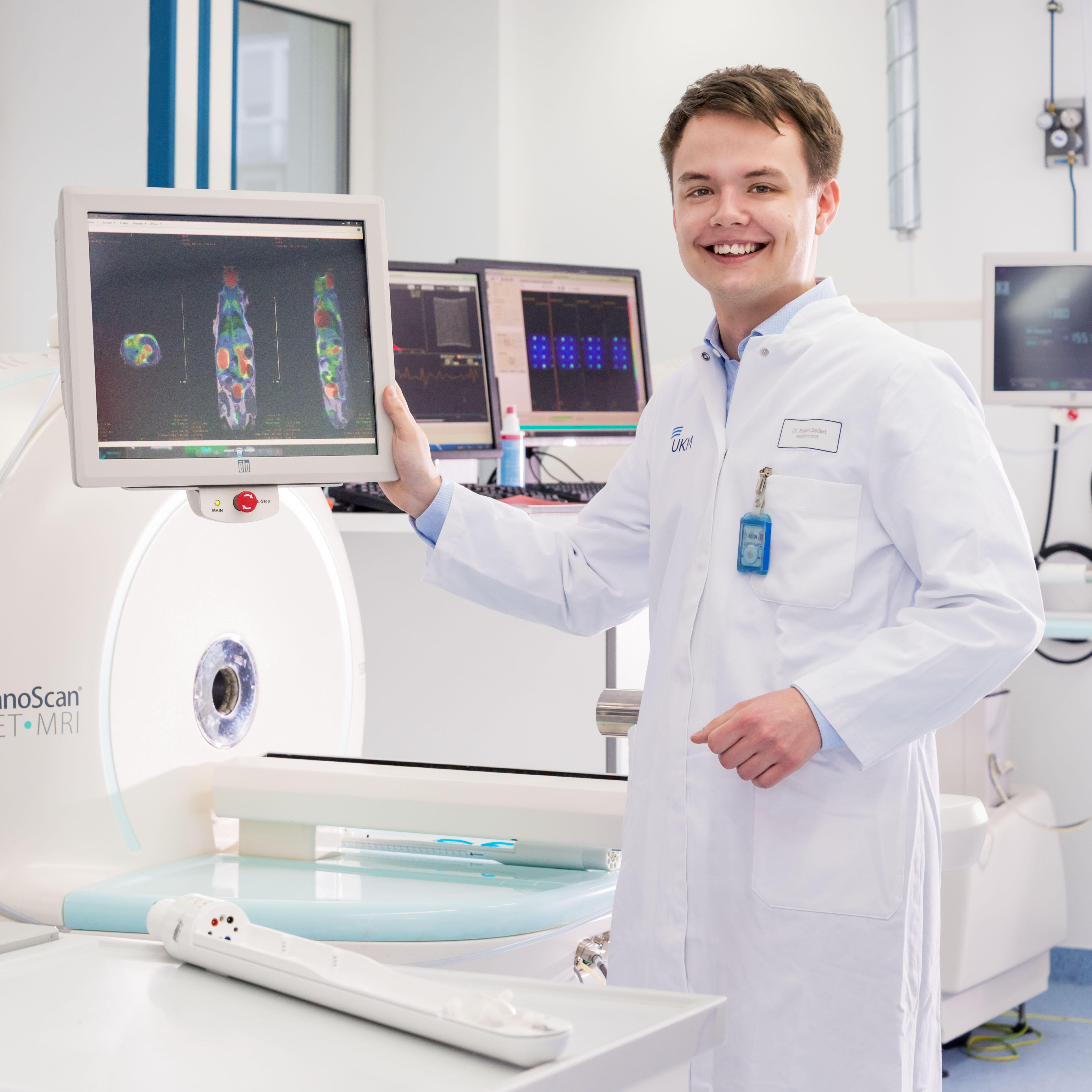
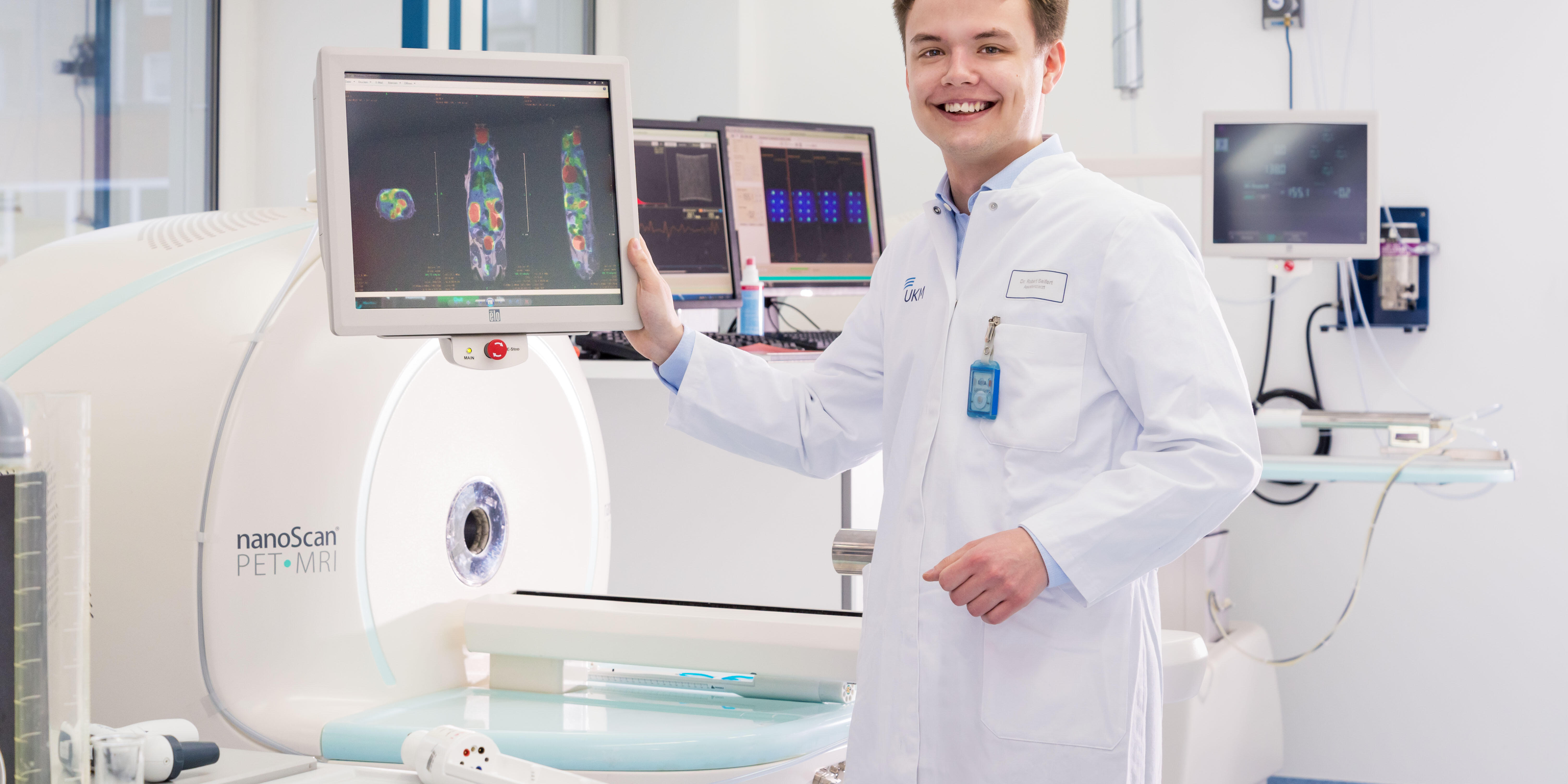
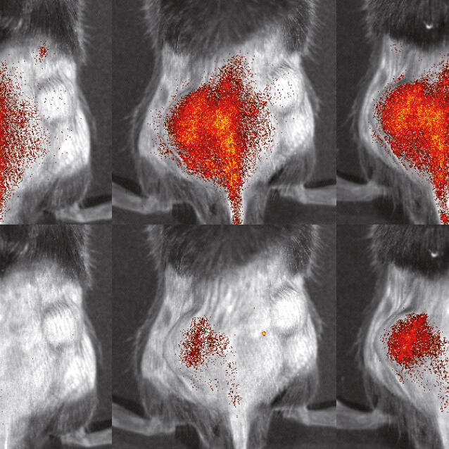
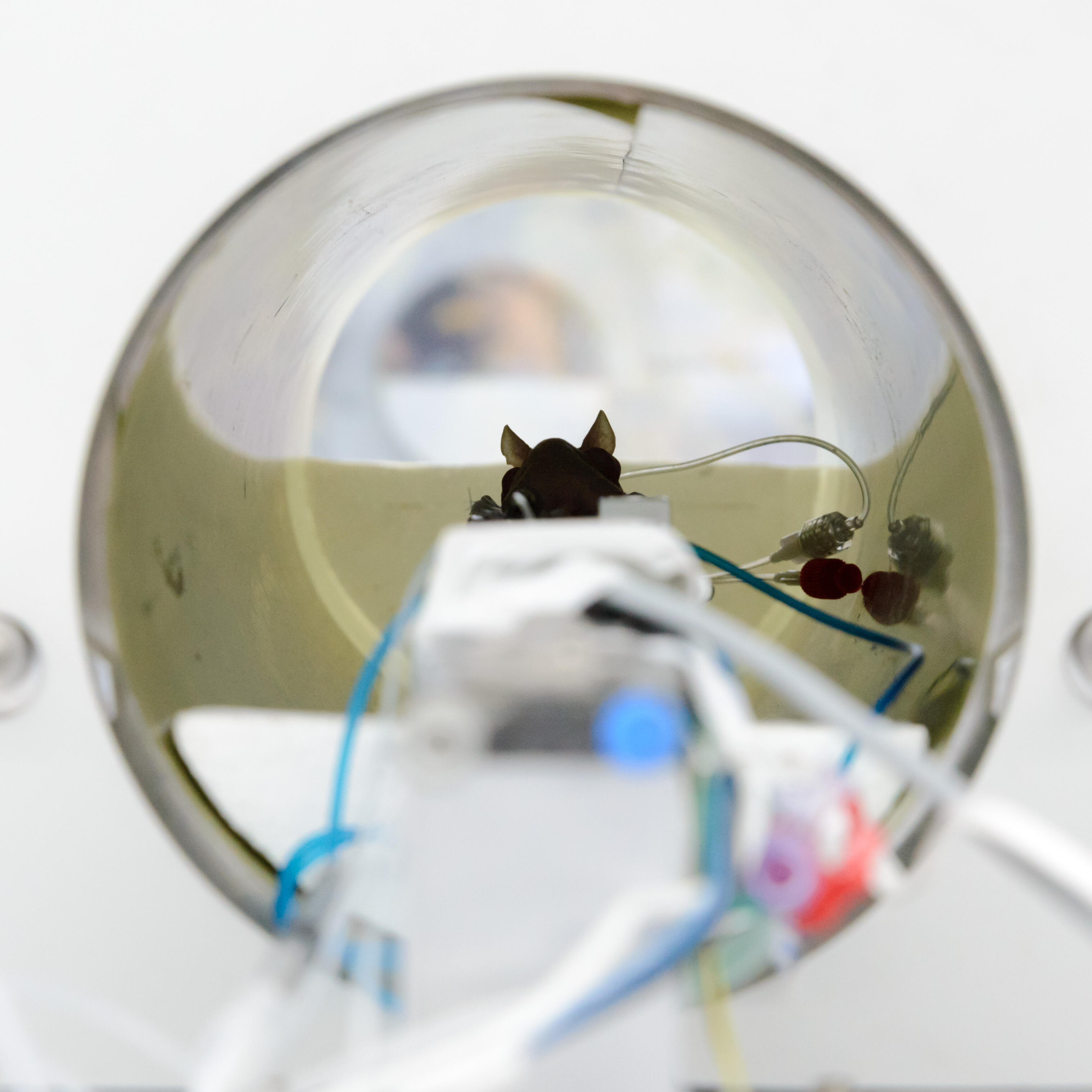
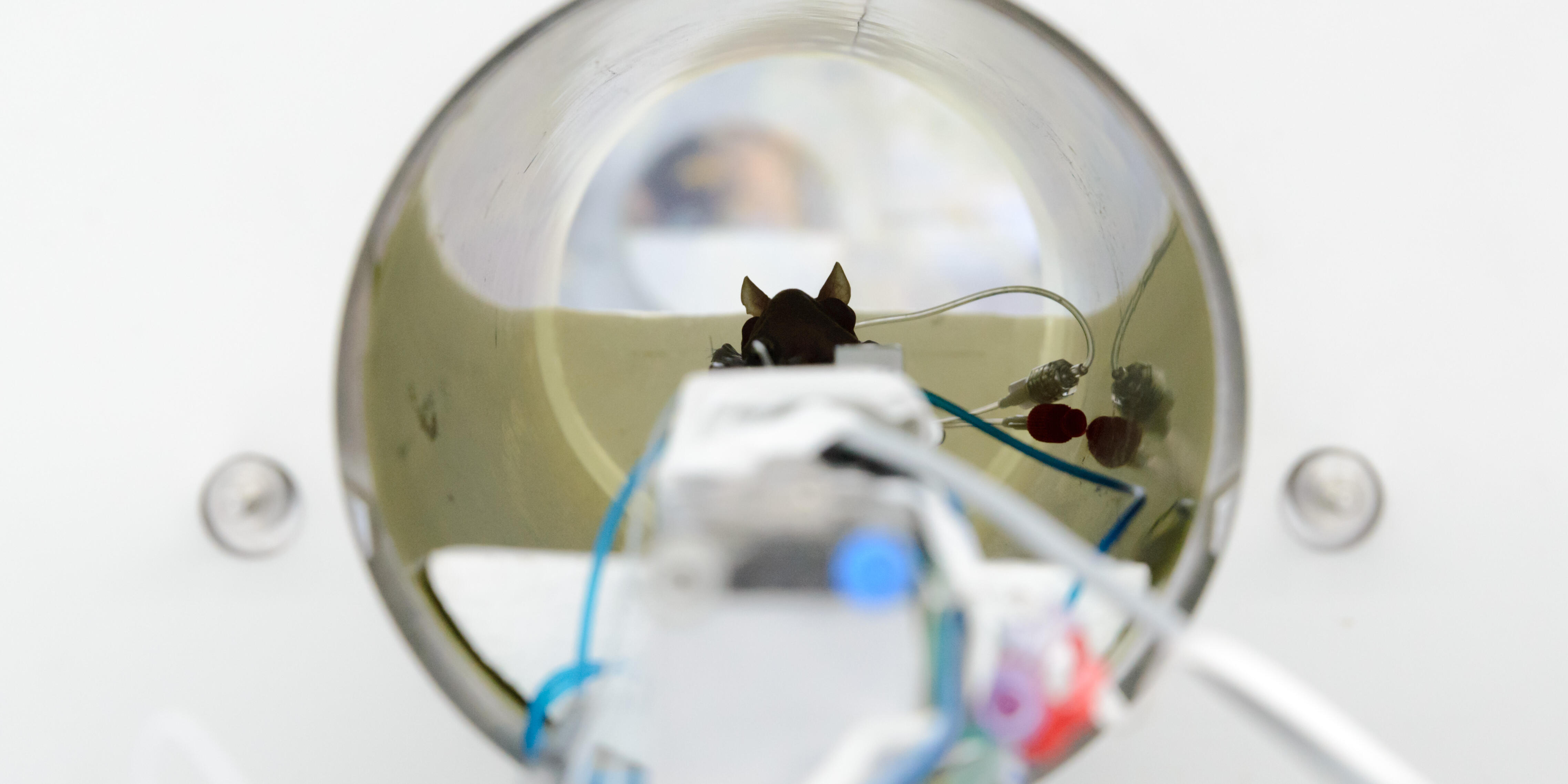
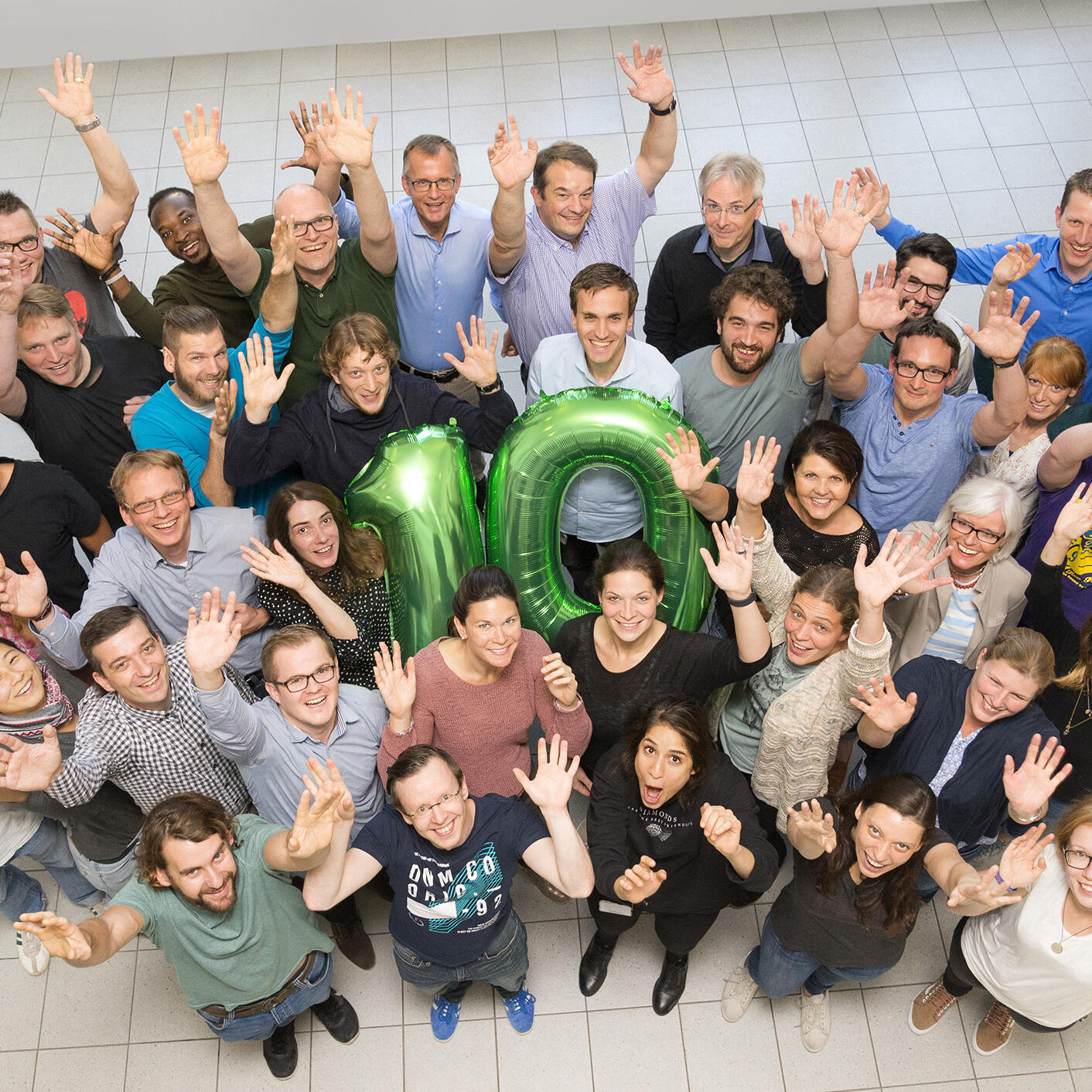
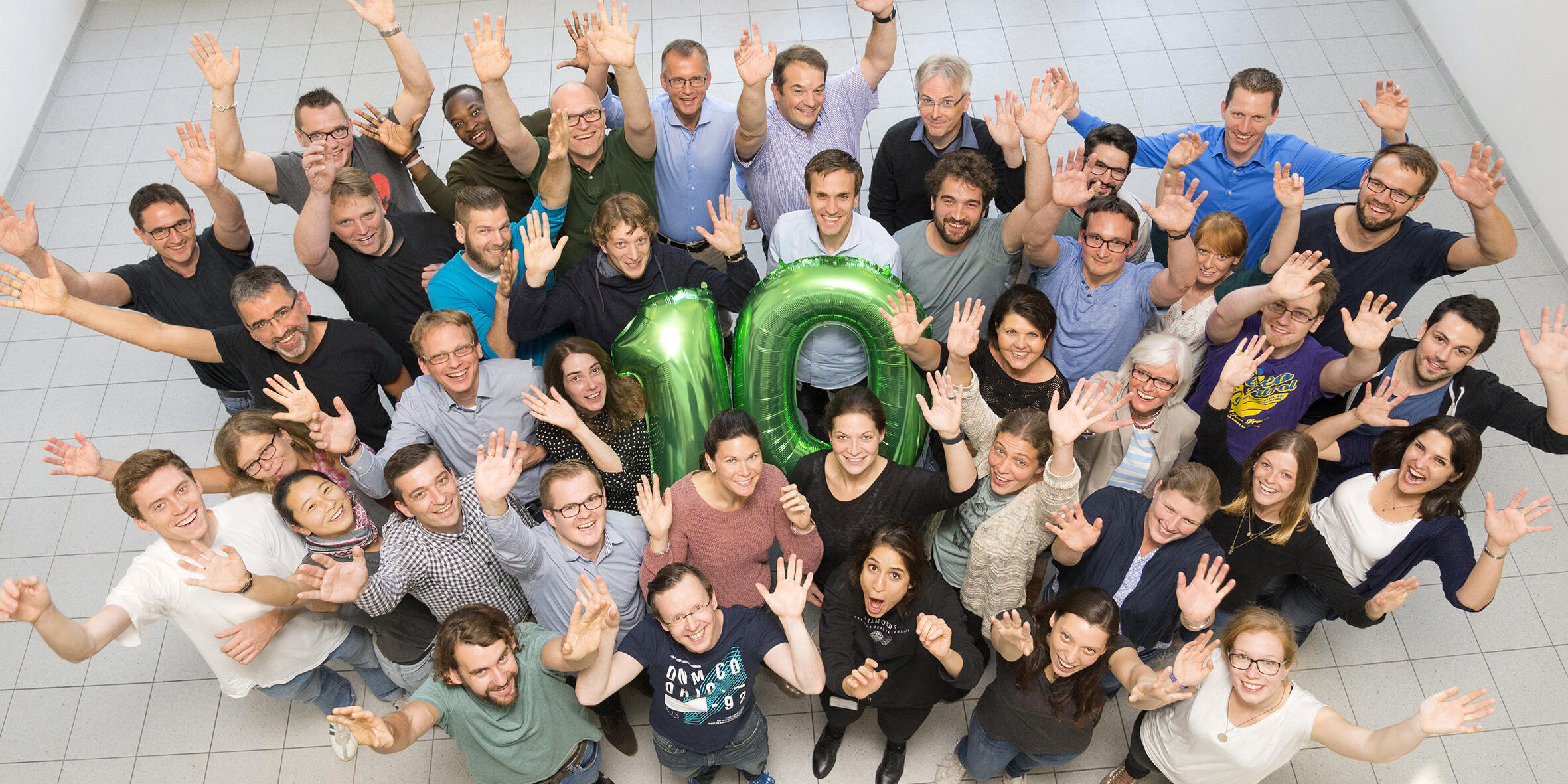
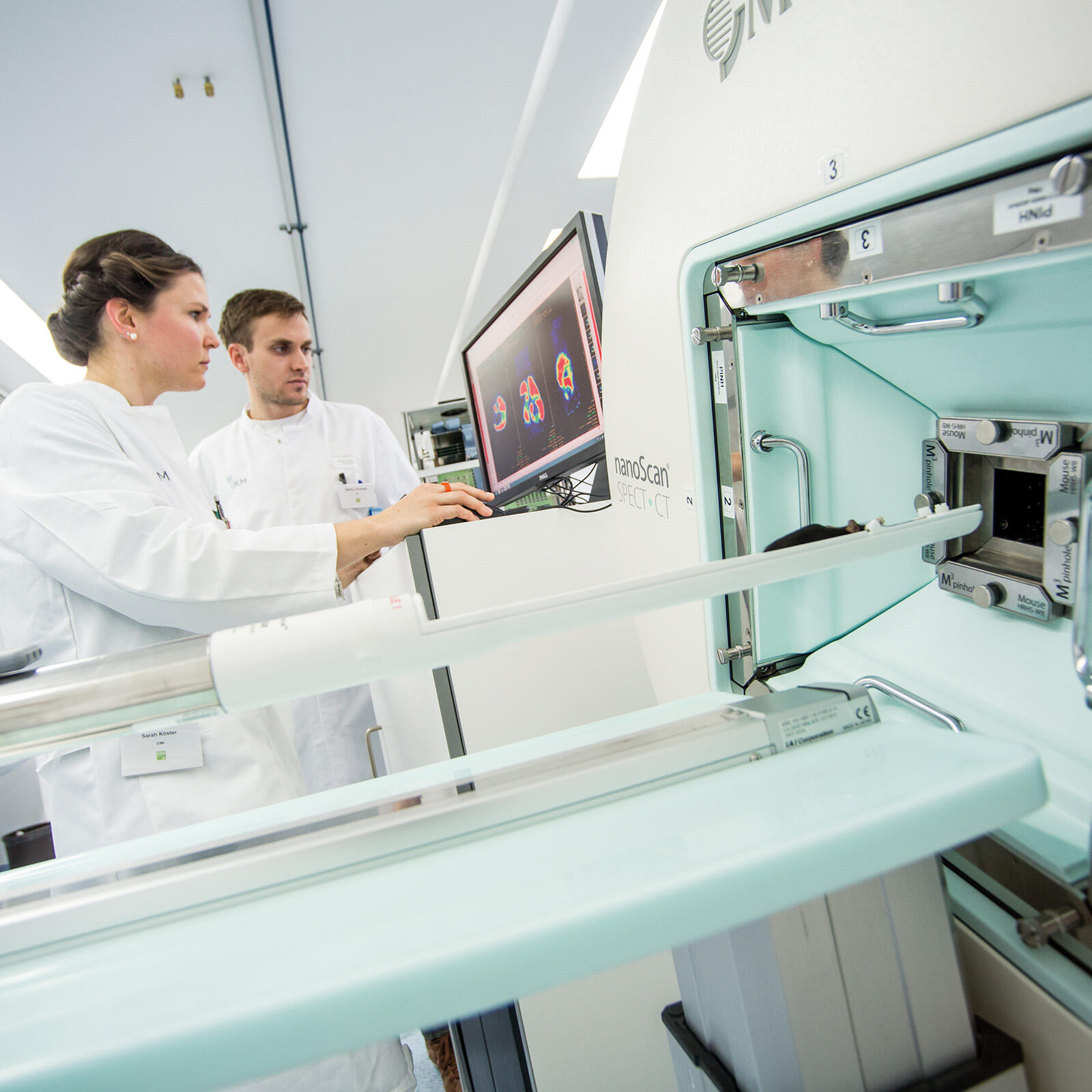
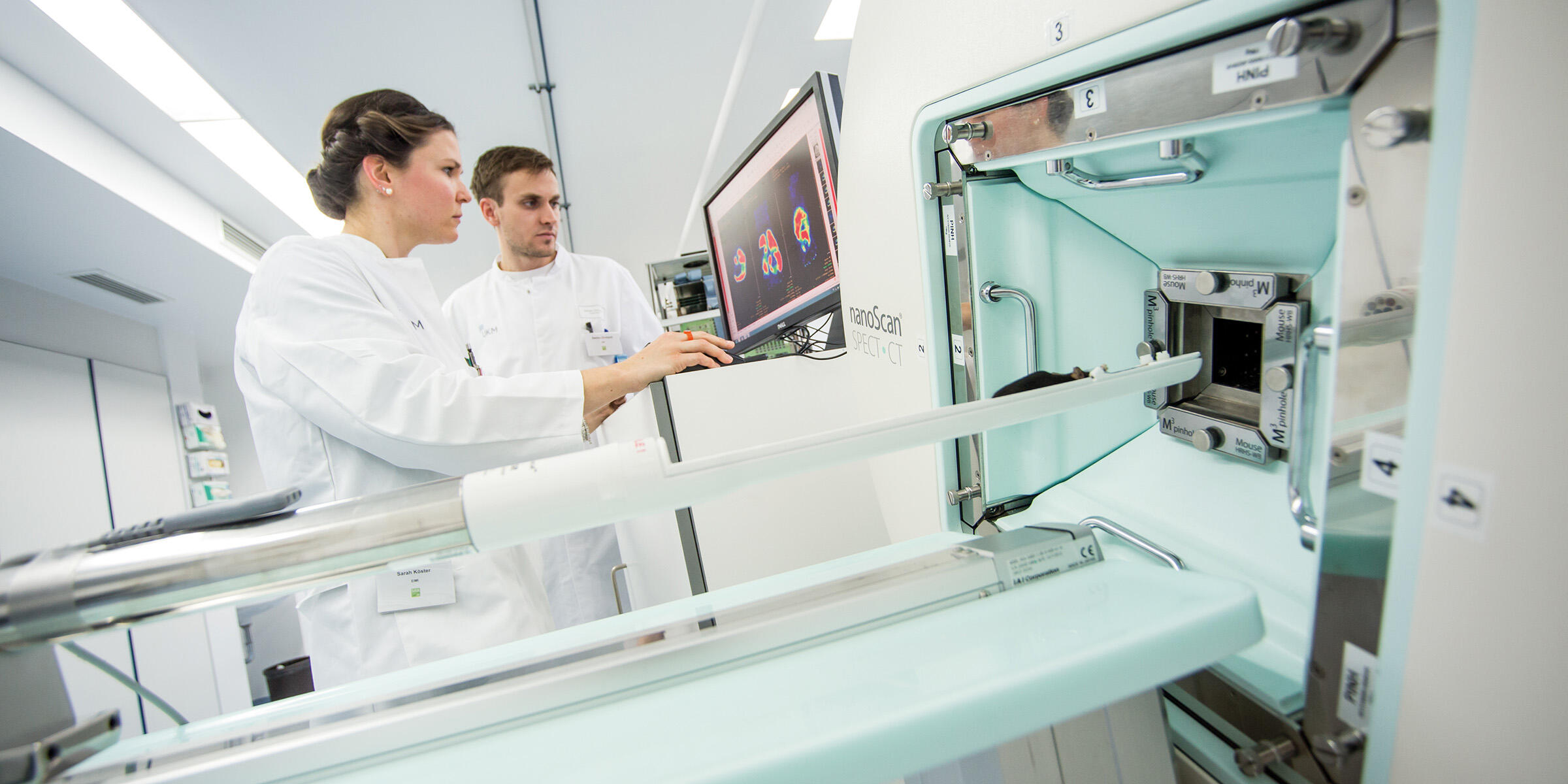
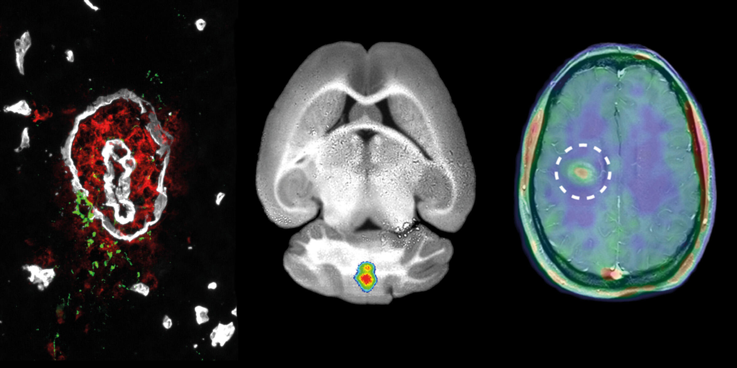
Scientists at the University of Münster use a broad spectrum of imaging techniques to investigate structures and processes in the body. Last week, they shared their knowledge with international junior researchers: The participants of the tenth annual Mouse Imaging Academy spent five days training on different methods for examining mice.
How do immune cells behave in the body? What happens during immunotherapy? To answer these questions, the European Union brings together leading experts from research and the pharmaceutical industry. The Europe-wide research project "Immune-Image", which is funded with 30 million euros over five years and in which scientists from Münster University are involved, started on 1 October.
The MD thesis of Dr. Robert Seifert, a physician, is based on an interdisciplinary cooperation, supported by the Cells-in-Motion Cluster of Excellence. He and his colleagues developed an algorithm for the precise analysis of image data. A WWU dissertation prize was awarded for this.
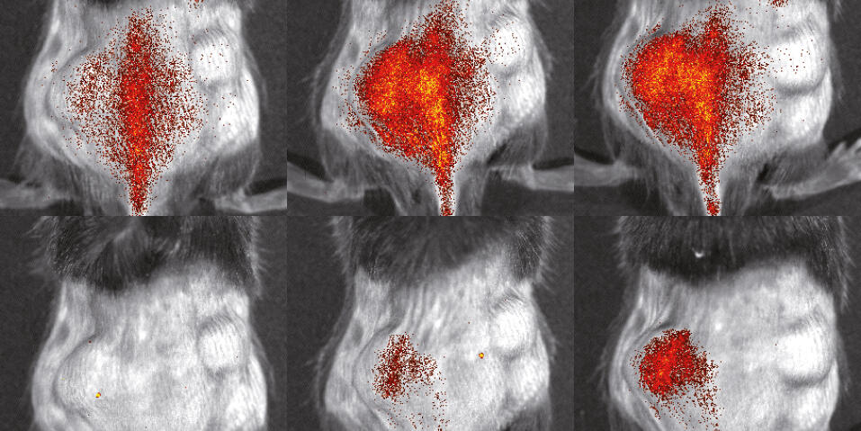
Immunologists and imaging specialists at the Cells-in-Motion Cluster of Excellence have jointly developed a method enabling them to better evaluate and study the activity of inflammatory cells in mice. The study has been published in the “Theranostics” journal.
In November, CiM researchers Prof. Michael Schäfers and Prof. Stefan Schlatt demonstrated experiments with mice to journalists and gave insights into how different animals are kept at Münster University. The University has compiled an experience report and a collection of links to press articles.
Prof. Friedemann Kiefer performs research at the Cells-in-Motion Cluster of Excellence, investigating how lymphatic vessels are formed and preserved. In his work, though, he constantly looks beyond the boundaries of his own field to develop new ideas. His overall aim is to make contributions of lasting value.
One part of the foundation of the Cells-in-Motion Cluster of Excellence celebrates its tenth anniversary: the European Institute for Molecular Imaging. How has the institute developed over the past ten years? What were special moments? In an interview, the EIMI Directors look back.
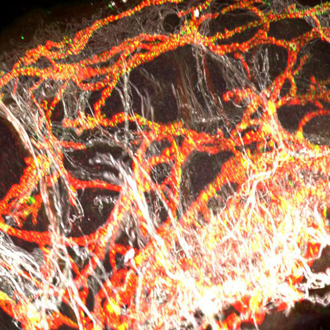
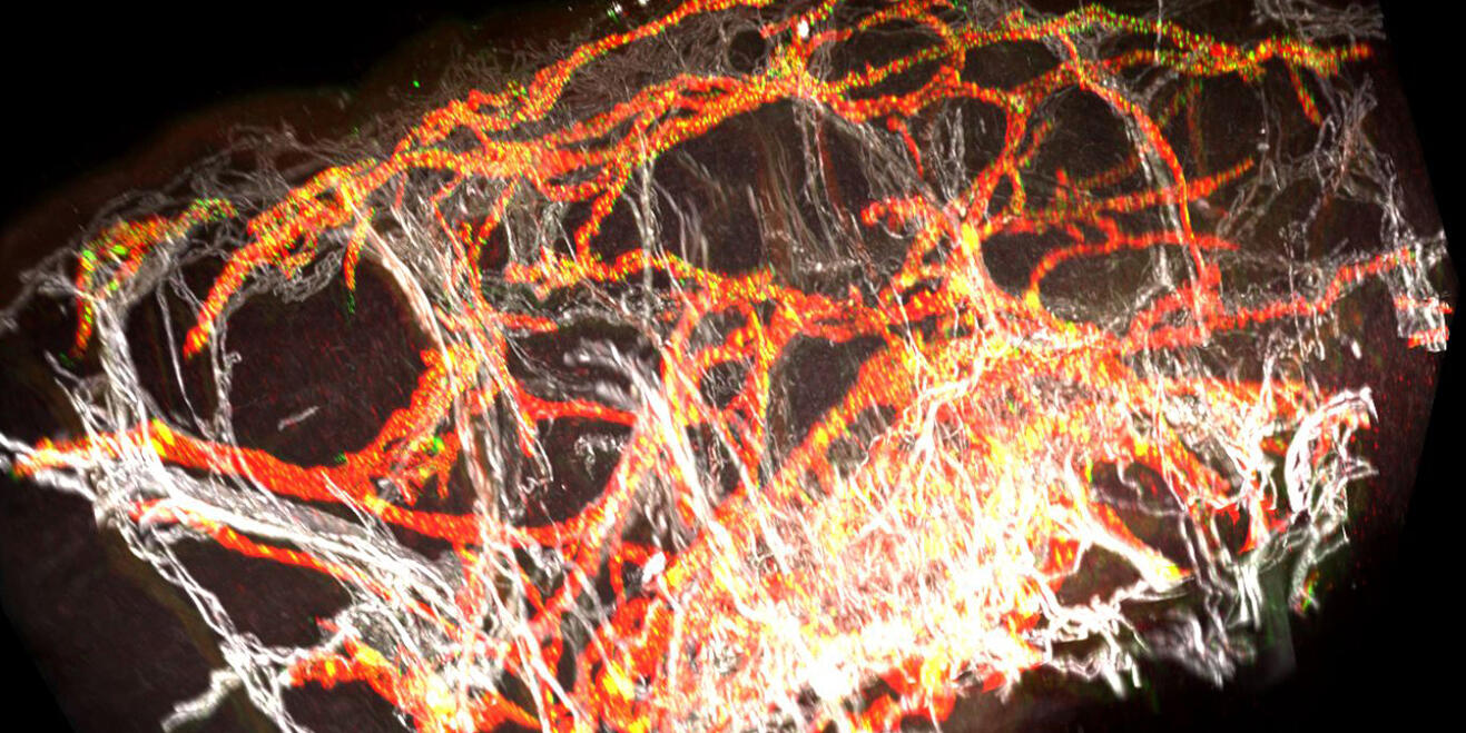
Researchers at the Cells-in-Motion Cluster of Excellence have developed a new method for producing digital 3D reconstructions of blood and lymphatic vessels from tissue samples and then creating images of them for analysis. The study has been published in the “JCI Insight” journal.
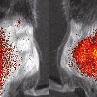
Prof. Michael Schäfers is fascinated by the possibilities offered by positron emission tomography, which enables to visualize molecular processes inside the body. With the help of this technology he would like to show inflammatory processes, e.g. to predict future heart attacks.
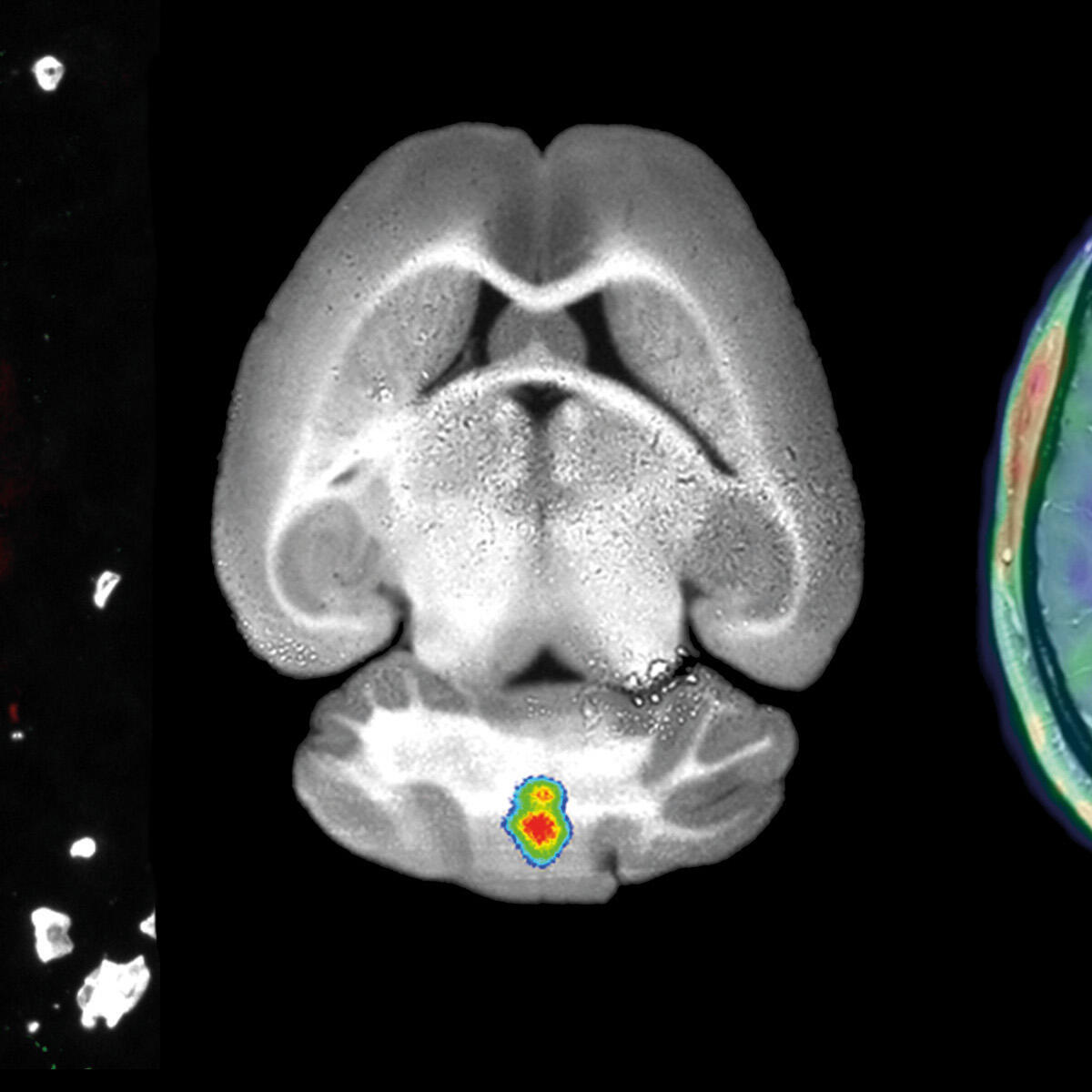
How can processes in the body that are normally hidden from the human eye, such as inflammation or disease, be made visible? To do this, scientists at the Cells-in-Motion Cluster of Excellence use a broad range of imaging technologies and work on developing innovative imaging strategies.
Researchers at the Cells-in-Motion Cluster of Excellence have succeeded in visualizing, for the first time, ongoing inflammation in the brain in patients suffering from multiple sclerosis. The study has been published in the prestigious journal "Science Translational Medicine".
Bacterial infections can have serious consequences – for example, when they colonize an artificial heart valve. There is especially problematic when the bacteria are resistant to several antibiotics. Researchers are looking for new methods of treatment as well as for ways to find centres of infection in the body, for example by means of special sugar molecules. Chemists, physicists, biologists and physicians were all involved in the study.
Without oxygen, cells cannot survive. Until now, scientists could not observe the effect that a reduced oxygen supply can have on individual cells. This was technically not feasible before. Scientists from Münster have now developed a reporter, which allows them to see an acute lack of oxygen of cells using light microscopy.
For Prof. Klaus Schäfers everything revolves around motion correction. He and his team aim to make clinical images even more exact. And they have no lack of ideas for improving high-tech equipment.
How can you take more sharply defined pictures from the body’s interior – even though motion comes in? Many of our scientists deal with this question in the labs of the Cells-in-Motion Cluster of Excellence. A newly developed programme now enables physicians to do away with the so-called respiratory belt in positron emission tomography (PET). This belt often has only limited uses in the case of people who are seriously ill.
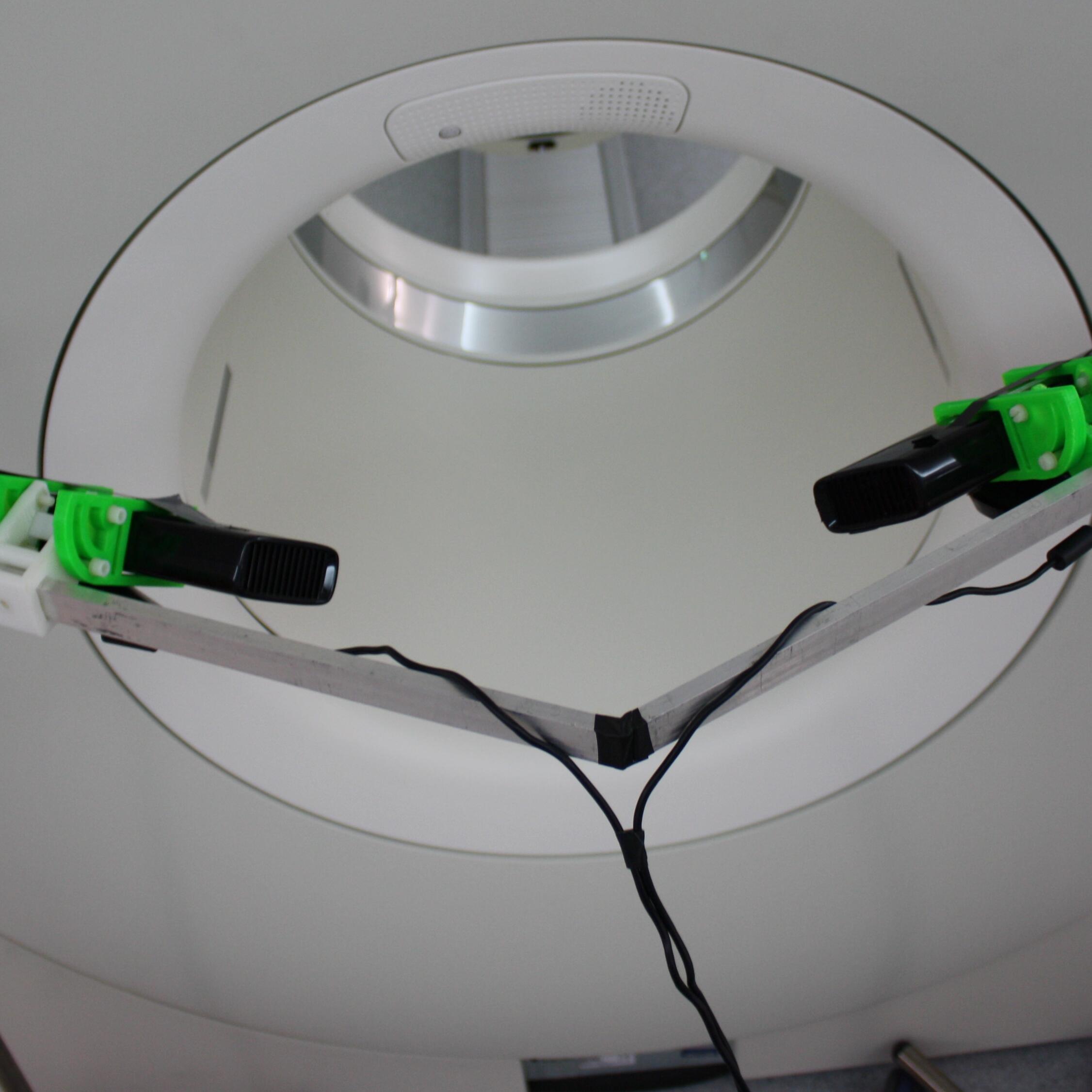
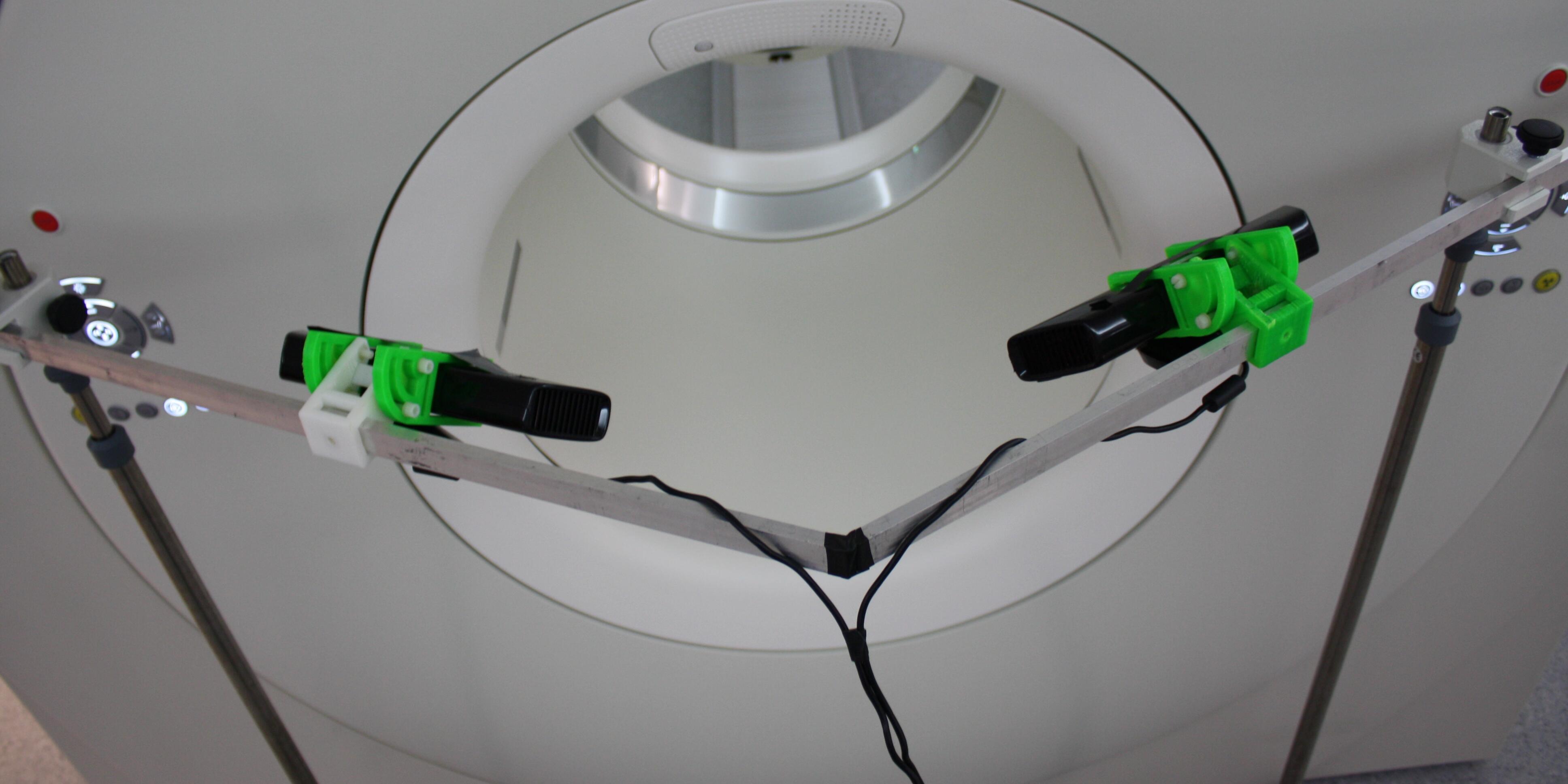
It does not always need to be the most expensive high tech product to optimize medical technology. A scientist from a research group in the Cells-in-Motion Cluster of Excellence used the Xbox technology for an experiment. With the help of this affordable technology Mirco Heß shows scientists a way to better understand clinical images of the inside from the outside.
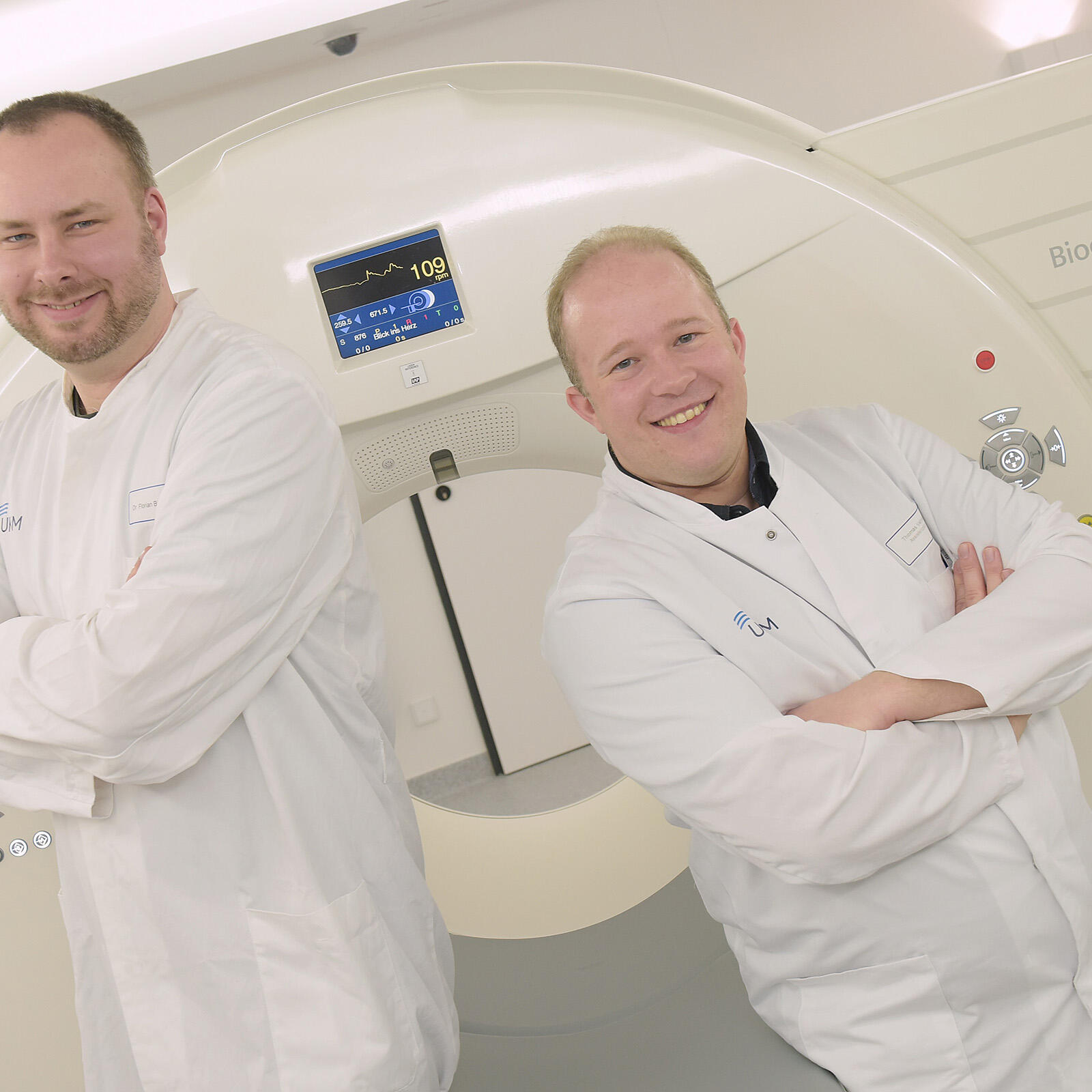
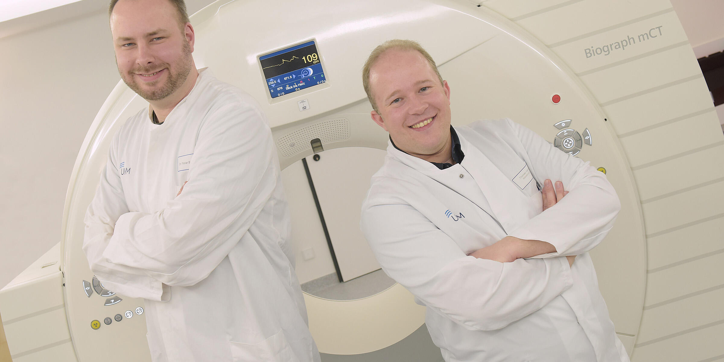
If you move during an examination with a whole body scanner the resulting images from inside the body will be blurred. A patient’s breathing is already enough to make tomographic images less precise. That is why medical physicist Dr. Florian Büther and nuclear medicine specialist Dr. Thomas Vehren develop new methods which shall improve the quality of medical images.
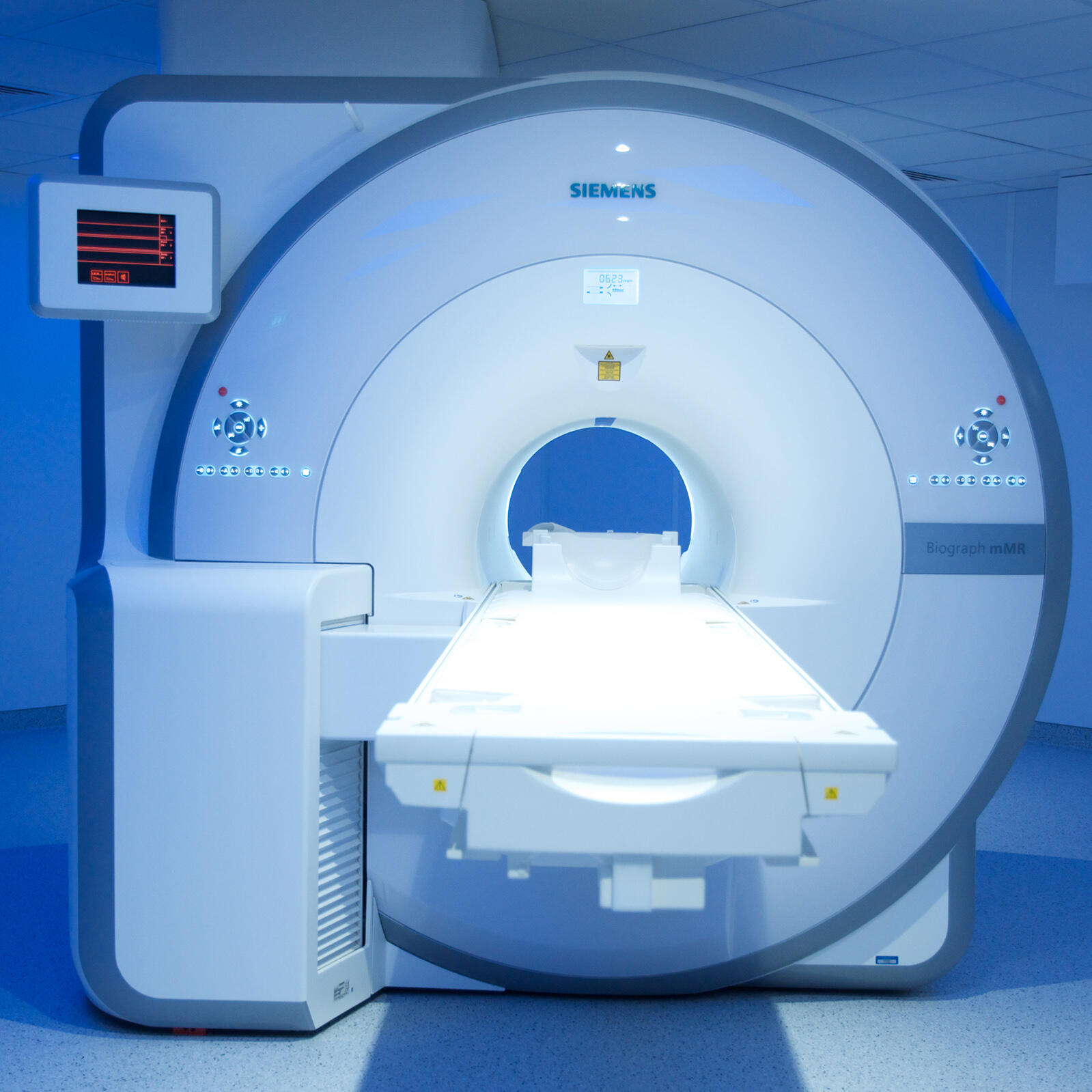
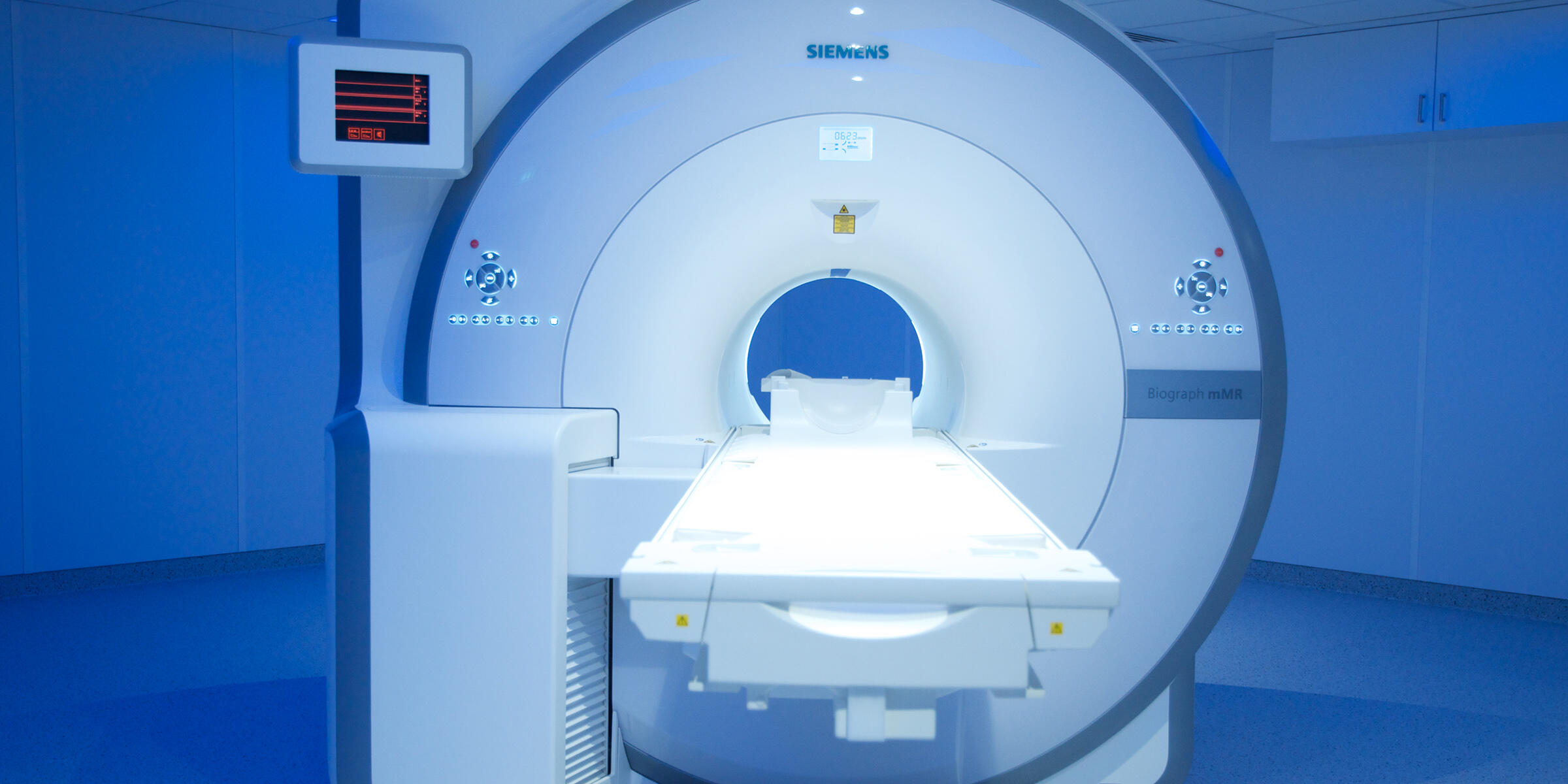
Scanning before dissection – to look inside the human body physicians nowadays use a set of imaging techniques. Just recently, the University Hospital Münster has installed an innovative hybrid system which combines PET and MRI. The high end scanner generates both unique images and challenges for the physicians.
When coronary arteries become inflamed, the vessel walls thicken and may rupture – leading to an acute heart attack. Dr. Thomas Vogl, Dr. Andreas Faust und Dr. Sven Hermann develop substances to visualise inflamed vessel walls with molecular imaging and to assess the individual risk.
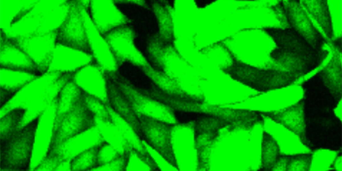
Cellular structures of different dimensions can be studied specifically by light microscopy – ranging from processes in the whole organism to the smallest components of individual cells. In the last thirty years, microscopes have benefitted from fluorescence and 3D-techniques and are extremely powerful, explain Prof. Jürgen Klingauf and Prof. Friedemann Kiefer.
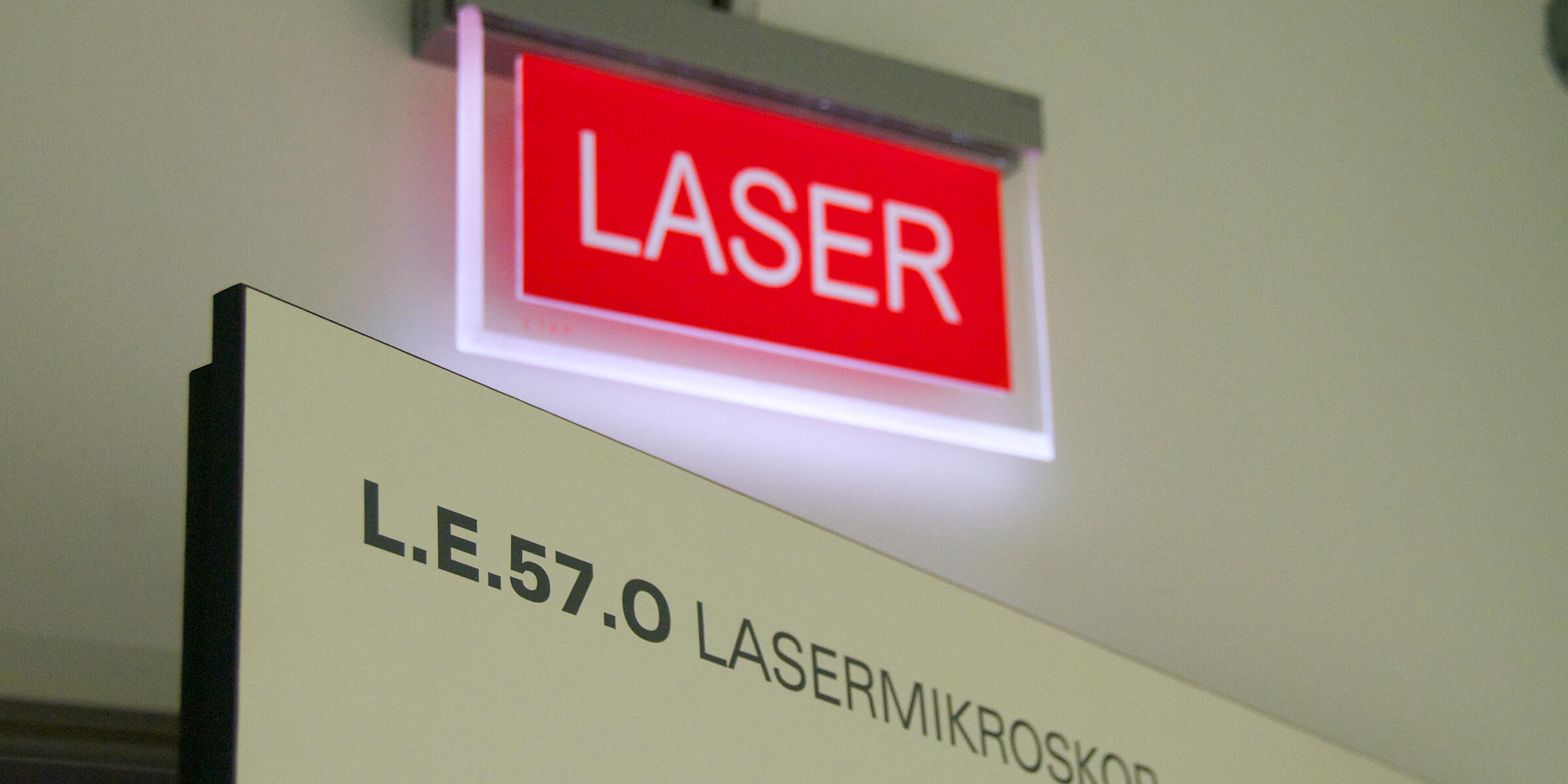
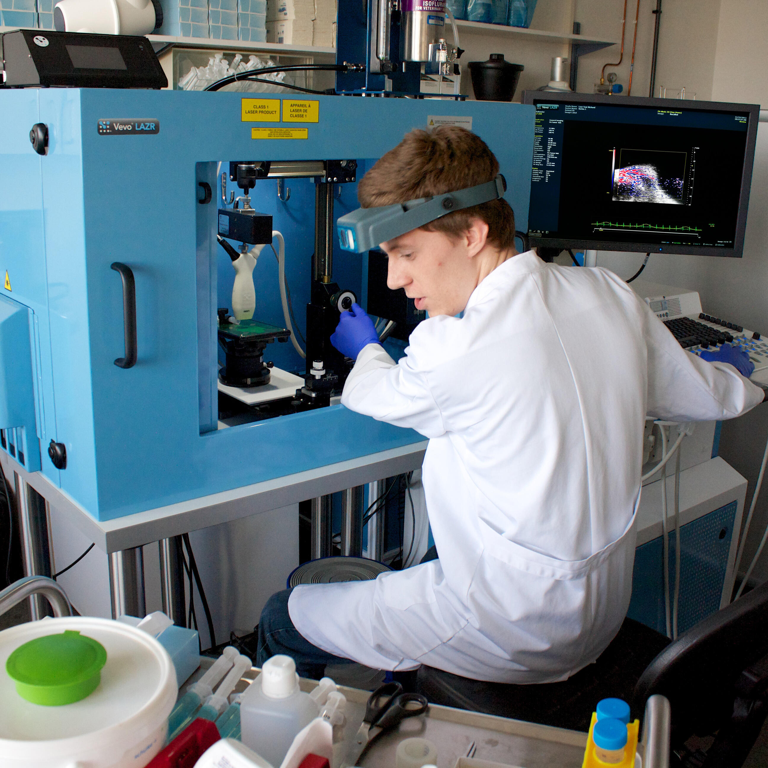
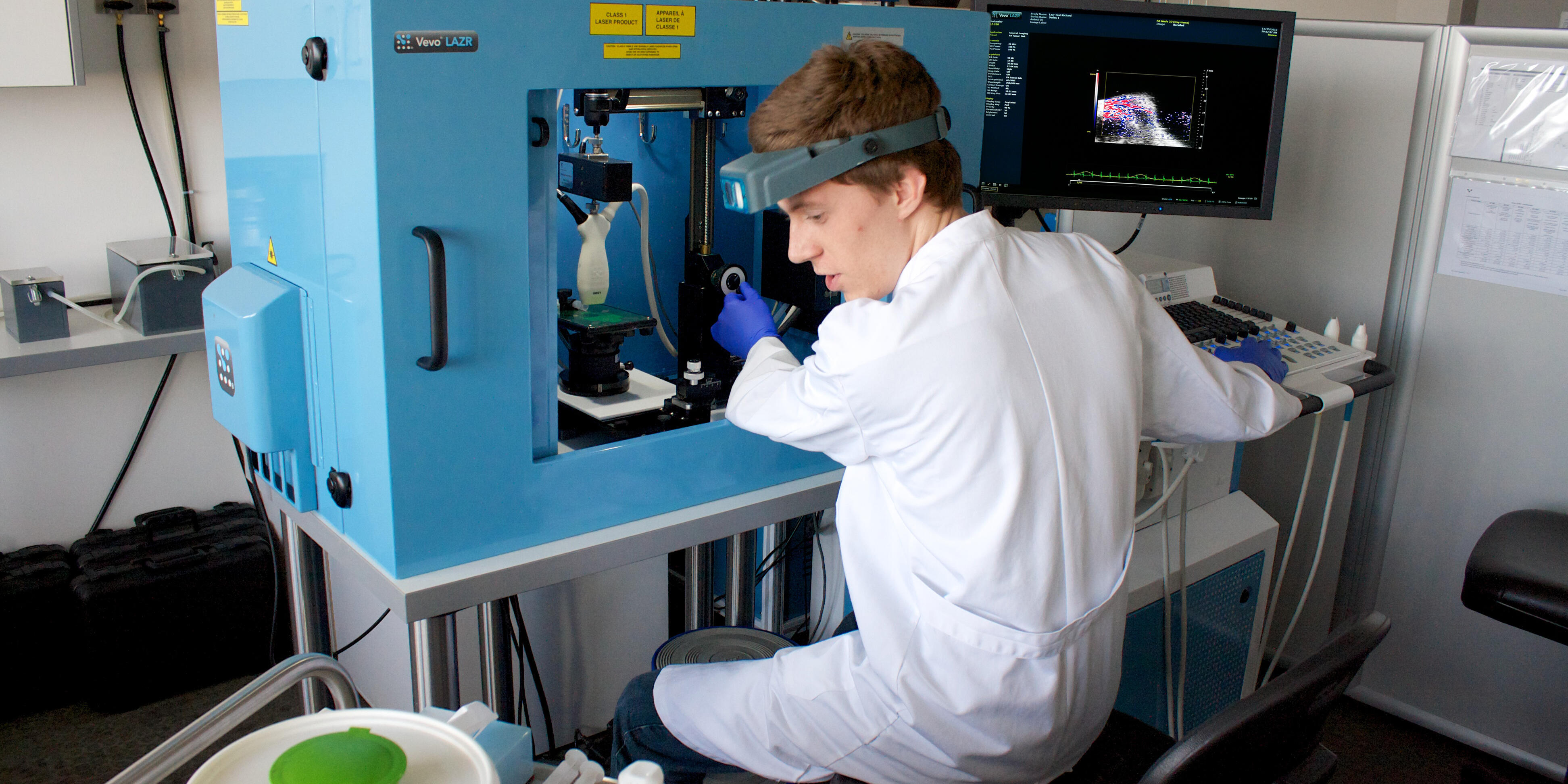
Just recently the Cells-in-Motion Cluster of Excellence (CiM) obtained a new device for 'photoacoustic imaging'. Prof. Michael Schäfers from the team of CiM coordinators explains in an interview with Christina Heimken why this prototype is especially important.