
“Already as a child I knew I wanted to be a scientist”
Prof. Sorokin, what research topic are you working on?
In my research group we focus on the extracellular matrix – these are protein structures that are secreted by cells and surround cells in tissues. Importantly, these structures not only form a tissue scaffold, but have a decisive influence on cellular functions. In the past ten years, there has been increasing awareness that the molecular and mechanical signals that stem from the extracellular matrix act in a Yin-Yang manner – meaning as opposing and complementary principles – to define tissue form and function.
We investigate these signals, focusing on a specialised extracellular matrix, the basement membrane. It is a thin two-dimensional sheet, rather like a sheet of paper but of course thousands of times thinner, that constitutes an integral part of all cellular barriers in the body. It acts to separate different tissue compartments in organs from each other. The outer wall of a capillary or small vessel, for example, is a basement membrane. It is connected to the endothelial cell layer – which forms the inner blood vessel wall – and separates it from the underlying tissue. When a tissue is inflamed, immune cells in the blood migrate through the endothelium and basement membrane to enter the tissue in order to combat the inflammation. We have already contributed significant findings showing how the basement membrane influences the behaviour of the immune cells and endothelial cells. We study these processes in mice and concentrate mainly on inflammation in the brain in order to find out more about the autoimmune disorder known as multiple sclerosis. Moreover, we also want to gain knowledge about how these inflammatory processes differ from those occurring in other organs.
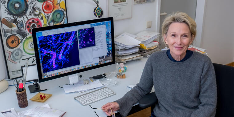
Video: Microscopy images of immune cells (pink) in the organism of a mouse that are migrating from the blood vessel lumen into the tissue.
© Jian Song, Lydia Sorokin
What inspires you during your daily work?
I am always excited by looking at cellular processes live. Using our intravital microscope, we can observe every step in the immune cell migration process and see how the cells change their shape as they migrate on and between the endothelial cells, and how they stay between the endothelial cell layer (in the video in green) and the basement membrane (not visible in the video) for quite a long time before pushing through the basement membrane into the tissue.
I also find it very exciting to try to simulate these situations in artificial tissue models in order to focus on specific steps. We have observed, for example, that immune cells migrating along the basement membrane beneath the endothelium push the endothelial cell layer upwards, towards the lumen, in a wave-like motion (as visible in the video) while the basement membrane does not deform – suggesting that spatially directed mechanical signals must be involved. We would never have realised this if we hadn’t seen it in vivo – in the living organism. If we could now mimic the situation in vitro such that the cells feel as if they were in the organism, we could analyse it in detail. However, this is a challenge because the basement membrane alone consists of 25 different components that have to be brought together in the right quantity and spatial arrangement.
What characterizes you personally as a scientist?
I think my main characteristics are my enthusiasm and my energy. At least that’s what I often hear after my presentations. I think people notice that I put my whole heart into my work. Already as a child I knew I wanted to be a scientist. I grew up in Western Australia where I had a lot of contact with nature and together with my father collected frogs and insects, for example. For a while, when walking to the institute in the morning, I saw two kingfishers that were nesting on the bank of the shore of the castle moat here in Münster. Things like that make me happy – I admire nature.
How has your work as a scientist changed during the Corona pandemic?
During the Corona pandemic, new ways have emerged to very efficiently inform oneself and to exchange knowledge about new developments within the international expert community. The icing on the cake for me are the digital “Global Immunotalks” every Wednesday afternoon – a friend from Sweden who is an immunologist gave me the tip. Digital lecture and meeting formats have also emerged in the field of vascular biology, offering opportunities to interact with colleagues via chat or in virtual rooms, for example. People are willing to share new things in these online formats that haven’t necessarily already been published. It keeps one up to date, and it does connect you – you wouldn’t otherwise see colleagues from the US so often, for example, or have the possibility of hearing talks given by very prominent scientists.
I also have the feeling that formats where you can see and listen are perhaps more accessible to younger people than pure reading material. Some content is permanently available as videos on the web. I also look through the tweets from such platforms every day, mainly while having breakfast, you’re quickly up to date – it’s really great. At our own events, too, we notice that we have more listeners from beyond Münster and Germany than before. For some time now, my team and I have been regularly exchanging information online with another working group here at the University of Münster, specifically on the topic of macrophages. Word of mouth got around and now groups from Bonn, France and even Israel are also taking part.