Cell dynamics and imaging

Short profile of our research

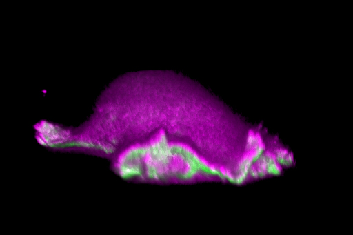
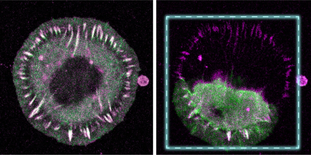
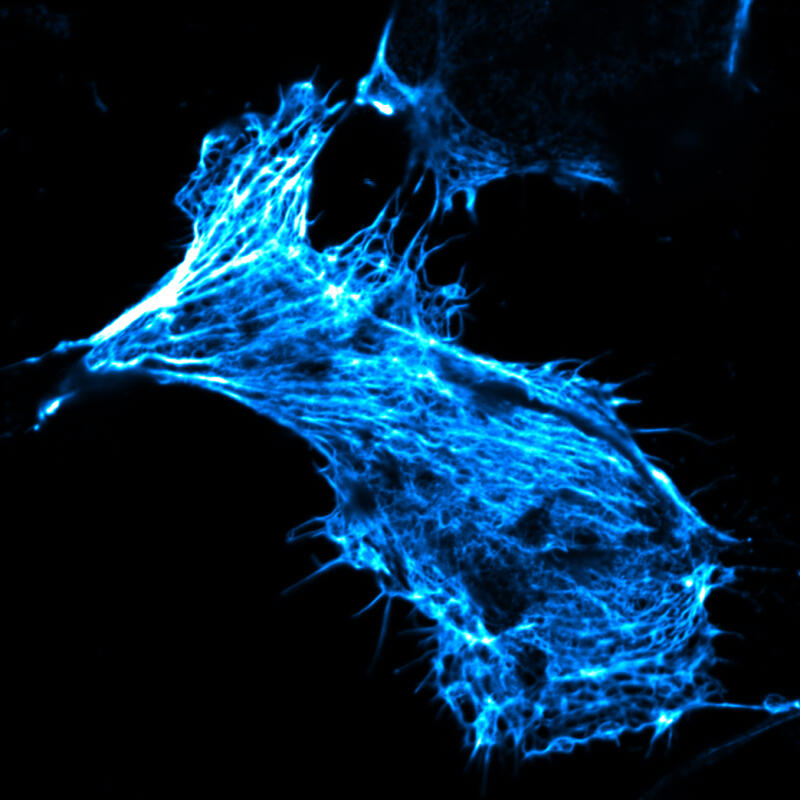
Members of our research network investigate how cells move and behave in organisms. To make processes in the body visible and to be able to analyse them, they employ and develop innovative imaging methods. Scientists from medicine, biology, chemistry, pharmacy, mathematics, computer science and physics work closely together in this field.

Our network is the centrepiece of the University of Münster’s research profile area “cell dynamics, inflammation and imaging”. We bring together researchers from various faculties working in this field and promote their cooperation, thus being an incubator for new interdisciplinary questions that contribute to further developing the research focus.

  • Cells in motion

    EXAMPLES

    A fibroblast from a mouse moves forward using a “lamellipodium”. Researchers have discovered that curvatures at the beginning of the lamellipodium trigger a self-organising system. As a result, cells can move in the same direction over a longer distance, forming search patterns. Visualisation using lattice light sheet fluorescence microscopy. Begemann I et al./Nature Physics 2019.
    A fibroblast from a mouse moves forward using a “lamellipodium”. Researchers have discovered that curvatures at the beginning of the lamellipodium trigger a self-organising system. As a result, cells can move in the same direction over a longer distance, forming search patterns. Visualisation using lattice light sheet fluorescence microscopy. Begemann I et al./Nature Physics 2019.
    © Isabell Begemann, Milos Galic
    • Fluorescence microscopy image of a connective tissue cell before and after exposure to mechanical forces. At first, the cell forms normal adhesion structures, which are important for attachment to the surrounding tissue (left). Irradiation with short laser pulses leads to a rupture of connections of the adhesion protein talin, without which the cell can no longer hold on to the underlying substrate and collapses (right). Researchers developed this method in which individual proteins can be broken with a high degree of temporal and spatial control to examine their mechanical role. Sadhanasatish T et al./Science Advances 2023.
      © AG Grashoff
    • Fluorescence microscopy image of epithelial cells enclosing a developing egg cell in the ovary of a fruit fly. Scientists found that, at the points where the membranes (pink) of three cells meet each other, the cells loosen their connections in an orderly way, thus allowing yolk-forming proteins to be transported through the intercellular spaces (green) into the egg. The results help understand how cell connections are dynamically remodelled and how, in this way, cells form barriers which protect tissue and control transport processes. Isasti-Sanchez J et al./Developmental Cell 2021.
      © Isasti-Sanchez et al./Dev Cell 2021
    • Optical cross-section through the brain of a fruit fly. A team of researchers has shown that, in addition to the already known blood-brain barrier, there is a second barrier in the brain of fruit flies. Here glial cells, too, ensure a spatial separation of different functional compartments, which becomes clear in the image. The green colouring indicates the distribution of the extracellular matrix protein "Trol", which is formed by all enveloping glial cells. Pogodalla N et al./Nature Communications 2021.
      © Nicole Pogodalla, Christian Klämbt
    • Scientists have succeeded in designing a synthetic hydrogel in which endothelial cells (pink cell nuclei) form new blood vessels that grow from a parent blood vessel (upright on the left). Here, the vessels were perfused with a liquid containing fluorescent beads (yellow). Using fluorescence microscopy the scientists showed that the beads flowed into the new vessels at a natural speed and that these actually formed cavities connected to the parent vessel. Liu et al./Nature Communications 2021.
      © Liu et al./Nat Comm 2021
    • Platelets (red) interacting with certain white blood cells – the regulatory T cells (yellow) – in the bacterially inflamed lung tissue of a mouse. Researchers found that these interactions play a significant role in resolving the inflammation. Confocal fluorescence microscopy. Rossaint et al./Journal of Experimental Medicine 2021.
      © Rossaint et al./JExpMed 2021
    • Which molecular mechanisms are at work when, in the case of inflammation, immune cells migrate from the blood vessel into the tissue? Researchers have found that the laminin 511 protein (red) inhibits the migration of immune cells (green) through the endothelial cell layer (blue) of the blood vessels. Immune cells migrate preferentially into the tissue at sites of low or no laminin expression (arrows). Intravital microscopy. Song et al./Cell Reports 2017.
      © Song et al./Cell Reports 2017

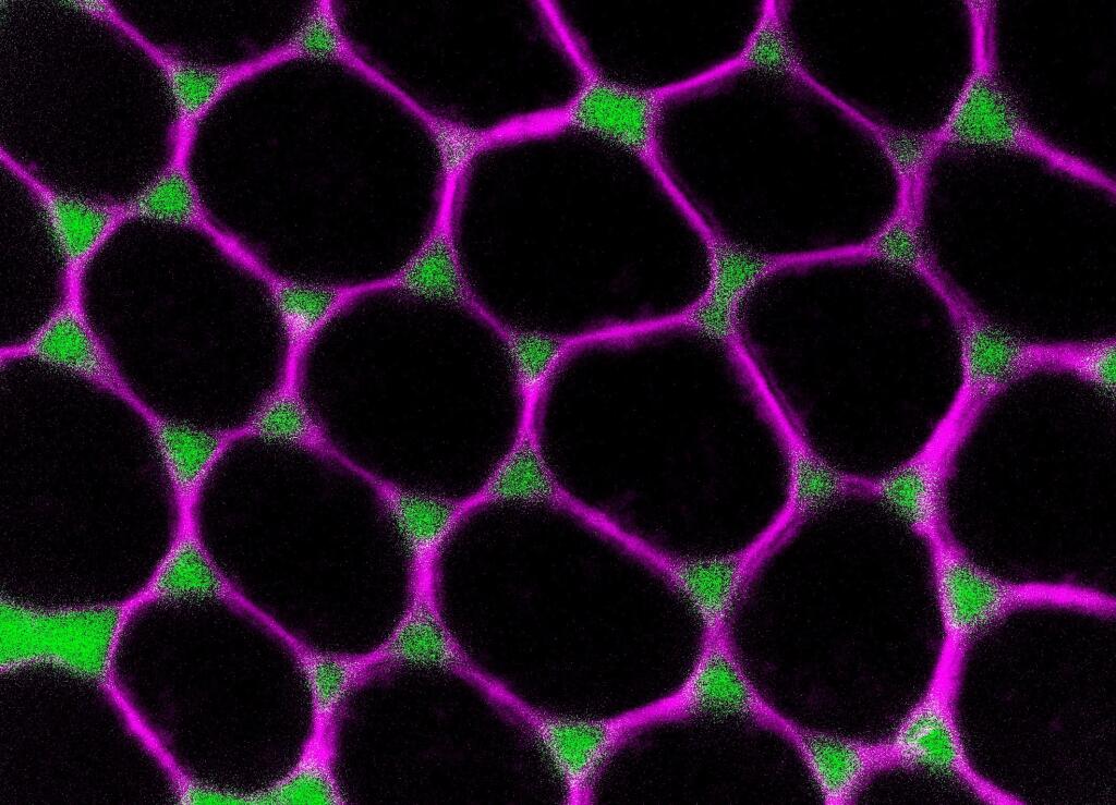
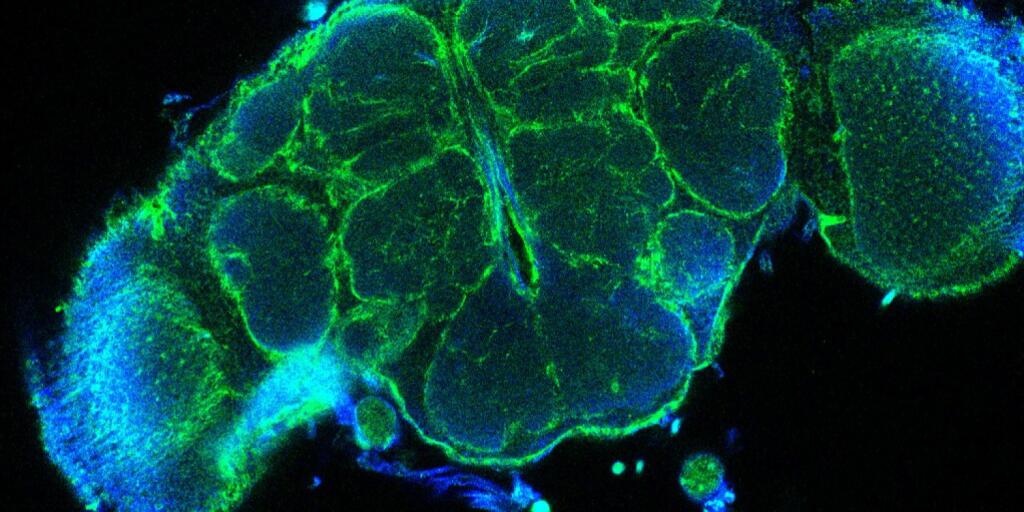
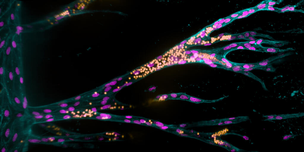
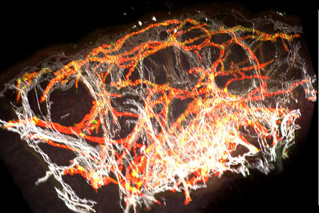
    Every process within every organism involves molecules and cells and their interactions. In order to understand these dynamic processes, we study not only relationships between the individual components within a cell but also interactions between cells. We investigate which biochemical and biophysical properties of a cell influence its behaviour, how these properties are determined by genes and how the dynamic cellular processes in an organism remain in healthy balance, which is called homeostasis. We concentrate mainly on cellular processes that occur in blood and lymphatic vessels and their impact on organ function – not only during vessel development but also when they are fully formed in the mature organism.

    By learning more about the molecular mechanisms that govern normal organ function in a healthy organism, we are able to draw conclusions as to what happens in the body in different diseases and analyse how healthy cellular systems can develop into diseased systems. Here, we are especially interested in inflammation, during which immune cells migrate out of blood vessels into tissues to fight infections or to repair tissue damage. However, inflammatory reactions may vary depending on the organ and the vessel type in which they occur, and in autoimmune diseases they are falsely directed against cells of the own organism. Studying mechanisms that explain these different scenarios is a focus of our research.

  • Innovative imaging

    EXAMPLES

    Distribution of immune cells (red) in the body of a mouse with an inflammatory skin disease, visualised using fluorescence reflectance imaging. Researchers developed a method enabling them to better evaluate and study the activity of these inflammatory cells: they succeeded in genetically modifying precursors of immune cells, then increasing their numbers in a test tube and finally tracking them spatially and temporally in living organisms. Gran S, Honold L et al./Theranostics 2018.
    Distribution of immune cells (red) in the body of a mouse with an inflammatory skin disease, visualised using fluorescence reflectance imaging. Researchers developed a method enabling them to better evaluate and study the activity of these inflammatory cells: they succeeded in genetically modifying precursors of immune cells, then increasing their numbers in a test tube and finally tracking them spatially and temporally in living organisms. Gran S, Honold L et al./Theranostics 2018.
    © S. Gran & L. Honold et al./Theranostics
    • Positron emission tomography (PET) imaging of tumours (dashed circles) in the body of a mouse (right in cross-section). Researchers used a newly developed radioactive substrate to label tumour cells in a living organism. Cells genetically modified to produce a so-called SNAP-tag enzyme took up the radioactive marker (orange), while cells without this enzyme did not. The research team thus adapted the SNAP-tag technology which is used for cell labelling in microscopy for use in whole-body imaging with PET for the first time. This opens up the prospect of examining cells with different imaging techniques and at different temporal stages. Depke DA, Konken CP, Rösner L et al./Chemical Communications 2021.
      © Depke et al.
    • Microscope image of a living human cancer cell in culture. Its actin cytoskeleton has been stained with fluorescently labelled phalloidin. Scientists delivered this toxic substance into the cell using newly developed nanocontainers. These containers made of sugar and protein components are taken up by cells through natural processes and can thereby transport substances that normally cannot penetrate the cell membrane – such as drugs or labelled substances for the investigation of cell functions – into cells. Kudruk S, Pottanam Chali S et al./Advanced Science 2021.
      © Kudruk & Pottanam Chali et al./Adv Sci 2021 (modified colours)
    • Using newly developed software, researchers digitally merged microscopic images of hundreds of zebrafish embryos. This enabled them to identify patterns in how primordial germ cells behave in the absence of the attractant receptor Cxr4b. They found that tissue along the longitudinal axis of the embryos acts as a physical barrier, influencing the direction of cell movement and hindering the cells from migrating through the tissue. Gross-Thebing, Truszkowski, Tenbrinck et al./Sci Adv 2020.
      © Gross-Thebing, Truszkowski, Tenbrinck et al. Sci Adv 2020;6: eabc5546/CC BY-NC
    • 3D computer reconstruction of a healthy human skin biobsy. Spatial arrangement of blood vessels (white) and lymphatic vessels (red) is distinctly visible. The visualisation is based on lightsheet microscopy images and was created using a newly developed method, which enables scientists to analyse changes of the blood and lymphatic vessels in lymphoedema in a more detailed way. Hägerling R et al./JCI Insight 2017.
      © JCI Insight

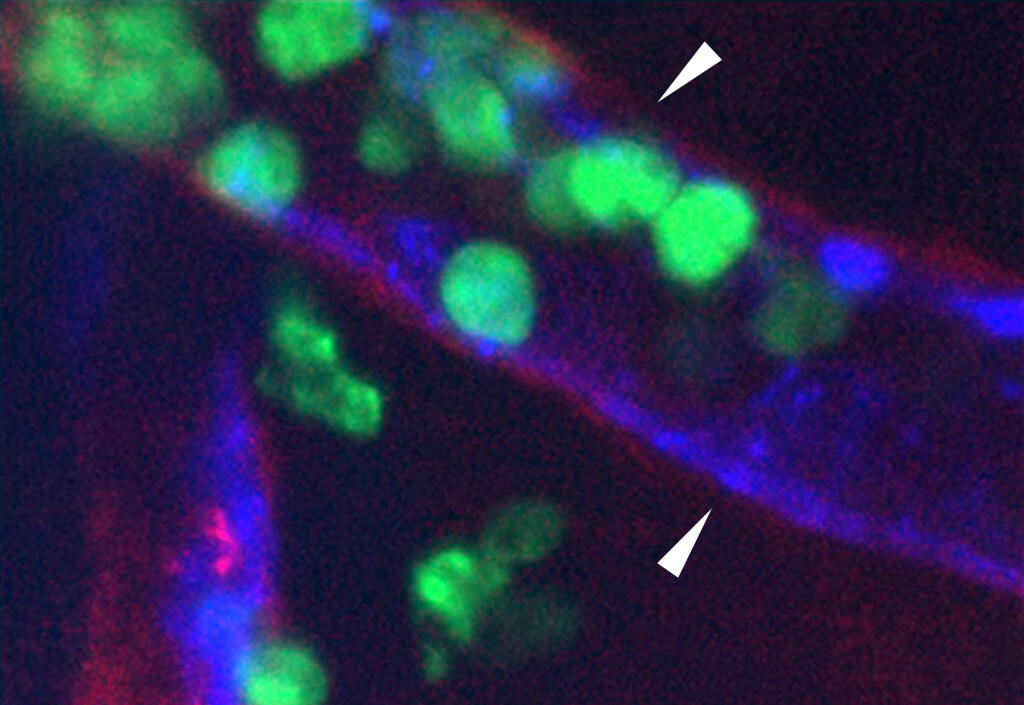
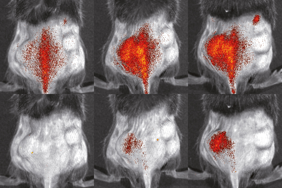
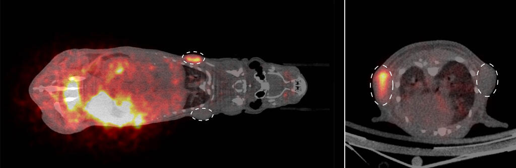
    To address biomedical questions, we systematically employ imaging methods to see cells and molecules in tissues and organs. These range from high-resolution light microscopy to whole-body imaging methods which reveal structures or processes in the entire organism. Commencing with a detailed analysis of the environment within and around cells, our field of view becomes ever broader as we “zoom out” to ask: How does the cell behave in tissues, in organs and in the entire organism? To bridge these questions and different spatial dimensions, we pursue a unique strategy in our science network: We develop chemical and mathematical methods that can be employed in different imaging methods with different resolutions. Only in this way is it possible to examine the same cell in various spatial dimensions over time, using a variety of imaging methods. This approach promotes the transfer of methods that are applied to model organisms – for example fruit flies, zebrafish and mice – to clinical imaging methods for patient diagnosis.

  • Interdisciplinary research

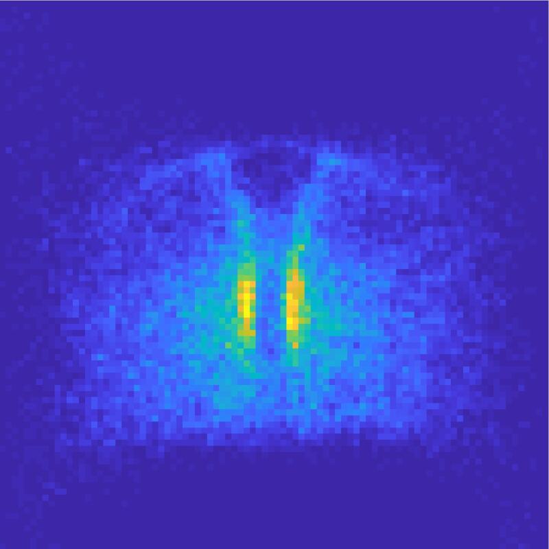
    In our scientific field, researchers from medicine, biology, chemistry, pharmacy, physics, mathematics and computer science work together. Research questions and results from the different disciplines drive and advance each other: Biologists and medical researchers, for example, identify molecules critical for specific cellular processes, and chemists develop new substances and signalers that bind to such molecules or cells to make them visible using imaging methods. Physicists develop detectors that capture specific signals for biomedical imaging as well as new measurement techniques that can analyse biophysical parameters, such as forces acting between cells. Mathematicians and computer scientists develop algorithms, some of which use artificial intelligence – so-called machine learning – to process and analyse the resulting large quantities of complex data. In this way, a computer may recognize specific patterns of cell behaviour in many different tissues that are not otherwise visible; thereby, biomedical questions can be answered and, at the same time, new research directions are identified.

    When researchers with different areas of expertise and approaches interact, this promotes scientific creativity and enables decisive progress. Our research network’s combination of scientific and clinical perspectives also facilitates the transfer of basic research results into clinical applications.