Researchers at the Cells-in-Motion Cluster of Excellence make ongoing inflammation in the human brain visible
Research from cells to patients / Findings published in "Science Translational Medicine"
The ultimate aim in biomedical research is the transfer of results from experiments carried out in animals to patients. Researchers at the Cells-in-Motion Cluster of Excellence (CiM) at the University of Münster have succeeded in doing so. For the first time, they have been able to image ongoing inflammation in the brain of patients suffering from multiple sclerosis (MS). This involved specialists from different disciplines working together in a unique way over several years, combining immunology, neurology and imaging technologies ranging from microscopy to whole-body imaging.
The consequences of an inflammation in the brain can already be shown using a clinically established process: magnetic resonance imaging (MRI). Making the inflammation itself visible too could, in future, help not only to more accurately diagnose multiple sclerosis patients but also to monitor therapies and apply them in a more targeted way. The study has been published in the prestigious journal "Science Translational Medicine".
Publication: Gerwien H*, Hermann S*, Zhang X, Korpos E, Song J, Kopka K, Faust A, Wenning C, Gross CC, Honold L, Melzer N, Opdenakker G, Wiendl H, Schäfers M*, Sorokin L*. Imaging Matrix Metalloproteinase Activity in Multiple Sclerosis as a Specific Marker of Leukocyte Penetration of the Blood-Brain Barrier. Science Translational Medicine, DOI: 10.1126/scitranslmed.aaf8020 (*equal contribution) Abstract
Disease progression in multiple sclerosis is associated with sporadic immunological “flares” that represent immune cell infiltration into the brain and damage. In this autoimmune disease, immune cells, i.e. cells from the body’s own defence system, target the very organism they are supposed to protect and attack the central nervous system. To do this, they have to pass through the blood-brain barrier, i.e. the border between blood vessels and nerve tissue that protects the brain from noxious substances or cells. CiM researchers investigate the neuroinflammation in mice using “experimental autoimmune encephalomyelitis”, a model that shares many similarities with multiple sclerosis in humans.
Several years ago, the group headed by CiM Spokesperson, Prof. Lydia Sorokin, discovered that certain proteins are pivotal in allowing immune cells to pass through the blood-brain barrier. These are the MMP-2 and MMP-9 enzymes, sub-types of the so-called matrix metalloproteinases (MMPs). The researchers have deciphered the function of these enzymes at the blood-brain barrier, showing that MMP-2 and MMP-9 activity are accurate and reliable markers of immune cell penetration of this barrier. They then started investigating methods to visualize this activity and thereby inflammation in the brain.
A New Tracer
The MMP labelling project was carried out together with chemists and nuclear medicine specialists in the working group of CiM Coordinator, Prof. Michael Schäfers. For some time, these scientists had been working on development of a molecular tracer, a chemical substance, that binds to MMPs. “We had synthesized a large number of tracer compounds capable of targeting MMPs and had accumulated a lot of data on these tracers,” explains Dr. Andreas Faust, a chemist. Initially, the tracers were tested in inflammatory vascular diseases such as atherosclerosis; now they wanted to try them out on inflammatory diseases of the central nervous system.
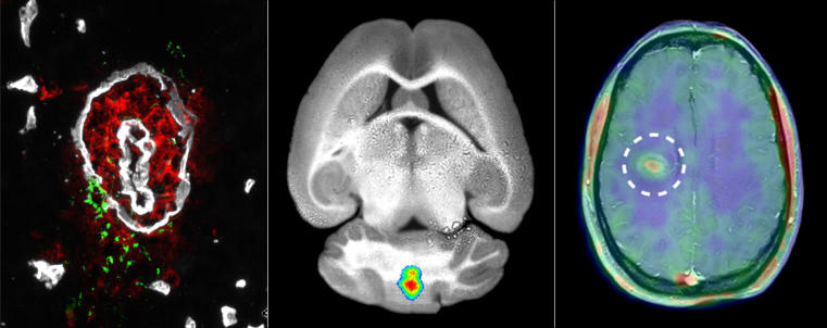
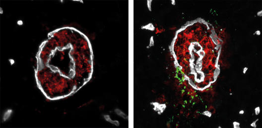
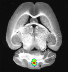
To make the tiny structures in a mouse’s brain visible, the chemists linked the MMP tracer to a fluorescent tag. The fluorescence light signal from such a tracer can be measured using optical imaging techniques. Dr. Hanna Gerwien, who has just completed her PhD in biology, injected the tracer into diseased mice and examined the animals using fluorescence reflectance imaging. This technique visualizes the fluorescent tracers in the near-infrared range in a living organism. The researchers discovered that the tracer accumulated in high concentrations in certain parts of the brain.
To investigate whether the tracer signal correlated with MMP activity at the blood-brain barrier and with immune cell infiltration, Hanna Gerwien removed the brain and examined it under the microscope. The result was that the localization of MMP activity and immune cells matched the fluorescence tracer signal. “We found that our observations of MMP activity provided precise information on where the immune cells penetrate the blood-brain barrier and where inflammation occurs in the brain,” says Hanna Gerwien.
First Case Studies with Patients
Next the researchers wanted to test whether the method could be transferred to humans. However, the fluorescent tracer could not be used because its light signal would not penetrate the skull of a patient. The researchers therefore modified the tracer again, this time adding a radioactive signal transmitter instead of a fluorescent dye. The radiation it emits can be measured and made visible using positron emission tomography (PET).
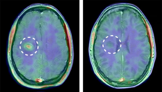
Nuclear medicine specialists and neurologists have now carried out the first case studies on multiple sclerosis patients. “We compared the PET measurements with the MMP tracer to measurements obtained using traditional magnetic resonance imaging,” explains Dr. Nico Melzer, a neurologist at Münster University Hospital.
The results were impressive. The MMP tracer accumulated in the human brain even before any damage to the blood-brain barrier could be detected using the traditional method. “It really is something special to be able to corroborate something in a patient that had been discovered doing basic research in animal experiments,” says Dr. Sven Hermann, an expert in nuclear medicine and small animal imaging, “it’s what every scientist dreams of”. The scientists also observed, as they predicted, that little or no tracer accumulated after the patients had undergone anti-inflammatory therapy.
The investigation described here is a pilot study. This process has not been used in clinical practice. The work was supported by the Cells-in-Motion Cluster of Excellence, the Collaborative Research Centre 656 “Molecular Cardiovascular Imaging” and the Collaborative Research Centre TR-128 “Multiple Sclerosis” at the University of Münster.