
When medicine and computer science join forces
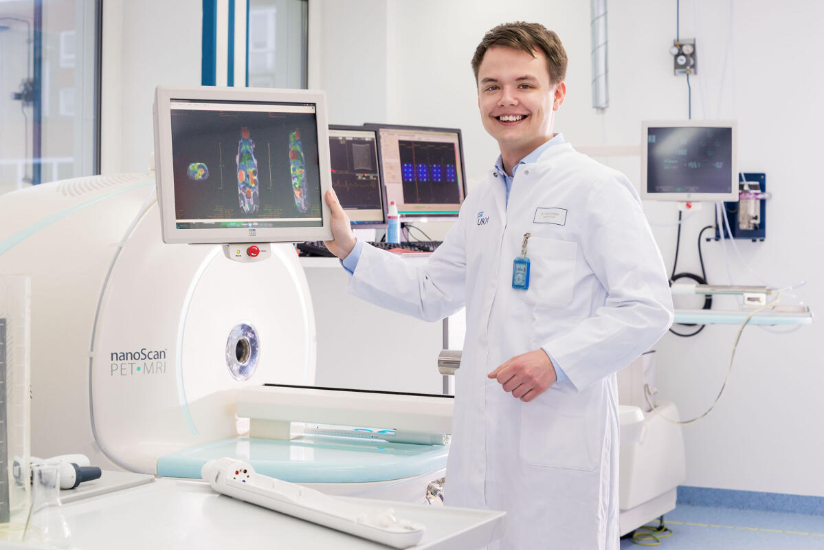
Medicine and Computer Science are two disciplines that many people would not automatically associate with each other. For Dr. Robert Seifert, however, they are, as he puts it, “a perfectly normal combination” and closely interlinked. Robert Seifert is a junior doctor at the Department of Nuclear Medicine at Münster University Hospital, handling images of the body’s interior is part of his daily work. The image data generated there are highly complex, which is why it is often difficult for doctors to understand, in purely visual terms, how severe a particular disease is. It is for this reason that they work together with computer scientists. This year, Robert Seifert received a dissertation prize from the University of Münster for a particularly successful collaboration. He and his colleagues succeeded in developing an algorithm that makes it possible not only to precisely analyse image data, but also in the future perhaps to recognise cardiovascular diseases more accurately and at an earlier stage.
This was the result of years of work. Back in 2012, Robert Seifert was a student of medicine and, under his PhD supervisor Prof. Michael Schäfers at the European Institute for Molecular Imaging at Münster University, he began on preliminary work for his project – initially with imaging experiments on mice. As a chronic inflammation of the blood vessels is often the cause of cardiovascular diseases, the researchers’ fundamental objective is to visualize molecular inflammation processes as well as they possibly can. “The problem, though, is that initially only limited information can be derived from the image produced,” says Robert Seifert. “Our idea was to take a look at the entire image and analyse the pattern of inflammation in the vessels.” He contacted computer scientist Dr. Aaron Scherzinger, who is a member of the research group headed by Prof. Xiaoyi Jiang and who already had some experience in projects with biomedical backgrounds. “We met regularly,” says Robert Seifert, “and worked out how to improve the visualization of vessels. Our aim was to define what we wanted from a medical point of view and to develop a suitable programme code.” This is how, step by step, an algorithm was created which can be used specifically to recognize highly inflamed regions in blood vessels.
First own application for research funding
It all sounds so easy, but the research environment that the researchers were working in helped a lot. The Cells-in-Motion Cluster of Excellence, to which their research groups belong, provided additional funding for a so-called pilot project. This funding line gives junior researchers an opportunity to start engaging in interdisciplinary collaborations and to submit what is often their first own application for research funding. “That was a fascinating experience – especially when we presented our project to the high-level experts of the approvals committee”, Robert Seifert remembers. The physicians and computer scientists used the funding primarily to continue developing their method and also to enable 3D versions of their images.
Since then, the results have already led to two joint publications. And, in addition, initial patient studies have been initiated, some of them in cooperation with the cardiologists at the University Hospital. The aim is to find out whether the new method can help in making the right decision for the treatment of patients who suffer from constrictions of the blood vessels in their legs. At present, it is not always easy for doctors to decide on a suitable treatment. A stent, for example, which keeps a vessel open, could help patients – but it could be counterproductive if there is acute inflammation present. This example shows, as Robert Seifert explains, that “the best case is when our research provides us with answers to the questions we have to deal with in our daily work in the hospital. The two areas – research and practical work as a doctor – are closely related.” Which is why he has already decided that after his training as a medical specialist he wants to continue working as a doctor but, at the same time, carry on with his research and embark on an academic career. “I want to continue working at the interface between biomedicine and computer science”, he says.