
Open lab day for families
Photos
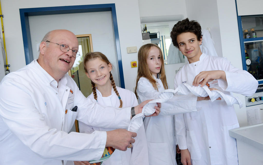
At the Institute of Physiological Chemistry and Pathobiochemistry the young researchers extracted DNA from puréed banana.© CiM - Wilfried Hiegemann 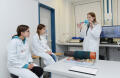
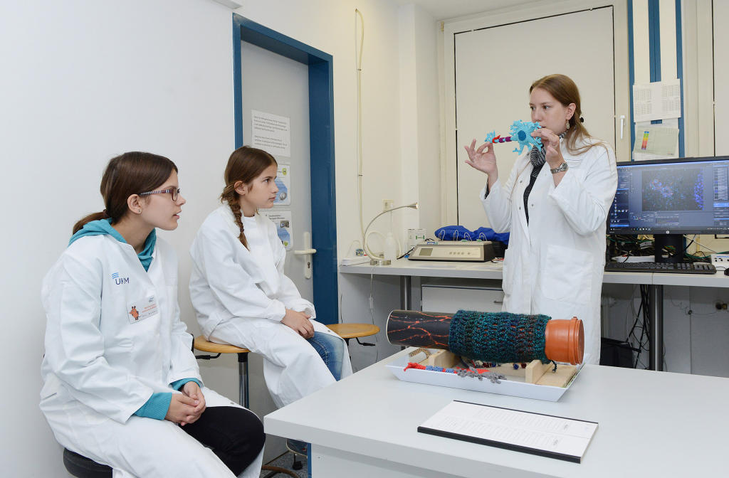
What happens in the autoimmune disease multiple sclerosis? CiM scientists explained it to children using crocheted cells.© CiM - Wilfried Hiegemann 
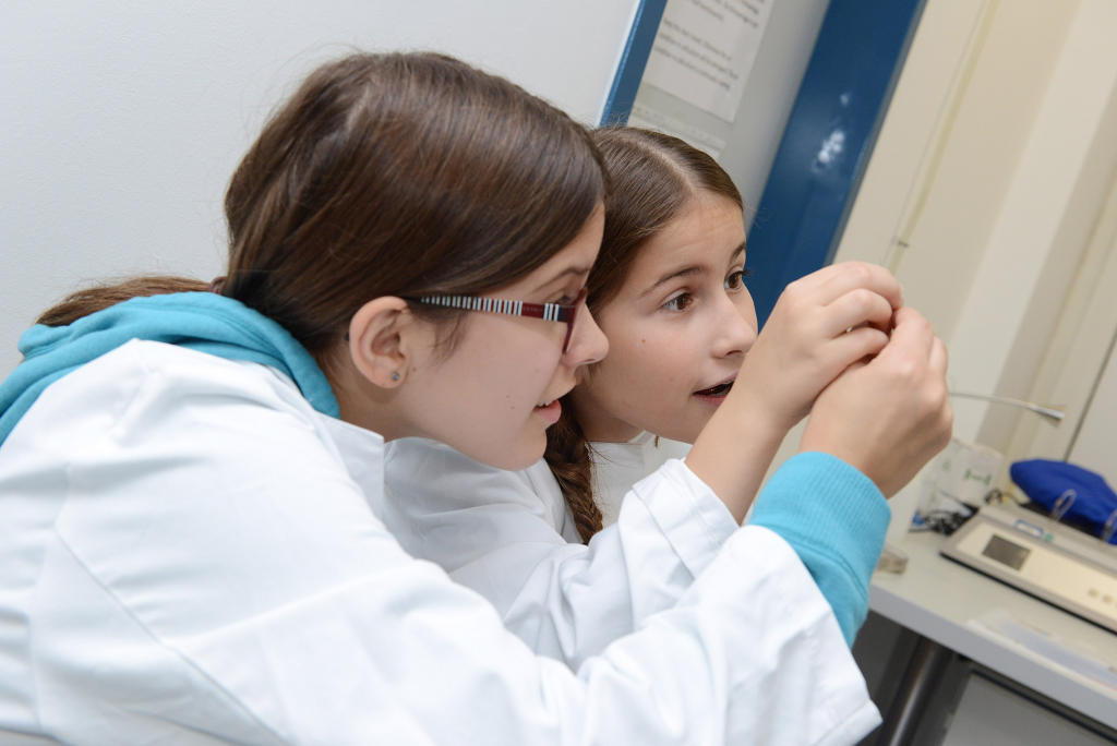
Astonishment at the results of experiments – that’s how fascinating research can be.© CiM - Wilfried Hiegemann 
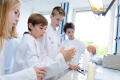
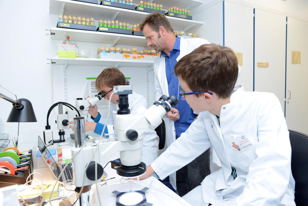
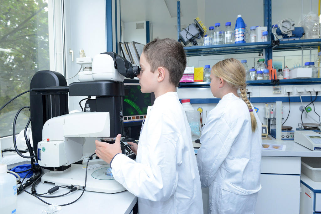
No visit to a laboratory is complete without looking through a microscope.© CiM - Wilfried Hiegemann 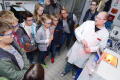
How much light is absorbed by a chemical element? Participants were able to see this at the Institute of Anorganic and Analytical Chemistry.© CiM - Wilfried Hiegemann 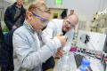
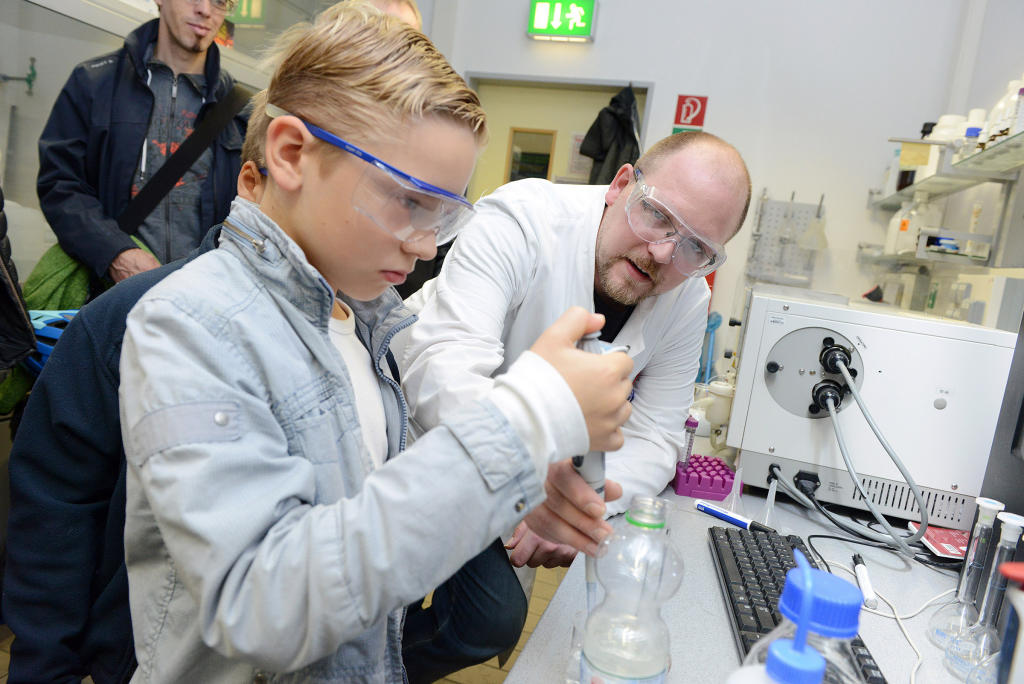
How hard is my tap water? The children were also able to analyse this using chemical methods.© CiM - Wilfried Hiegemann 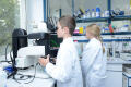
Fly larvae under the microscope. Not something you see every day.© CiM - Wilfried Hiegemann 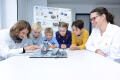
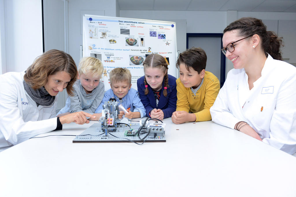
Using an atomic force microscope made of Lego, participants were able to scan the surface of a coin at the Institute of Physiology II.© CiM - Wilfried Hiegemann 
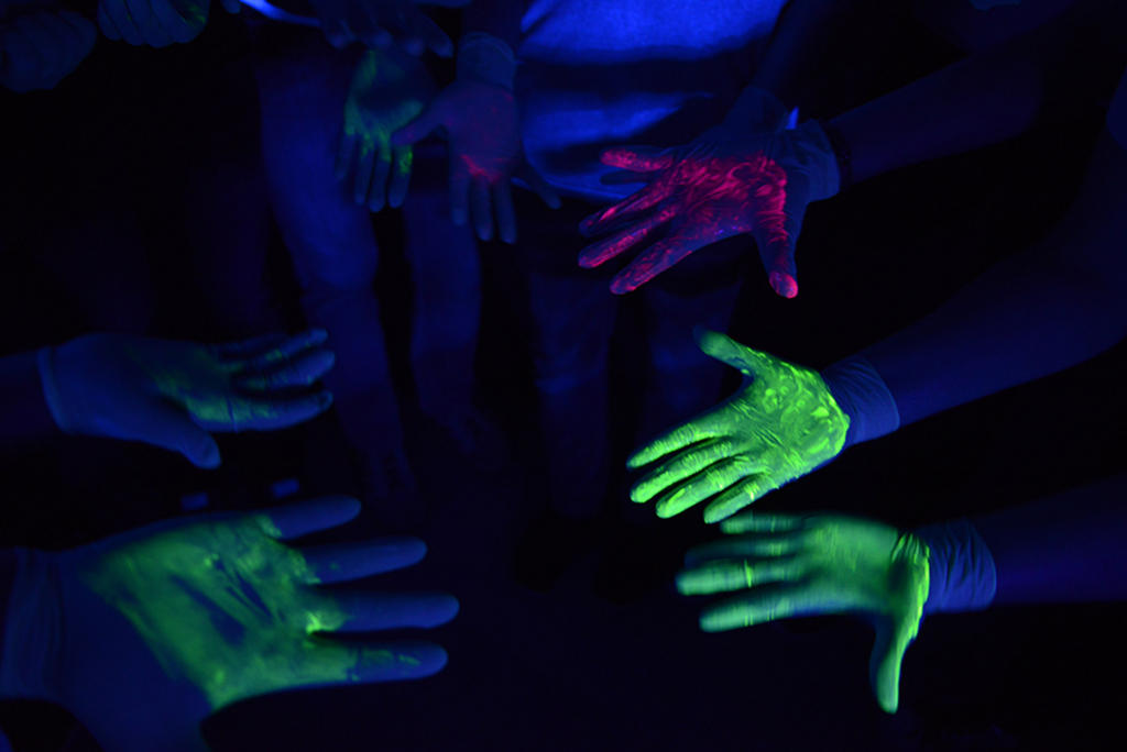
How are germs transmitted by shaking hands? Black light was used to show how, at the Institute of Molecular Virology.© CiM - Wilfried Hiegemann 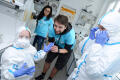
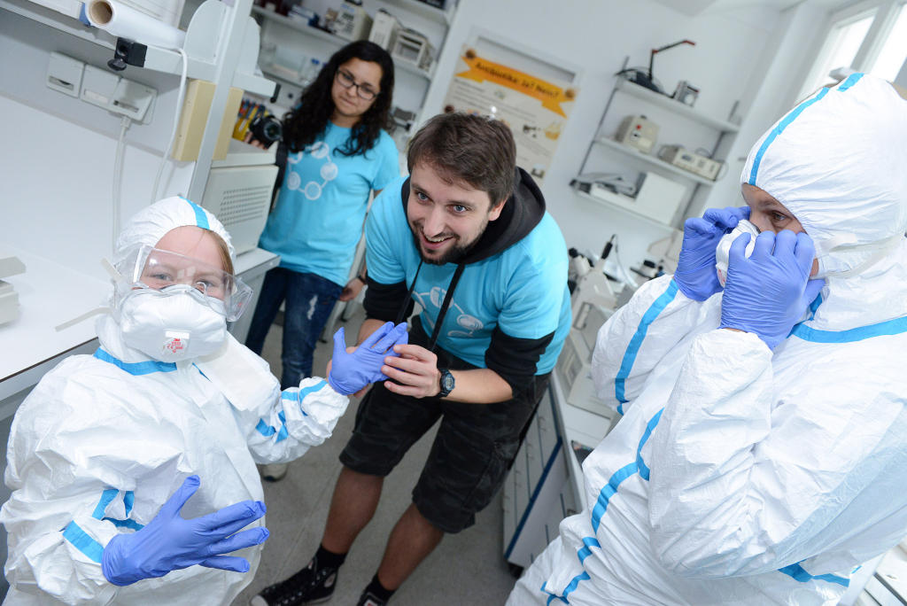
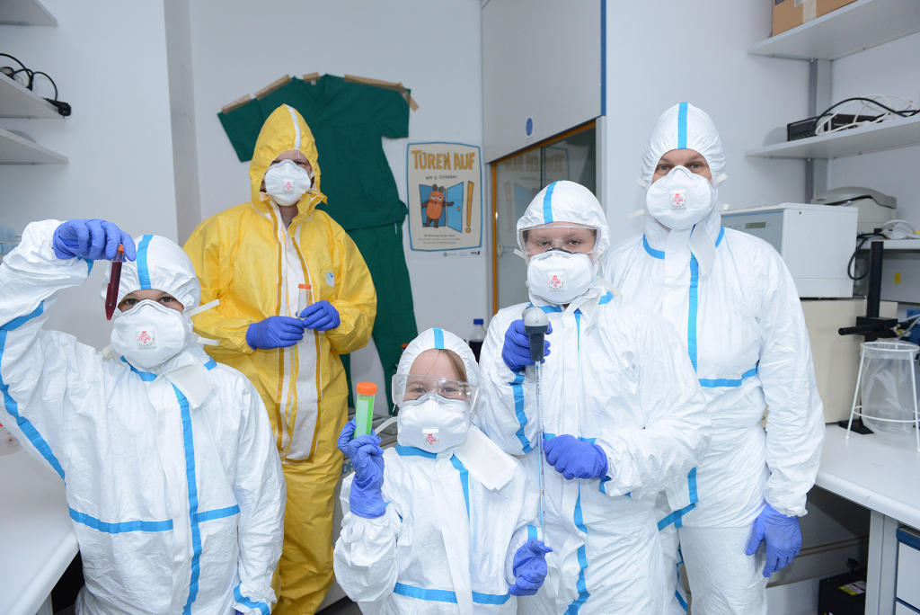
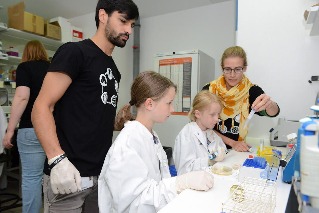
To be a proper virus researcher, of course, protective clothing had to be worn…© CiM - Wilfried Hiegemann 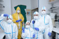
… done! The experiments can now begin.© CiM - Wilfried Hiegemann 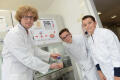
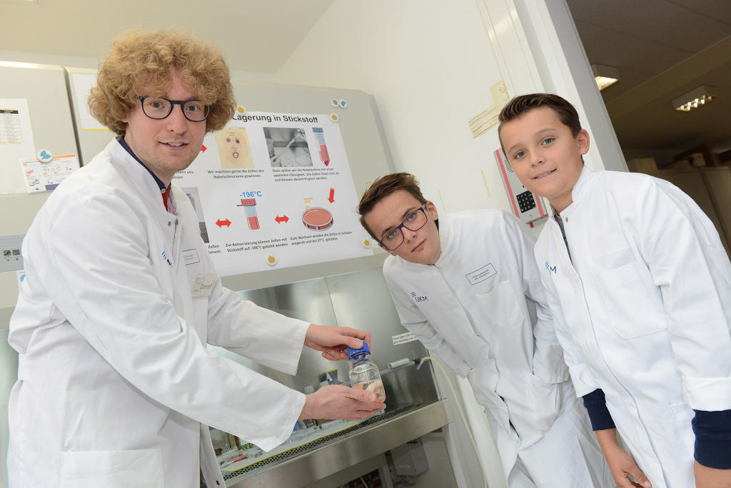
One of the things tomorrow’s scientists saw at the Institute of Medical Biochemistry was a human umbilical cord.© CiM - Wilfried Hiegemann 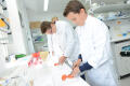
Looks just like in a kitchen – but it’s part of a biochemical experiment: DNA can be extracted from tomatoes.© CiM - Wilfried Hiegemann 
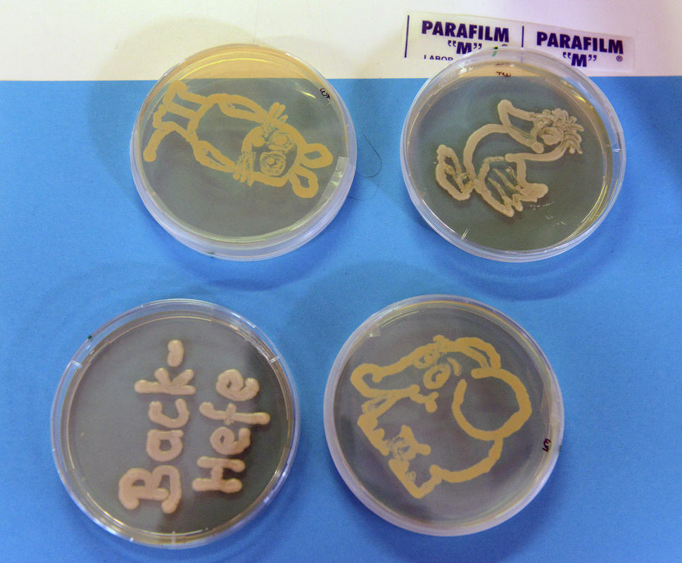
Yeast cultures in petri dishes: at the Institute of Cell Dynamics and Imaging the children were able to “paint” their own cultures…© CiM - Wilfried Hiegemann 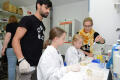
…and scientists from the Institute explained how to do it.© CiM - Wilfried Hiegemann 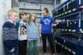
Visiting a fish laboratory: at the Institute of Cell Biology, CiM researchers use zebra fish to study how a body grows and develops.© CiM - Wilfried Hiegemann 
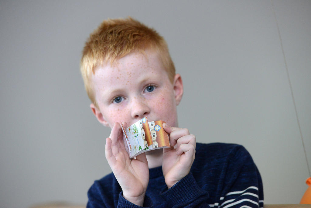
A small souvenir of the Open Day: each child was given a flip book to take home with them.© CiM - Wilfried Hiegemann


Laboratories of the Cells-in-Motion Cluster of Excellence opened their doors for the "Türöffner-Tag" on 3rd October for families. There was a lot to discover, with the young researchers taking a look through microscopes, taking part in chemical experiments or extracting DNA.
Every year, on October 3rd, the children's TV show "Die Sendung mit der Maus" which has been making science fun for almost 45 years promotes a nationwide open day. Several labs from the Cells-in-Motion Cluster of Excellence regularly take part in this event.