
Over a Million Euros of Funding for Interdisciplinary Research
The “Cells in Motion“ (CiM) Cluster of Excellence at Münster University is providing 1.1 million euros of funding for no fewer than twelve new so-called flexible funds projects. These are research projects which established scientists apply for and then jointly implement. What is special about the projects is that the project partners in each case devote themselves to research questions across faculty boundaries, uniting laboratories and clinics from the Departments of Biology, Chemistry, Physics, Mathematics and Medicine. “The range of topics and the creative approaches to the projects are unique in this form,” says immuno-biologist Prof. Klaus Ley from the La Jolla Institute of Allergology and Immunology in the USA, who is one of the external advisors to the Cluster of Excellence. Together with other top international researchers from the fields of natural sciences and medicine, he made a thorough examination of the new project ideas. So far the CiM, with its interdisciplinary orientation, has provided funding for a total of 39 flexible funds projects.
Three examples of projects: Of sperm, nano-capsules and high-performance scanners
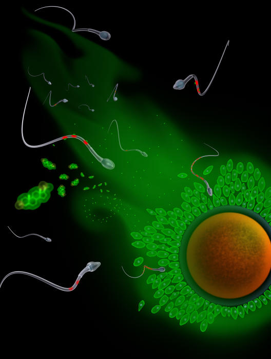
Sperm in a flow channel
In their flexible funds project, biochemist Prof. Timo Strünker and physicist Prof. Carsten Fallnich want to research into a fundamental principle of fertilization. The question they are looking into is: how do sperm find the egg cell? “Sperm are guided by the flow of a fluid which is produced in the ovary and flows off towards the womb,” Timo Strünker explains. “To get to the egg cell, the sperm always flow ‘up-river’, like salmon. We know that the sperm also react to messenger substances which are emitted by the egg cell into the flowing fluid. To understand how sperm actually track down the egg cell we need to decipher the interplay between messenger substances and fluid during the sperm’s navigation.” For this purpose, the scientists use a model consisting of tiny glass tubes in which they can analyse the swimming behaviour of the sperm in a fluid – with and without an egg-cell messenger substance. Using as a basis the analysis of professional swimmers’ techniques, Carsten Fallnich would also like to develop a kind of counter-current system for sperm which would make it possible to hold on to an individual sperm, using optical tweezers. In this way the researchers want to study how the flagellum – the long tail and propeller of the sperm – moves under various conditions.
Docking onto cells with nano-capsules
By contrast, CiM biochemists deal for example with the membranes of cells in our bodies. Cell membranes consist of a patchwork of proteins and lipids, in other words water-insoluble fats. These pass on external signals into the interior of the cell, and thus tell the cells where they should move to. Making lipids visible and undertaking research on them is difficult, though. In another of the flexible funds projects CiM scientists are now aiming to infiltrate lipids marked with fluorescent substances into cells. “From natural hydrocarbon compounds we want to produce tiny nano-capsules which can dock onto certain cells,” says Prof. Bart Jan Ravoo, a chemist. What is special about this is, he says, “that the release mechanism in the nano-capsules only becomes active when a cell has absorbed them. Then for example lipids with fluorescent substances leak out which we can see by means of microscopy.”
More precise diagnosis through hybrid imaging device
In another project a group of doctors and researchers want to use the PET-MRT, which combines two high-resolution imaging procedures, to make a precise, detailed diagnosis of CNS lymphomas in patients. CNS lymphomas are malignant tumours which affect the central nervous system, in other words the brain and the spinal cord. The PET-MRT at Münster University Hospital combines the strengths of positron emission tomography (PET) and magnetic resonance tomography (MRT). “Using the MRT images we can precisely diagnose changes in the tissue,” says Prof. Georg Lenz, who is both a physician and a basic researcher. “Sometimes, however, we cannot assess whether a piece of tissue is still active after a therapy. But by combining it with the PET this will probably be possible. Also, by using the PET-MRT we want to try to assess the individual prognosis of patients affected.”