
How to Build an Organ?
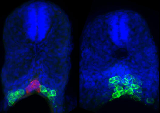
During embryonic development, cells migrate to position themselves in the correct place where they later form an organ. Abnormal cell positioning can lead to pathological consequences such as organ fusion and malfunction. To investigate which factors control progenitor cell positioning, cell biologists Azadeh Paksa and Prof. Erez Raz from the Cells-in-Motion Cluster of Excellence at the University of Münster studied the primordial germ cells in zebrafish embryos, cells that eventually give rise to eggs or sperm. They found that three factors control progenitor cell positioning: chemical cues, physical barriers and cell adhesion. Also involved in the project was an international team of scientists from Israel, France and the USA; the study was published in the journal Nature Communications.
In previous studies, cell biologists from Prof. Erez Raz’s lab in the Center for Molecular Biology of Inflammation focused on the cellular mechanisms that guide progenitor cells to the region where they participate in building the gonad. “The current work deals with events occurring following cell arrival at this region, the position where the cells are maintained and interact with other cell types”, says Azadeh Paksa. “We found that physical barriers and repulsive tissues restrict the movement of the cells away from the developing gonad region. The progenitor cells keep a distance from repulsive tissues, while they come in contact with tissues that function as physical barriers. When cells hit a physical barrier, they migrate in the other direction.” Additionally, a proper level of cell adhesion among the progenitor cells determines cell cluster size and position.”
To make some of the study’s key observations, University of Münster scientists collaborated with different research institutes that contributed advanced imaging techniques. Dr. Philipp Keller’s group from the Howard Hughes Medical Institute (USA) made advanced light sheet microscopy available for the project and assisted in processing the resulting data. Dr. Nadine Peyriéras’s group from the French National Centre for Scientific Research in Paris assisted in two-photon microscopy. Finally, Prof. Nir Gov’s group from the Weizmann Institute of Science (Israel) performed mathematical modelling based on the data acquired in Münster.
In follow-up studies the scientists intend to understand the mechanisms responsible for the observed cell responses, as Azadeh Paksa states. “What happens inside the cells that hit a physical barrier for example, such that they change their migration direction?” The mechanisms that keep cells confined to their normal position are also important in the context of tumor formation; when those mechanisms fail, cancer cells can metastasize. Sometimes, this can happen when cells “disobey” chemical signals or break through physical barriers.